
Table I
The oligonucleotide sequences for the primer, crRNA, and ssDNA reporter.
| Name Sequence (5’-3’) | NoV-GII.2-F1 AGGTGCYAATGCAATAAATCAGAGGGCAGA |
|---|---|
| NoV-GII.2-F2 | CAATGGGCTYAGTTCAYTRATYAATGCAGG |
| NoV-GII.2-R1 | CACCTCTGGCTGCATCAGCRGGGGAAAAGC |
| NoV-GII.2-R2 | TGTTTTATAGCCATCATRTCTGCCTGCAGC |
| Pre-NoV-GII.2-crRNA-F | GAAATTAATACGACTCACTATAGGGTAATTTCTACTAAGTGTAGATAATCATGATAAGGAGATGTT |
| Pre-NoV-GII.2-crRNA-R | AACATCTCCTTATCATGATTATCTACACTTAGTAGAAATTACCCTATAGTGAGTCGTATTAATTTC |
| ssDNA reporter | 6-FAM-TTATTATT-Biotin |
| NoV-GII.2-qPCR-F | GAGGGCAGAATTTGATTTTAATC |
| NoV-GII.2-qPCR-R | CCTTGTTTTATAGCCATCATG |
| NoV-GII.2-qPCR-P | FAM-TTGCCTGAATCTGAGCCTGC-BHQ1 |
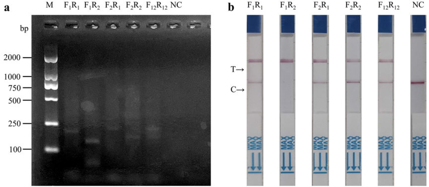
Fig. 1.
Screening of the optimal primer pair for RT-RPA for detection of norovirus GII.2.
a) The amplification efficiency of RT-RPA with various primer combinations was assessed using gel electrophoresis; b) the amplification efficiency of RT-RPA with various primer combinations was evaluated using LFS, driven by the cleavage activity mediated by the complex of Cas12a and the target amplified product. M – DL2000 marker; F1R1 – primer pairs of NoV-GII.2-F1/NoV-GII.2-R1; F1R2 – primer pairs of NoV-GII.2-F1/NoV-GII.2-R2; F2R1 – Primer pairs of NoV-GII.2-F2/NoV-GII.2-R1; F2R2 – primer pairs of NoV-GII.2-F2/NoV-GII.2-R2;
F12R12 – primer pairs of NoV-GII.2-F1/NoV-GII.2-F2 and NoV-GII.2-R1/NoV-GII.2-R2; NC – negative control. In the context of LFS, T denotes the testing line, while C represents the quality control line.
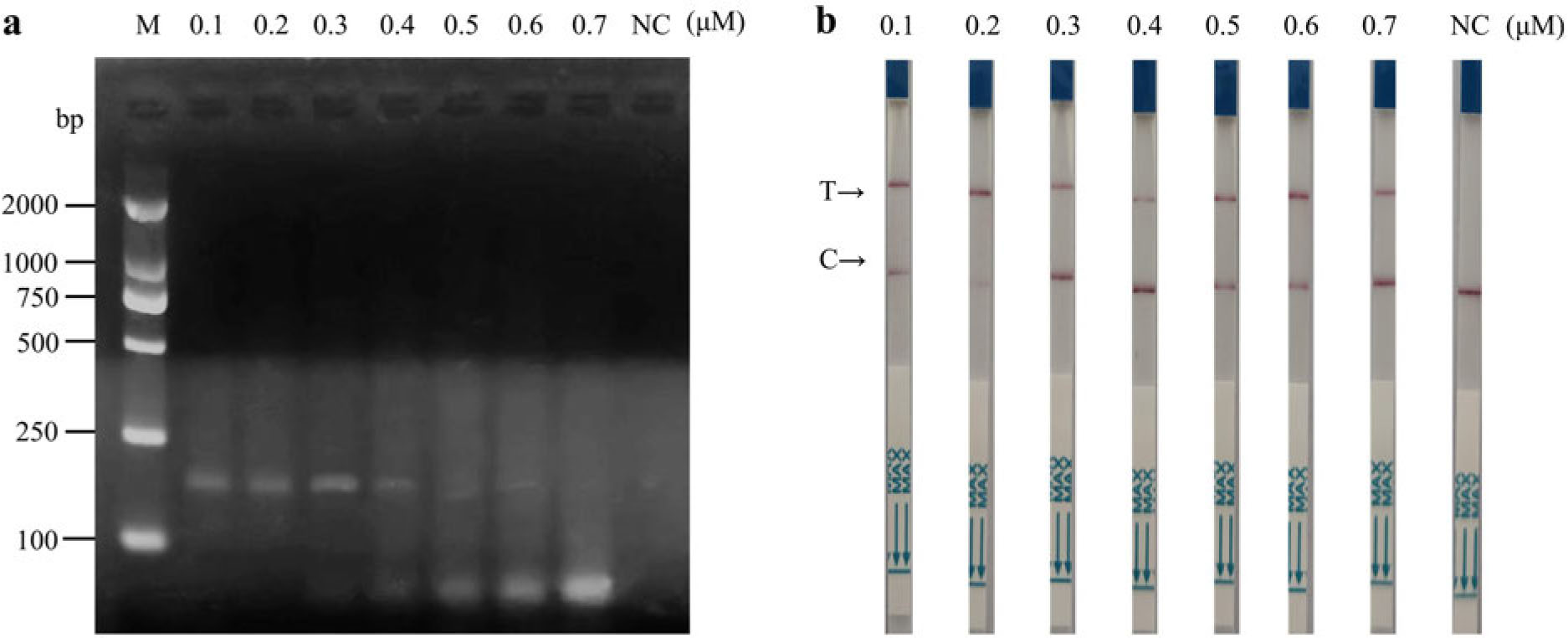
Fig. 2.
Screening of the optimal concentration of the primer pair for RT-RPA-Cas12a-LFS to detect norovirus GII.2.
a) The amplification efficiency of RT-RPA with different primer concentrations was examined using gel electrophoresis;
b) the amplification efficiency of RT-RPA with various primer concentrations was assessed using LFS, facilitated by the cleavage activity mediated by the complex of Cas12a and the target amplified product. In the context of LFS, T denotes the testing line, while C represents the quality control line.
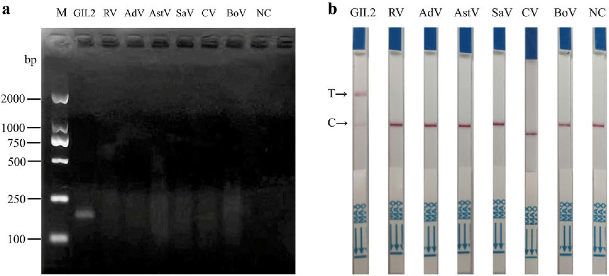
Fig. 3.
Screening of the optimal reaction time of RT-RPA for RT-RPA-Cas12a-LFS to detect norovirus GII.2.
a) The amplification efficiency of RT-RPA with different reaction times was determined using gel electrophoresis;
b) The amplification efficiency of RT-RPA with different reaction times was analyzed using LFS, facilitated by the cleavage activity mediated by the complex of Cas12a and the target amplified product. In the context of LFS, T denotes the testing line, while C represents the quality control line.
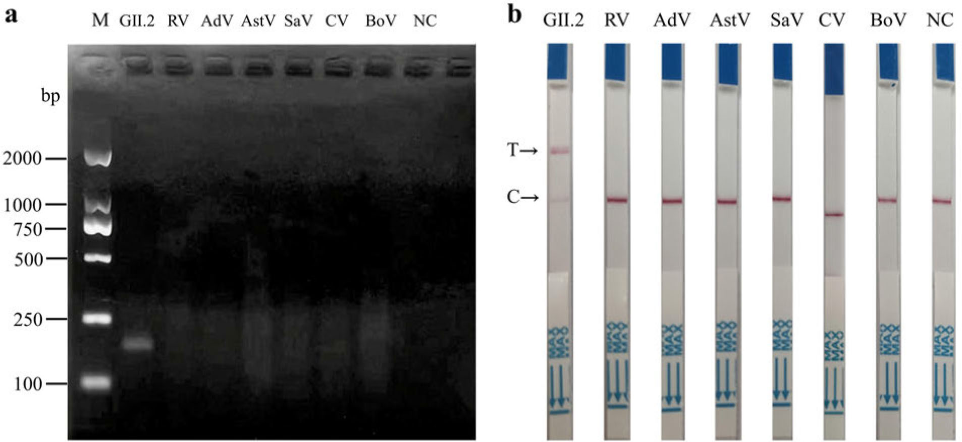
Fig. 4.
The specificity analysis of RT-RPA-Cas12a-LFS with various viral genome samples was assessed using gel electrophoresis (a) and LFS (b). In the context of LFS, T denotes the testing line, while C represents the quality control line.
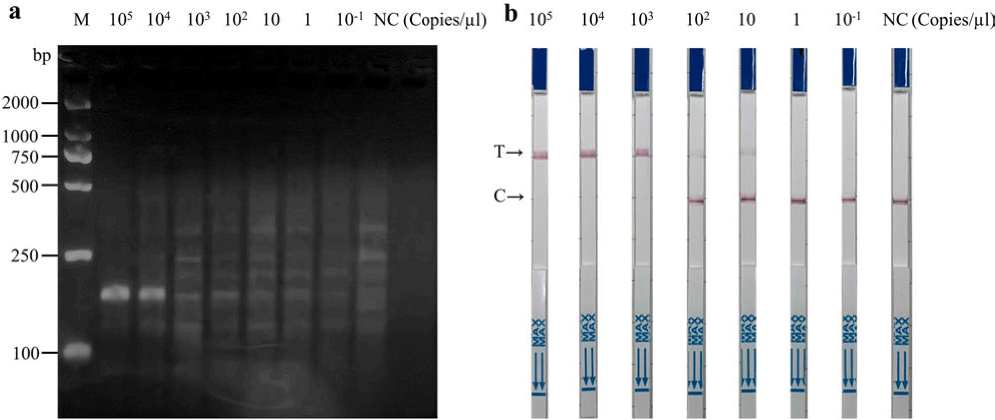
Fig. 5.
The sensitivity analysis of RT-RPA-Cas12a-LFS for detection of norovirus GII.2.
The sensitivity analysis of RT-RPA-Cas12a-LFS with various concentrations of standard RNA was assessed using gel electrophoresis (a) and LFS (b). In the context of LFS, T denotes the testing line, while C represents the quality control line.
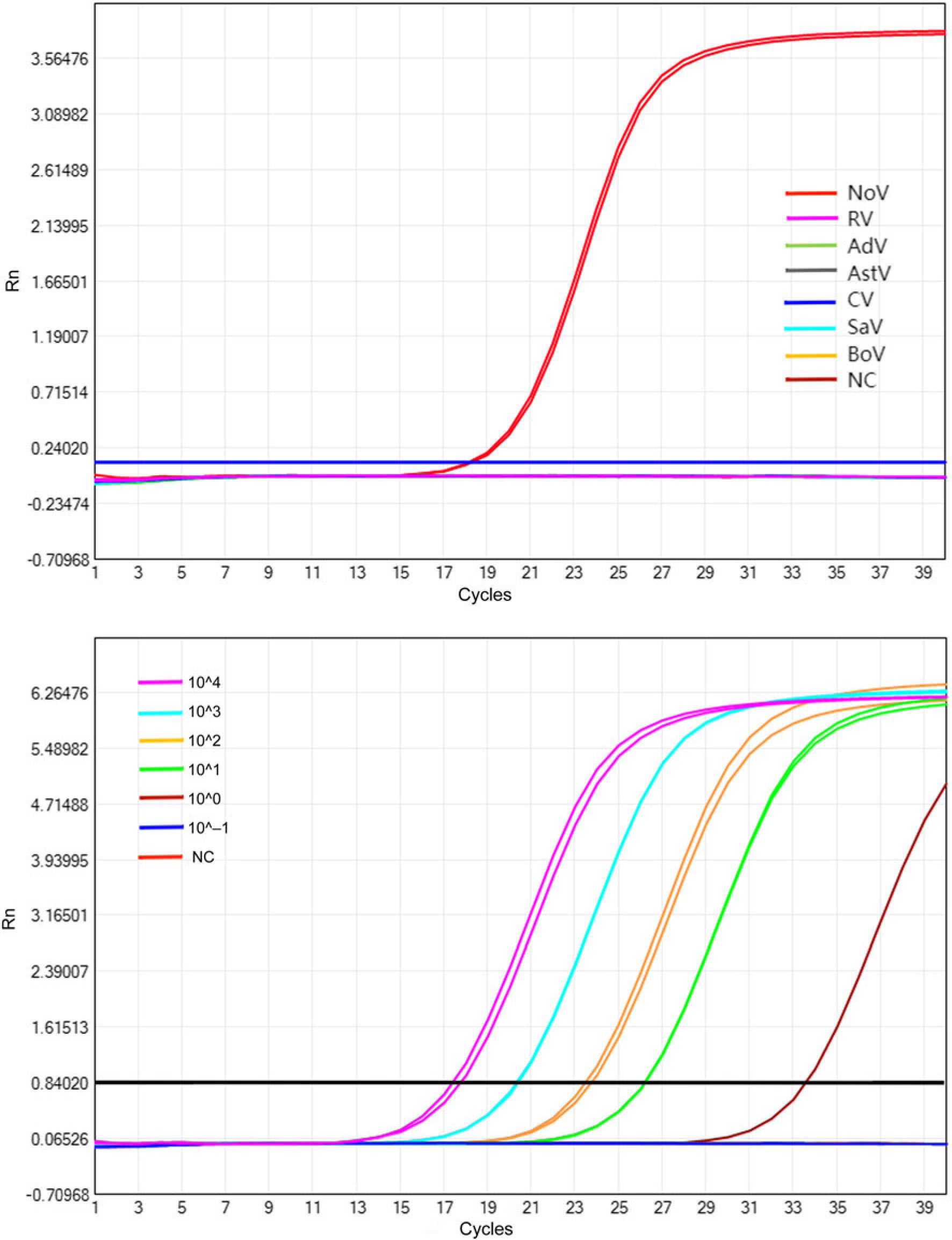
Fig. 6.
The specificity and sensitivity analysis of qRT-PCR for detection of norovirus GII.2.
The specificity analysis of qRT-PCR was conducted with various viral genome samples (a), while the sensitivity analysis of qRT-PCR was performed with different concentrations of standard RNA (b), and the results were evaluated based on the fluorescence curve. NoV – norovirus GII.2 subtype, RV – human rotavirus, AdV – adenovirus, AstV – astrovirus, CV – coxsackievirus, SaV – sapovirus, BoV – bocavirus, NC – ddH2O.
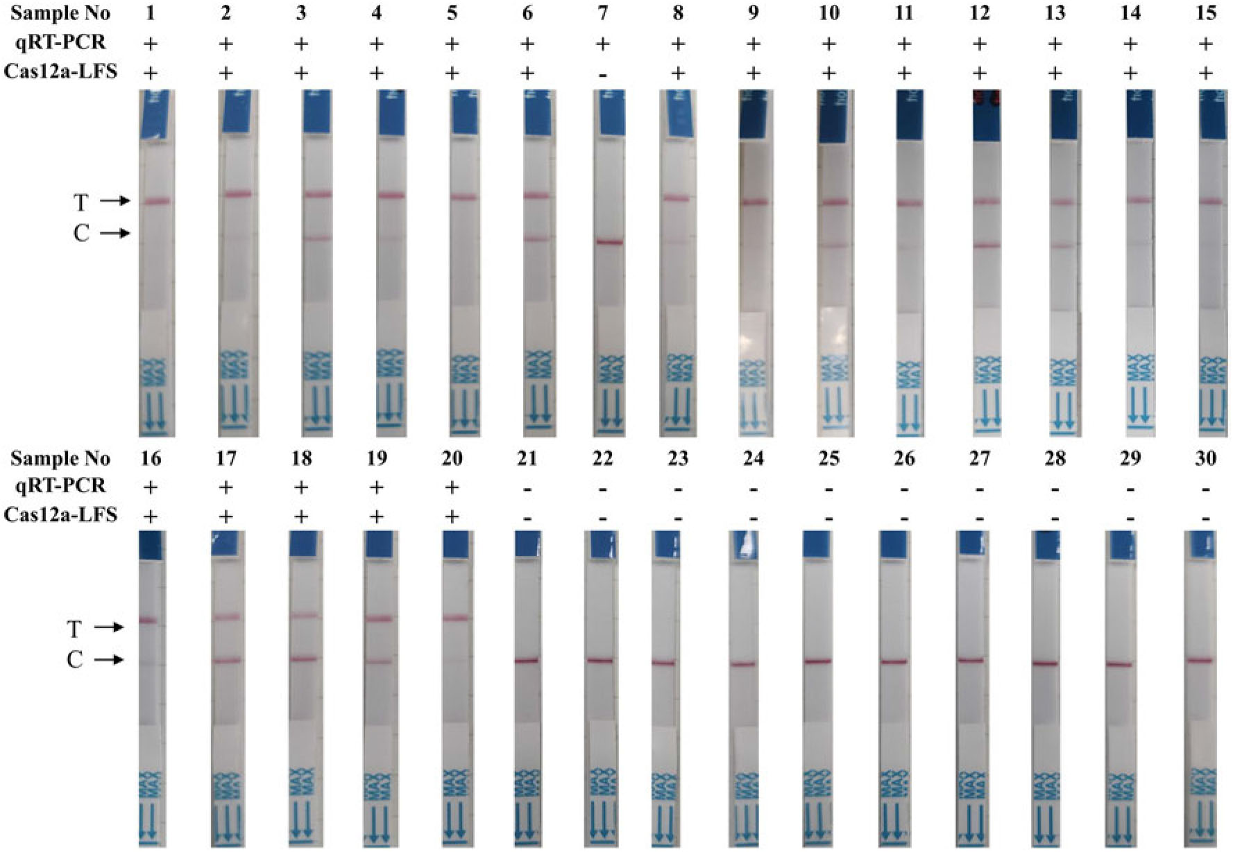
Fig. 7.
The performance analysis of RT-RPA-Cas12a-LFS for detecting norovirus GII.2, compared with qRT-PCR, was evaluated using simulated clinical samples. 30 representative results out of 60 samples are displayed.