
Table I
PCR primers used in the experiments.
| Primer | Sequence | Product (bp) |
|---|---|---|
| ΔInlG-F1 | CGGGATCCGCTTATGATACAGTAGAAGAA (BamHI) | 633 |
| ΔInlG-R1 | CCTCCGATGAAAAGCGTTCCTAAAATAGTAGGAATAATTCCCAAGATAGCTGTCACT | |
| ΔInlG-F2 | ATGTTTTACTTGTAGTGACAGCTATCTTGGGAATTATTCCTACTATTTTAGGAACGC | 596 |
| ΔInlG-R2 | GCTCTAGAAGATAATTCAAGTTCTGTTA (XbaI) | |
| D-F | TGTCCGCAACAGCTAGCCCAG | 1,644/3,100 |
| D-R | GCAAGTGGGGTTAAATCACTT | |
| Hly-F | GATGCATCTGCATTCAATAA | 1,510 |
| Hly-R | TTATTCGATTGGATTATCTAC |
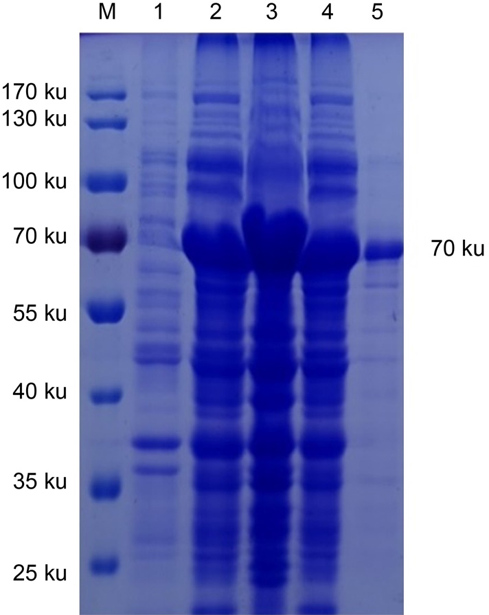
Fig. 1
Analysis of the recombinant protein. M – Protein marker, 1 – bacteria before the induction with IPTG, 2 – the induced bacteria, 3 – supernatant of lysed induced bacteria, 4 – precipitation of induced bacteria, 5 – purified InlG recombinant protein, 6 – uninduced bacteria reaction with positive serum, 7 – induced bacteria reaction with positive serum.
Table II
Determination of the median lethal dose of the bacteria (BACT) in mice.
| BACT (CFU) | Death/Total | Mortality (%) |
|---|---|---|
| 109 | 9/10 | 90 |
| 108 | 8/10 | 80 |
| 107 | 6/10 | 60 |
| 106 | 4/10 | 40 |
| 105 | 2/10 | 20 |
| 0 | 0/10 | 0 |
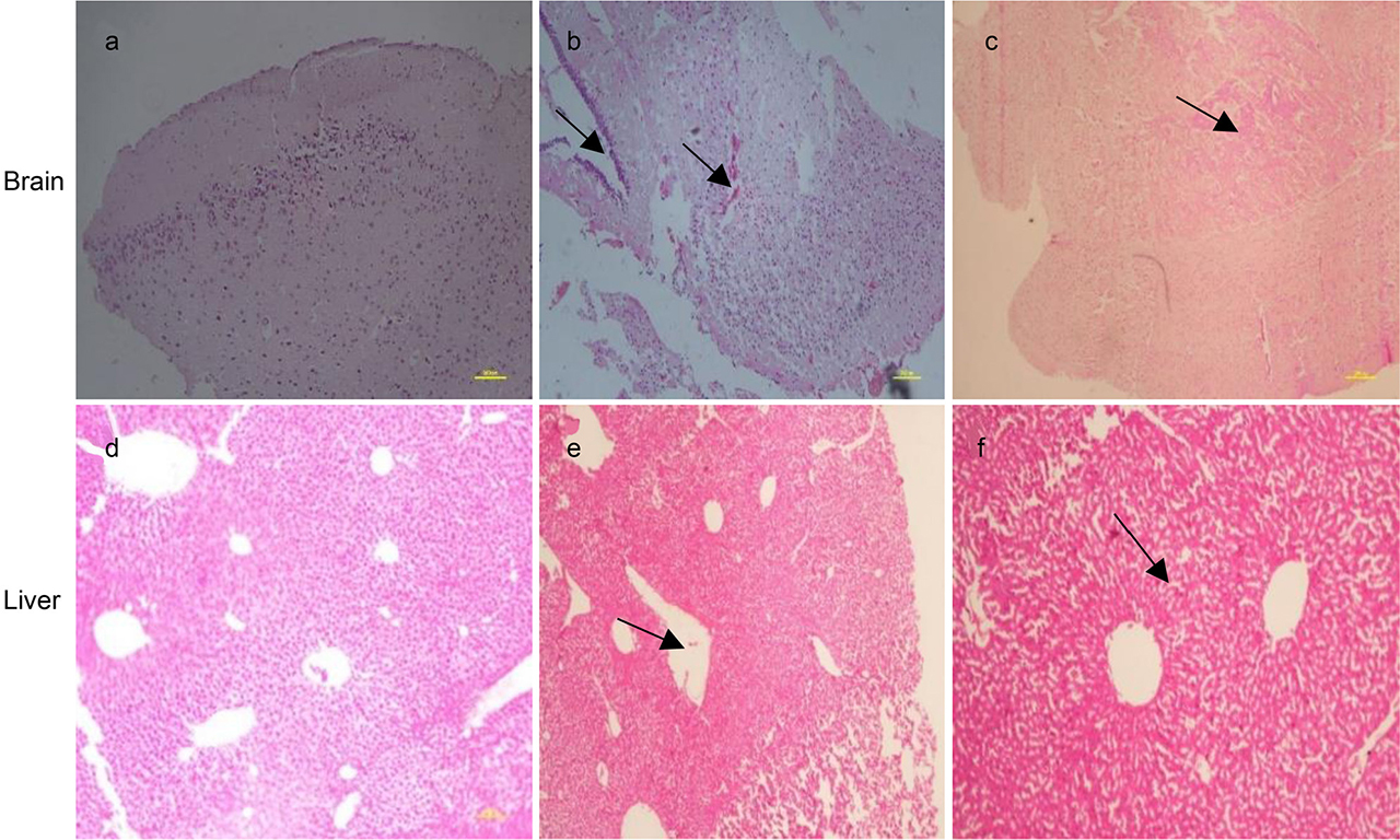
Fig. 2
Pathological changes in the tissue of infected mice stained with hematoxillin-eosin. a/d – Normal mouse, b/e – the mice immunized with normal saline, c/f – the mice immunized with protein and challenged with bacteria.
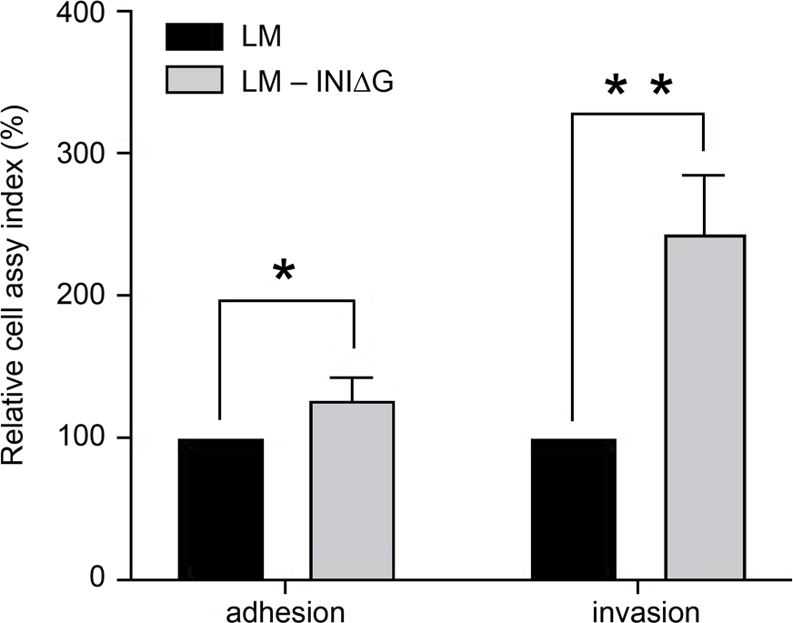
Fig. 3
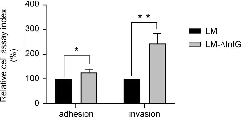
Relative adhesion and invasion of LM and LM-ΔInlG in Caco-2 cells. This test was done in triplicate in each run and repeated for three times. The cell assay rates of LM19111 were set at 100%. * p < 0.05; ** p < 0.01.
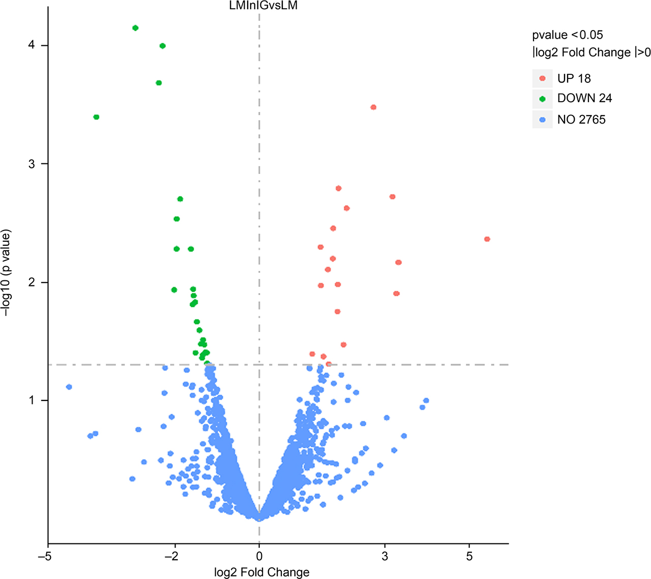
Fig. 4
Differentially expressed genes in LM-ΔInlG compared to LM. The abscissa indicates the fold change of gene expression, and the ordinate indicates the significance of the gene difference. The red dots indicate the up-regulated genes, the green dots indicate the down-regulated genes, and the blue dots indicate the genes that are not significantly different.
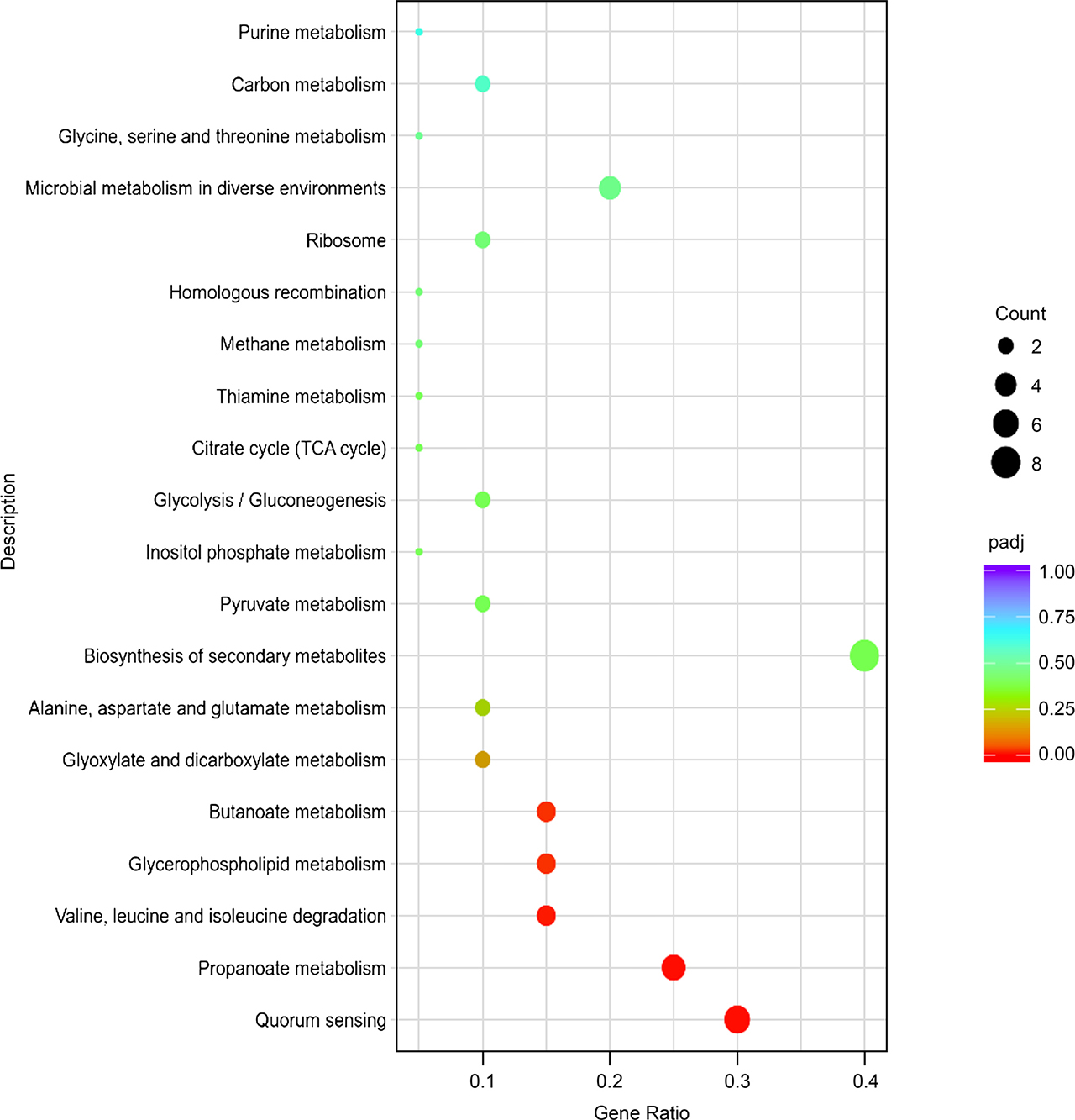
Fig. 5
KEGG pathway analysis of differentially expressed genes in LM-ΔInlG compared to LM. The enriched KEGG categories are on the vertical axis. The ratio of the enriched DEGs in the KEGG category to the total genes in that category is shown on the horizontal axis.
Table III
Differentially expressed genes in related KEGG pathways in LM-ΔInlG compared to LM.
| Term | Gene ID | Name | Log2FC | Type | Description |
|---|---|---|---|---|---|
| Quorum sensing | lmo0205 | plcB | 1.89 | up | phospholipase C |
| lmo0202 | hly | 1.77 | up | listeriolysin O precursor | |
| Novel00001 | no | 1.76 | up | thiol-activated cytolysin | |
| Novel00002 | no | 1.64 | up | thiol-activated cytolysin beta sandwich domain | |
| lmo2363 | no | –1.35 | down | glutamate decarboxylase | |
| lmo0447 | no | 1.66 | up | pyridoxal-dependent decarboxylase conserved domain | |
| Propanoate metabolism | lmo1373 | no | –1.86 | down | transketolase, pyrimidine binding domain |
| lmo1153 | no | 1.88 | up | propanediol dehydratase subunit alpha | |
| lmo2720 | no | –2.02 | down | AMP-binding enzyme C-terminal domain | |
| lmo1374 | no | –1.52 | down | biotin-requiring enzyme | |
| lmo1371 | no | –1.28 | down | pyridine nucleotide-disulphide oxidoreductase | |
| Valine, leucine, nd isoleucine degradation | lmo1373 | no | –1.86 | down | transketolase, pyrimidine binding domain |
| lmo1374 | no | –1.52 | down | biotin-requiring enzyme | |
| lmo1371 | no | –1.28 | down | pyridine nucleotide-disulphide oxidoreductase | |
| Glycerophospholipid metabolism | lmo0205 | plcB | 1.89 | up | phospholipase C |
| lmo1176 | eutC | 3.18 | up | ethanolamine ammonia-lyase small subunit | |
| lmo1175 | eutB | 1.87 | up | ethanolamine ammonia-lyase large subunit | |
| Butanoate metabolism | lmo1369 | no | –2.94 | down | phosphate acetyl/butaryl transferase |
| lmo2363 | no | –1.35 | down | glutamate decarboxylase | |
| lmo0447 | no | 1.66 | up | pyridoxal-dependent decarboxylase conserved domain |