Introduction
The importance of the human papillomavirus (HPV) as an etiological factor in cervical cancer (CC) has been recognized since the 1980s (Muñoz et al. 1994). In developing regions, CC is the second most common type of cancer. In 2018, it caused approximately 311,000 deaths worldwide (WHO 2020). In America, 3.8 million cases were diagnosed in the same year, and about 1.3 million died (www.paho.org). According to GLOBOCAN (https://gco.iarc.fr), 4,121 women died from CC in Mexico in 2018, 1.3% of all CC-related deaths worldwide.
HPV belongs to the family Papillomaviridae, which comprises non-enveloped viruses with a double-stranded DNA genome of approximately 8,000 base pairs (bp). There are 228 different types of papillomaviruses registered in the International HPV Reference Center (www.hpvcenter.se) (Bruni et al. 2019). Papillomaviruses are classified into low-risk (LR) and high-risk (HR) types based on their association with cancer (Egawa and Doorbar 2017). It is thus crucial to identify the viral genotype with which a patient is infected. Since people who are immunosuppressed due to infection with the human immunodeficiency virus (HIV) have a high incidence of neoplasms (Goedert et al. 1998; Frisch et al. 2001), immunosuppressed women infected with some type of oncogenic HPV have a greater probability of developing cervical cancer (Clifford et al. 2005).
Several studies have shown that HIV-infected women co-infected with LR-HPV and HR-HPV have a two- to seven-fold greater risk of developing low- and high-grade neoplastic intraepithelial lesions, and even CC, compared to HIV-negative women (Mbulawa et al. 2009; Yamada et al. 2008; Videla et al. 2009). In 2018, Hispanic women with HIV were reported to have a higher incidence of HPV-associated CC compared to other ethnic groups (Ortiz et al. 2018). The present study aimed to identify the presence of genetic variants of HPV in a group of HIV-positive Mexican women undergoing antiretroviral therapy. The polymorphisms of the most prevalent genotype were identified. In some areas of Mexico, HPV51 predominates over other genotypes (Gallegos-Bolaños et al. 2017; Jácome-Galarza et al. 2017; Campos et al. 2019).
Experimental
Materials and Methods
Study population. It was a cross-sectional and descriptive study. It was approved by the review committee of the Hospital General de Puebla and participating patients signed an informed consent form (143/2009). Forty female patients from the Centro Ambulatorio para la Prevención y Atención en SIDA e Infecciones de Transmisión Sexual, in Puebla, Mexico (CAPASITS-SSA), were selected through their clinical records. It was done by considering the last CD4+ lymphocyte count and the last HIV viral load measurement, as long as they were not taken more than six months before enrollment in the study. The HIV viral load measurement and the CD4+ lymphocyte count were performed by the hospital’s clinical analysis service per the corresponding diagnosis, treatment, and follow-up guidelines of the World Health Organization (WHO) and the country’s health authorities (WHO 2009; 2010). HIV-positive patients were classified into two main groups based on the number of CD4+ cells, one with >350 cells/mm3 and the other with < 350 cells/mm3. This threshold was the main criterion for the initiation of antiretroviral therapy and was significantly associated with more rapid HPV clearance (Sabin and Phillips 2009; Kang and Cu-Uvin 2012).
Biological material. Two endocervical exfoliation samples were taken. One for cervical cytology analysis (a thin prep) and one for DNA extraction and PCR amplification.
Cervical cytology. A clean, non-lubricated vaginal mirror and an Ayre spatula were used to collect endocervical secretions. The smears were placed on previously labeled glass slides and fixed with 95% ethanol. After staining using the Papanicolaou method, the smears were analyzed in the Cytopathology Department of the Medical School of the Benemérita Universidad Autónoma de Puebla. Possible cellular abnormalities found by the thin prep were analyzed and classified according to the Bethesda classification system as Type I (negative), negative for intraepithelial lesion or malignancy; Type II (inflammatory process), reactive cellular changes associated with radiation, intrauterine contraceptive device, glandular cells status post hysterectomy, cellular changes consistent with viral activity, Trichomonas vaginalis, fungal organisms, etc.; Type III (LSIL): low-grade squamous intraepithelial lesions; Type IV (HSIL), high-grade squamous intraepithelial lesions (Nayar and Wilbur 2015).
HPV Amplification Assays by Polymerase Chain Reaction. For the collection of cervical samples and DNA extraction, the QIAamp Fast DNA tissue kit® with dacron swabs and transport buffer (Qiagen®) was used following the manufacturer’s instructions. For the amplification reaction, the QIAGEN Multiplex PCR kit® was used following the manufacturer’s instructions, with the following general primers: MY09/11 and GP5 +/GP6 +. These primers amplify conserved sequences of the HPV L1 gene of both high and low-risk types. The primers for the actin gene were used as an internal control (Manos 1991; Qu et al. 1997). For the identification of HPV types in the amplified samples, the PCR products were sequenced with the dideoxy method using an Abi Prism 310 Sequencer (Applied Biosystems) and the primer GP5+ in all sequencing reactions. The sequences obtained were aligned using the Basic Local Alignment Search Tool (BLAST) of the NCBI platform to determine the similarity of the regions of interest (Madden 2008).
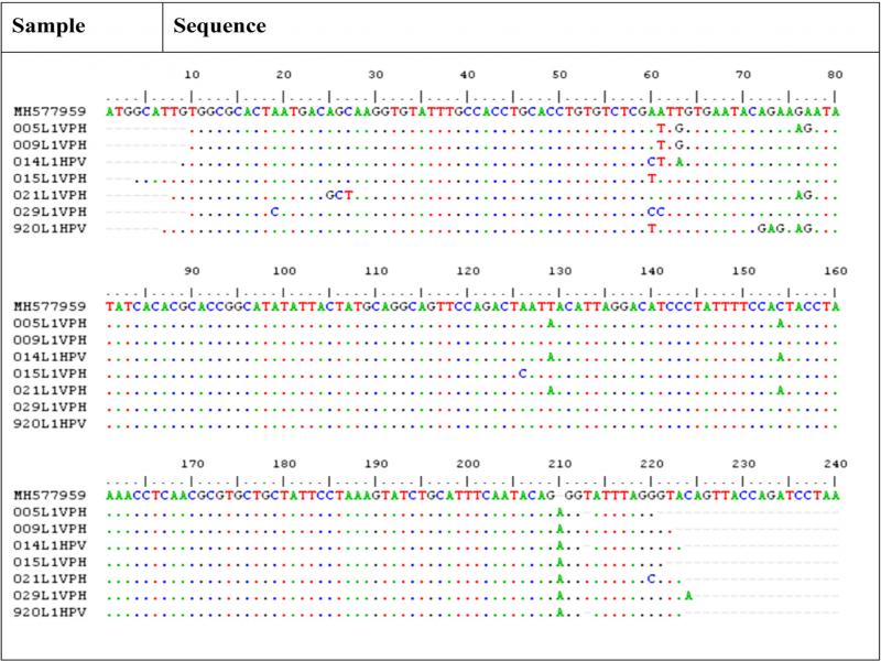
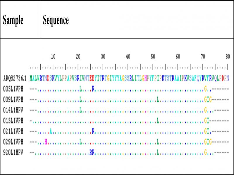
Nucleotide and amino acids sequence analysis. The nucleotide and amino acid sequences of positive samples were compared against GenBank using Blast. A multiple alignment of these sequences was performed (ClustalW Multiple Alignment v1.4) using BioEdit Sequence Alignment Editor version 7.0.9.0. (Hall 1999): For the analysis of nucleotides, the sequence MH577959.1 (Xu et al. 2019) was used as a reference. For the analysis of amino acid sequences, the sequence ARQ82736.1 was used (Oliveira et al. 2017). See the attached key resources table.
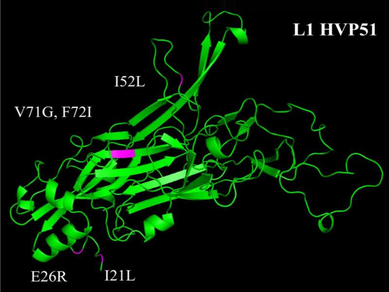
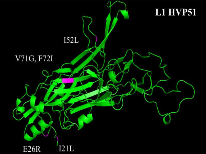
Homology modeling. The L1 structure of HPV51 was generated from amino acid sequences ARQ82736.1 in the I-TASSER platform (Roy et al. 2012; Yang and Zhang 2015). The preliminary sequence alignments were performed using the local meta-threading server of I-TASSER (Wu and Zhang 2007) to generate a list of templates for modeling (i.e., 3IYJ, 3OFL, 2R5K, and 1DZL). In addition to the sequence alignment, I-TASSER uses the TM-align structural alignment program to match the first I-TASSER model to all structures in the Protein Data Bank (PDB) library. For this monomer, the PDB codes used were 3IYJ, 1DZL, 2R5I, and 2R5K. The model validation outcome on the same website gave no hints of bad/unusual geometrical features. The visualization was performed using the program PyMOL (TM) 1.7.4.5 Molecular Graphics System, Version 2.0 Schrödinger, LLC.
Statistical analysis. Descriptive statistics were used for quantitative and categorical variables. The Pearson’s chi-squared test was used to check whether the prevalence of HPV infection increased with age or the presence of certain genotypes. The age of the patients was stratified into groups of < 45 years, 45–54 years, and > 54 years (Lazcano-Ponce et al. 2001; Tharcisse et al. 2020). The interaction between high-risk genotypes in each group of patients was also analyzed. Spearman’s Rho or Pearson’s tests were used to assess the correlation between the variables of viral load, CD4+ cell count, and low and high-risk subtypes of HPV. The statistical analysis was performed using IBM® SPSS® Statistics version 25.0. See the attached key resources table.
Results
Presence of HIV and immunological status of the patient. Clinical features. The analysis of the patients’ clinical history showed they had been infected with HIV for an average of 4.6 years after AIDS diagnosis and had been under antiretroviral treatment for an average of 3.46 years. Table I describes the age and the last measurement of CD4+ cells.
Table I
Clinical characteristics of the patients.
| Patient age | HIV viral load (copies/ml) | CD4+ (cell/ml) | Pap cytology1 | HPV type2 |
|---|---|---|---|---|
| Group I3 | ||||
| 45 | < 50 | 744 | I and II | 90 (LR) |
| 30 | < 50 | 726 | I and II | ND |
| 23 | 5,840 | 655 | I | 11 (LR) |
| 30 | < 50 | 602 | I and II | 97 (LR) |
| 17 | 90,800 | 580 | I and II | 51 (HR) |
| 42 | < 50 | 521 | I | ND |
| 44 | < 50 | 506 | I | 66 (HR) |
| 50 | 400 | 503 | I and II | 16 (HR) |
| 35 | < 50 | 485 | I | 16 (HR) |
| 27 | < 50 | 481 | I and II | 51 (HR) |
| 37 | < 50 | 410 | IV | 51 (HR) |
| 42 | < 50 | 405 | I | 11 (LR) |
| 60 | 57 | 388 | I | 58 (HR) |
| 29 | < 50 | 380 | I and II | 70 (LR) |
| 31 | 4,940 | 364 | I and II | ND |
| Group II3 | ||||
| 35 | < 50 | 317 | I and II | ND |
| 35 | < 50 | 294 | IV | ND |
| 24 | 1,170 | 282 | I and II | ND |
| 30 | < 50 | 280 | III | 54 (LR) |
| 42 | < 50 | 258 | I and II | 102 (LR) |
| 49 | < 50 | 255 | I and II | 6 (LR) |
| 35 | < 50 | 208 | I | 84 (LR) |
| 32 | 369 | 180 | I | 51(HR) |
| 36 | 13,900 | 175 | I and II | 51 (HR) |
| 37 | < 50 | 161 | III | 81 (LR) |
| 28 | 67 | 160 | I and II | ND |
| 24 | 74,620 | 146 | I and II | ND |
| 51 | < 50 | 132 | III | 51 (HR) |
| 37 | 73 | 127 | IV | 81 (LR) |
| 48 | 5560 | 104 | I and II | 6 (LR) |
| 43 | < 50 | 103 | I and II | 86 (LR) |
| 37 | ND | 102 | I | 56 (HR) |
| 31 | < 50 | 76 | III | 52 (HR) |
| 23 | > 100,000 | 70 | I and II | 6 (LR) |
| 34 | 822 | 65 | III | 58 (HR) |
| 63 | 451 | 63 | I and II | ND |
| 44 | 5,140 | 60 | I and II | 51 (HR) |
| 25 | 95200 | 30 | I and II | 81 (LR) |
| 41 | ND | 22 | III | 33(HR) |
| 31 | > 100,000 | 16 | I and II | 70 (LR) |