
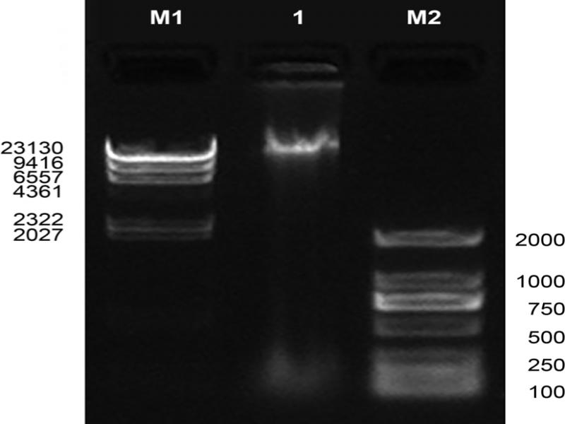
Fig. 1.
Electrophoresis pattern of Pestalotiopsis kenyana PG52 genome. Agarose concentration (%): 1; voltage: 180 V; time: 35 min.; molecular weight standard name: M1: λ-Hind Ш digest (Takara), M2: D2000 (Tiangen); sample volume: M1: 3 μl, M2: 6 μl.
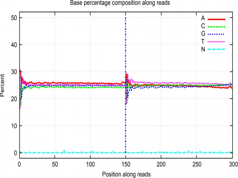
Fig. 2.
Pestalotiopsis kenyana PG52 base composition distribution map. The X axis represents the position on reads, and the Y axis represents the percentage of bases.
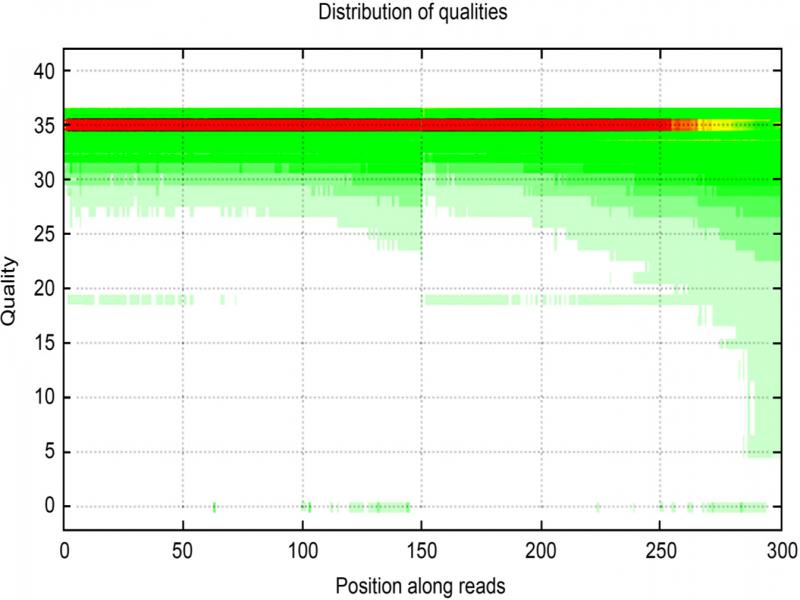
Fig. 3.
Pestalotiopsis kenyana PG52 base mass distribution map. The X axis is the position of the base in reads, and the Y axis is the base quality value. Each point in the figure represents the total number of bases at this position that reach a certain.
Table I
The comparison of Pestalotiopsis genome sequences.
| PG52 | FICI | NC0098 | |
|---|---|---|---|
| Assembly size (Mb) | 55 | 52 | 46.61 |
| Scaffold N50 (Mb) | 6.6 | 4.0 | 5 |
| Coverage (fold) | 335.0 | 24.5 | 24 |
| GC content (%) | 53.30 | 48.73 | 51.28 |
| Protein-coding genes | 20,023 | 15,413 | 15,180 |
| Gene density (genes per Mb) | 345.22 | 296.90 | 327.08 |
| Exons per gene | 3.13 | 2.76 | 2.83 |
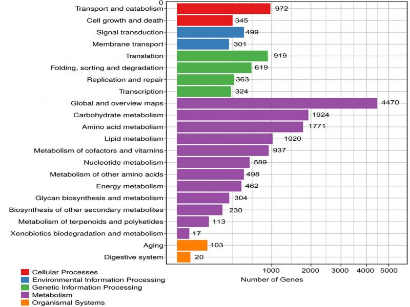
Fig. 4.
KEGG function analysis.
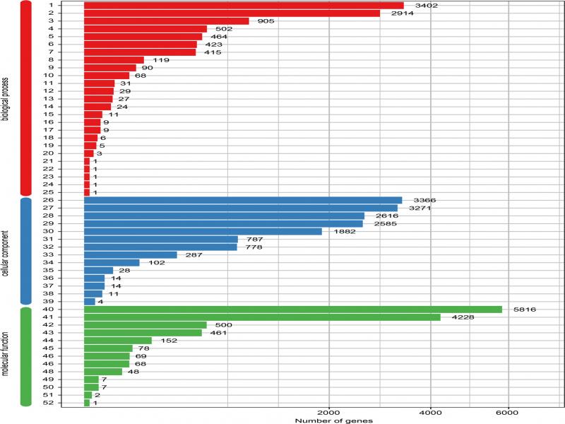
Fig. 5.
GO functional classification map of all unigenes with GO annotation.
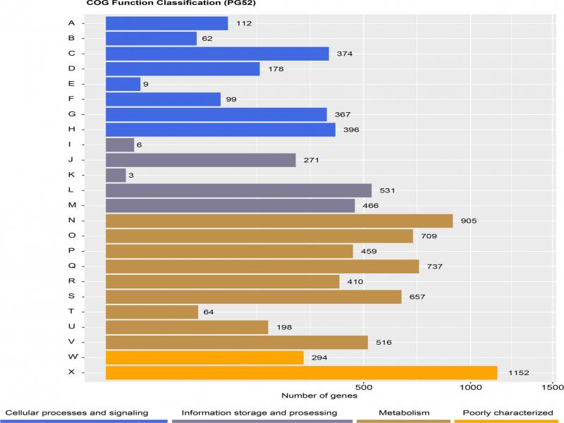
Fig. 6.
COG function analysis.
Table II
Glycosyl hydrolase families related to mycoparasitic in mycoparasites.
| Species | GH18 | GH75 | GH17 | GH55 | GH64 | GH81 |
|---|---|---|---|---|---|---|
| Pestalotiopsis sp. PG52 | 3 | 6 | 7 | 19 | 3 | 2 |
| Trichoderma harzianum | 20 | 4 | 4 | 5 | 3 | 2 |
| Trichoderma atroviride | 29 | 5 | 5 | 8 | 3 | 2 |
| Trichoderma virens | 36 | 5 | 4 | 10 | 3 | 1 |
Table III
The number of polyketide synthases and nonribosomal peptide synthetases of P. fici, Pestalotiopsis sp. NC0098 and mycoparasites.
| Secondary metabolites | Pestalotiopsis sp. PG52 | P. fici | Pestalotiopsis sp. NC0098 | T. harzianum | T. atroviride | T. virens |
|---|---|---|---|---|---|---|
| NRPS | 13 | 12 | 12 | 17 | 16 | 28 |
| PKS | 102 | 27 | 21 | 27 | 18 | 18 |
| Total | 115 | 39 | 33 | 44 | 34 | 46 |