
1. Introduction
The families Sparidae (sea breams and porgies) and Nemipteridae (threadfin breams) are diverse groups of ray-finned fishes with significant ecological and economic importance. Sparidae comprises approximately 155 species across 38 genera, predominantly inhabiting shallow temperate and tropical coastal waters, reefs, and sandy bottoms (Froese & Pauly, 2024; Nelson et al., 2016). These demersal carnivores feed on invertebrates and small fish (Smith & Heemstra, 1986). They are characterized by deep, compressed bodies, small mouths, and strong molariform teeth adapted for crushing hard-shelled prey (Carpenter & Niem, 2001). While most sparids are carnivorous, some have adapted to herbivory, reflecting the family’s commercial and recreational fisheries value (Heemstra & Randall, 1993). Phylogenetic studies confirm Sparidae as monophyletic (Chiba et al., 2009), though traditional classifications based on dentition and feeding strategies are non-monophyletic due to convergent evolution (Day, 2002), with three major clades identified (Herrán et al., 2001) and morphological plasticity contributing to adaptive radiation and taxonomic confusion (Carpenter & Johnson, 2002). Similarly, Nemipteridae comprises diverse genera and species adapted to Indo-West Pacific marine environments, characterized by distinctive body shapes, elongated fins, and vibrant coloration (Froese & Pauly, 2024). Nemipterids inhabit shallow coastal waters, including coral reefs and sandy substrates, and are ecologically and economically important in local fisheries (Mora et al., 2011), acting as both predators and prey (Matsuura, 2016). Molecular research has identified distinct lineages, indicating a complex evolutionary history influenced by geographic and habitat factors (Hossain et al., 2020), with the family diverging from other berycoid fishes during the late Cretaceous (Froese & Pauly, 2024). Genera such as Nemipterus and Scolopsis exemplify niche adaptations (Deng et al., 2019), and ongoing integrative studies continue to refine the phylogenetic framework of Nemipteridae, clarifying subfamilial relationships and revealing convergent evolution.
Taxonomic and phylogenetic studies of fish species rely on various features, including morphometric and meristic data (e.g., Bektas & Belduz, 2009; Ben Labidi et al., 2021; Neves et al., 2023), morphological descriptions (e.g., Jawad, 2008), coloration patterns (e.g., Karcher et al., 2012; Marshall et al., 2018), and genetic/molecular analyses (Bani et al., 2013; Francisco et al., 2022). While molecular techniques provide valuable insights, they often require specialized laboratory setups, extended processing times, and substantial financial investment (Anderson & Thompson, 2002). In contrast, hard structures such as scales, skeletal elements, and otoliths (calcium carbonate deposits in the inner ear of fish) offer a cost-effective and reliable alternative for species identification, ecological studies, and age determination (Chilton & Beamish, 1982; Panfili et al., 2002).
Among these structures, otoliths are particularly valuable due to their metabolic inertness, preventing reabsorption (Campana & Neilson, 1985), unlike scales (Simkiss, 1974). Otoliths exhibit growth increments (daily to annual), providing high-resolution temporal data on fish life history (Black et al., 2008; Campana, 1999; Elsdon et al., 2008; Morales-Nin, 2000). Given that some fish species live for decades, otolith analysis serves as a crucial biological archive, offering insights into growth, migration, and environmental conditions (e.g., Black et al., 2008; Campana, 1999).
Otolith morphology is also species-specific, making it a powerful tool for taxonomic identification (Aguirre & Lombarte, 1999; Assis, 2003; Battaglia et al., 2010, 2015; Jawad, 2005, 2007, 2008; Tuset et al., 2003, 2006). Among the three otolith types in teleost fish, the sagitta (saccular otolith), being the largest, is most frequently studied (Echreshavi et al., 2021; Mehraban et al., 2023; Neves et al., 2023). Research on otoliths spans diverse applications, including age and growth estimation (Green et al., 2009), migration tracking (Hanson et al., 2022), larval fish identification (Lord et al., 2010), paleobiological and systematic studies (Gierl et al., 2013), and habitat and population assessments (Avigliano et al., 2014; Campana & Thorrold, 2001).
Ontogenetic studies indicate that both genetic and environmental factors influence otolith shape and size (Hüssy, 2008; Lombarte et al., 2003). However, juvenile otoliths may lack diagnostic features, limiting their use in species identification until adulthood (Hüssy, 2008; Jawad et al., 2018a, 2018b). Additionally, otolith morphology aids in paleontological reconstructions from fossilized specimens (Nolf, 1985, 2013).
This study focuses on two species: (1) B. boops (Linnaeus, 1758). A demersal marine fish inhabiting depths of 0–350 m (Sanches, 1991), widely distributed in the Eastern Atlantic and Mediterranean. It reaches up to 400 mm in length (Crec’hriou et al., 2013) and has an omnivorous diet (Frimodt, 1995) and (2) N. randalli (Russell, 1986). A benthic marine species found at 20–450 m depths in the Western Indian Ocean, with a maximum length of 270 mm (Mehanna & Farouk, 2021).
Despite their ecological and commercial importance, detailed otolith descriptions for these species remain scarce (Çiçek et al., 2021; Lin & Al-Abdulkader, 2019; Tuset et al., 2008; Veen & Hoedemakers, 2005; Yazici, 2022), with most studies embedded in broader biological investigations rather than focused morphological analyses.
Scanning electron microscopy (SEM) is indispensable in otolith studies due to its ability to: capture high-magnification, high-resolution images of microstructural details beyond the limits of light microscopy; reveal fine-scale morphological variations critical for distinguishing closely related species; analyze surface textures (e.g., growth patterns, erosional features) that aid in ecological and taxonomic differentiation; and document diagnostic features consistently, supporting phylogenetic and paleontological studies (Jawad et al., 2023).
The objectives of this study are: (1) to provide a morphological description and iconography of otoliths from B. boops and N. randalli collected in Antalya Bay (Mediterranean coast of Türkiye), a region with limited existing otolith data; and (2) to identify taxonomically informative features using SEM to enhance species identification and ecological understanding. By leveraging SEM’s capabilities, this work will contribute to a more comprehensive understanding of otolith morphology in these species, supporting future taxonomic, ecological, and fisheries research.
2. Materials and methods
2.1. Study area and fish sampling
Two teleost species B. boops and N. randalli were experimented for this investigation, encompassing a total of 138 specimens (56 of B. boops and 82 of N. randalli) (Fig. 1). The sampling conducted in Antalya Bay along the Mediterranean coast of Türkiye (36° 49′ 42″ N–36°45′ 21″ N; 30 50′ 22″ E–31° 20′ 05″ E) (Fig. 2) from October 2020 to September 2021. The total length (TL) of the fish ranged from 138–225 mm for B. boops and 126–240 mm for N. randalli. The mean lengths and standard deviations were 177. 9 ± 2.449 mm and 175.52 ± 2.786 mm, respectively. The fish were caught using a commercial bottom trawl net with a 44 mm mesh size (22 mm in the cod end). The fishing depth ranged from 40 m to 160 m. Later, the specimens were stored on ice until coming to the laboratory, where they were identified according to Bauchot and Hureau (1986) for B. boops and Russell (1990) for N. randalli. The TL, measured from the snout to the end of the upper and lower caudal fin lobes (combined), was recorded to the nearest millimeter. Sex determination of the specimens was performed through macroscopic and histological examination of the gonads following dissection. In adult individuals, sex was identified macroscopically based on differences in the color, shape, and texture of the gonads (Brown-Peterson et al., 2011; Holden & Raitt, 1974). For juvenile (immature) or resting-phase individuals whose sex could not be distinguished macroscopically, gonadal tissues were extracted and fixed in 10% buffered formalin solution. Following standard histological procedures, these tissues were stained with hematoxylin and eosin (Suvarna et al., 2018), examined under a light microscope, and sex was verified at the cellular level. Sexual size dimorphism was assessed by comparing TL between female and male specimens of B. boops and N. randalli. Before analysis, data were tested for normality using the Shapiro–Wilk test and for homogeneity of variances using Levene’s test. An independent-samples *t*-test was used to compare mean TLs between sexes for each species; in cases where assumptions of normality were violated, a non-parametric Mann–Whitney U test (Chicco et al., 2025) was applied as an alternative. All statistical analyses were performed using [insert software, e.g., SPSS, R, IBM, etc.], and statistical significance was set at *p* <0.05.
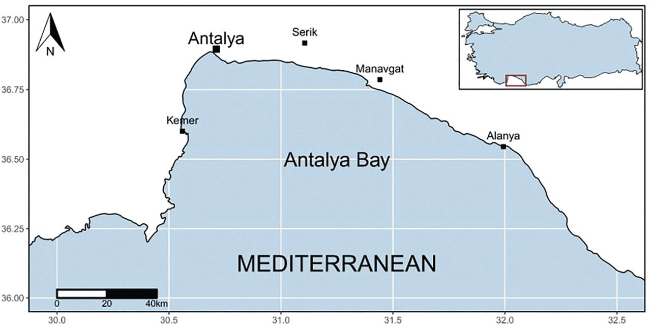
Figure 1
Map showing the location of fish samples collection from the Antalya Bay, Mediterranean coast of Türkiye.
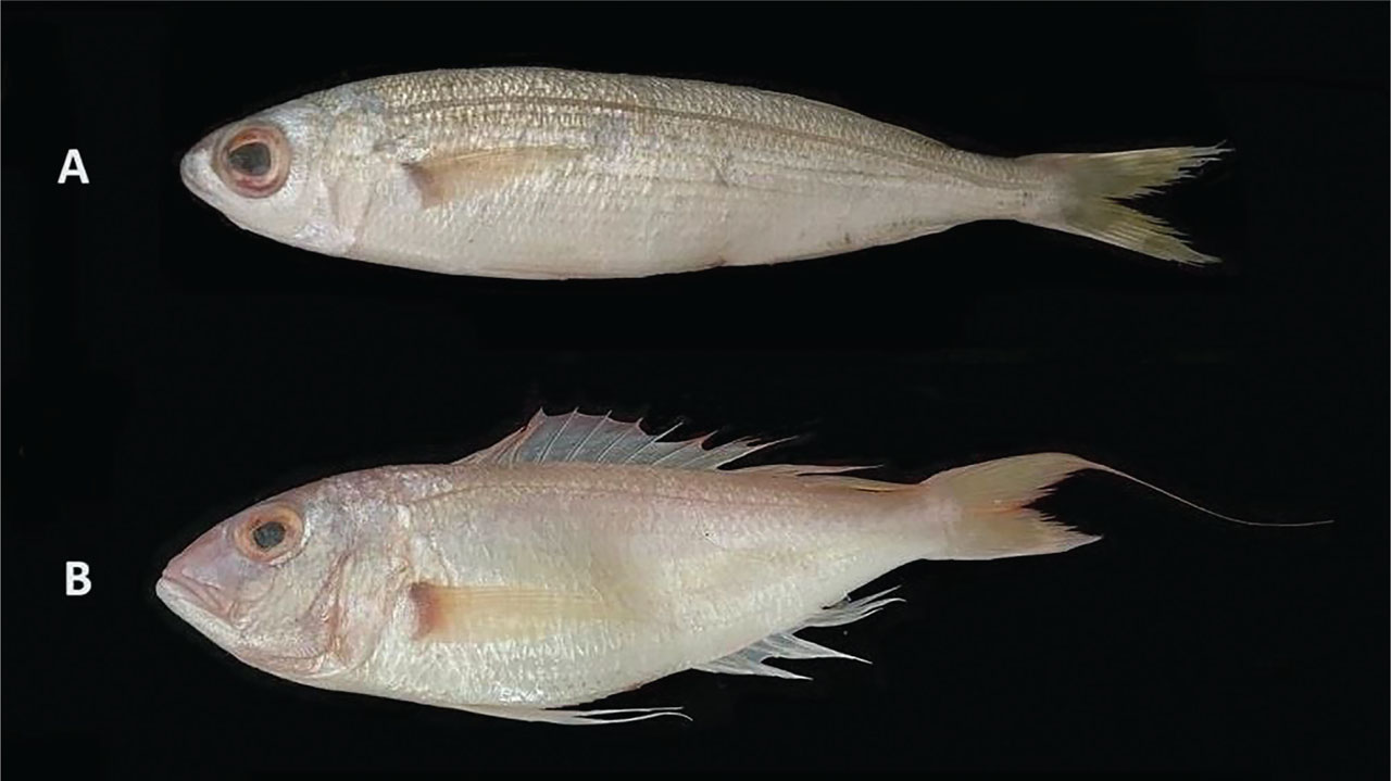
Figure 2
Images of (A) B. boops, 161 mm TL; (B) N. randalli, 231 mm TL. TL, total length.
2.2. Otolith sampling
The otoliths were isolated from the left and right sides of the head by making a gap at the top of the cranium. The otic capsules were separated, and the otoliths were carried away with a pair of fine forceps. They were cleaned in 70% ethanol and dried. To inspect the otolith surface outer features, they were split into groupings corresponding to the fish TL. The species B. boops was grouped into five length groups (GI, 130–150 mm TL; GII, 151–170 mm TL; GIII 171–190 mm TL; GIV, 191–210 mm TL; and GV, 211–230 mm TL). The species N. randalli was grouped into four length groups (GI, 120–150 mm TL; GII, 151–180 mm TL; GIII, 181–210 mm TL; and GIV, 211–240 mm TL). All otoliths were verified and elected specimens from all length groups were afterward imaged via SEM. In planning for SEM, otoliths were air cleaned and mount up on an aluminium stub utilizing double-sided carbon tape. Otoliths were examined in SEM device model FEI (Thermo Fisher Scientific) Quanta FEG 250. No coating was employed to the sample surfaces to ensure conductivity. As an alternative, conductivity was achieved using the device’s low vacuum mode, and the chamber pressure was set to 60 pa. SEM exhibits in detail the surface of otoliths, permitting the record of morphological descriptions on the mesial and lateral faces of the otoliths. The morphological terminology of the otoliths is grounded on Smale et al. (1995) (Fig. 3).
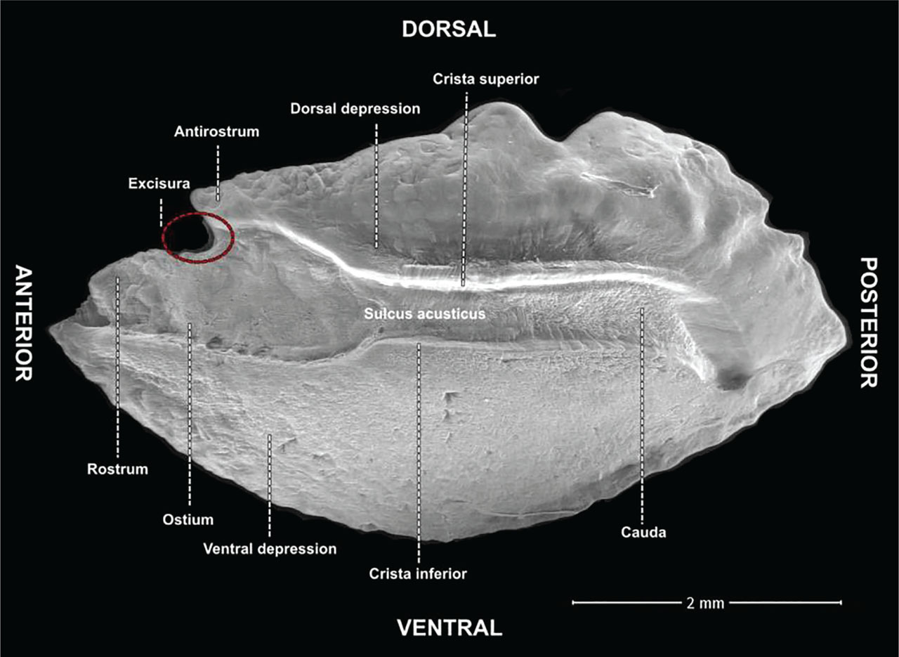
Figure 3
Right otolith of B. boops specimen with 175 mm TL male showing the terminology used in the text for the anatomical features in the morphological descriptions of the otolith. TL, total length.
3. Results
Sexual size dimorphism was not observed in either species. For N. randalli, mean TL for females (17.82 ± 1.98 cm, *n* = 12) was nearly identical to that of males (17.76 ± 2.42 cm, *n* = 21), with no significant difference detected by independent samples *t*-test (*t* = 0.07, df = 31, *p* = 0.944) or Mann–Whitney U test (U = 126.5, *p* = 0.987). Similarly, for B. boops, mean TL for females (18.52 ± 2.52 cm, *n* = 18) was slightly greater than that of males (17.39 ± 2.27 cm, *n* = 23), but again the difference was not significant (*t* = 1.51, df = 39, *p* = 0.139; Mann–Whitney U = 154.5, *p* = 0.163). These findings indicate that neither N. randalli nor B. boops exhibit significant length-based sexual dimorphism in the studied samples.
3.1. Developmental variation
In this study, 21 otolith features were examined and compared across different size groups of B. boops and N. randalli collected from Antalya Bay, along the Mediterranean coast of Türkiye (Tables 1 and 2). This section specifically describes the surface morphology of otoliths for each size group. Detailed SEM images reveal the surface structure of the otoliths, enabling the identification and documentation of their morphological characteristics.
Table 1
Otolith characteristics of five size classes of B. boops collected from the Antalya Bay, Mediterranean coast of Türkiye.
| Characters | A | B | C | D | E |
|---|---|---|---|---|---|
| Otolith shape | Oblong, rectangular, elliptical | Elongated, elliptical, oblong, rectangular | Elliptical, hexagonal, oblong, rectangular | Elliptical, elongated, oblong, rectangular | Elliptical, hexagonal, oblong, rectangular |
| Otolith width | Wide | Wide | Wide | Wide | Wide |
| Otolith thickness | Thick | Thick | Thick | Thick | Thick |
| Mesial surface | Convex | Convex | Convex | Convex | Convex |
| Lateral surface | Concave | Concave | Concave | Concave | Concave |
| Shape of the dorsal margin | Coarsely lobate posteriorly, high, finely wavy posteriorly, low | Raised posteriorly, smooth anteriorly, irregular anteriorly, coarsely irregular anteriorly | Irregularly or smoothly raised posteriorly, irregular or smooth anteriorly | Irregularly raised posteriorly, irregular or smooth anteriorly | Irregularly or smoothly raised posteriorly, smooth or irregular anteriorly |
| Shape of the ventral margin | Finely serrated, smooth | Smooth, finely serrated | Smooth or finely serrated | Smooth or finely serrated | Smooth or finely serrated |
| Shape of the posterior margin | Short, long, uniformly rounded, irregular | Irregular, short, long, smoothly rounded, broadly pointed | Long or short, irregular, smooth, pointed, rounded | Long or short, broadly or finely pointed, irregular | Long or short, irregular, finely pointed, irregularly rounded |
| Sulcus acusticus | Heterosulcoid, ostial | Heterosulcoid, ostial | Heterosulcoid, ostial | Heterosulcoid, ostial | Heterosulcoid, ostial |
| Ostio-caudal differentiation | Absent | Absent | Absent | Absent | Absent |
| Ostium | Deep, shallow, long, short | Deep, shallow, long, short | Long or short, deep or shallow | Long or short, deep or shallow, flared or narrow anteriorly | Flared or narrow anteriorly, deep or shallow |
| Cauda | Deep, shallow, deep or shallow posteriorly | Deep, shallow, long posteriorly, short posteriorly | Deep or shallow, straight anteriorly, constricted anteriorly, deep or shallow posteriorly | Deep or shallow, straight or curved, deep or shallow posteriorly | Deep or shallow, straight, deep or shallow posteriorly |
| Crista superior | Well-developed thick or thin | Well-developed thick or thin, absent | Absent, well-developed thick or thin | Well-developed thick or thin | Well-developed thick or thin |
| Crista inferior | Well-developed thick or thin | Well-developed thick or thin, absent | Absent, well-developed thick or thin | Well-developed thick or thin | Absent, poorly developed |
| Shape of the dorsal depression | Narrow, deep, or shallow | Narrow, shallow, deep | Narrow, deep, oval shape, elongated, curved posteriorly | Narrow, deep or shallow, elongated, curved downward | Narrow or wide, irregular, shallow, oval shape |
| Shape of the ventral depression | Broad and shallow | Broad and shallow | Broad and shallow | Broad and shallow | Broad and shallow |
| Shape of the rostrum | Broadly or finely pointed | Broadly or finely pointed | Broadly or finely pointed, straight or upward curved tip | Broadly or finely pointed, straight or curved upward | Broadly or finely pointed, straight or curved upward |
| Size of the rostrum | Long or short | Long or short | Long or short | Long or short | Long or short |
| Thickness of the rostrum | Thick | Thick | Thick | Thick | Thick |
| Antirostrum | Long, short, absent, pointed, rounded | Poorly developed, short or long, absent | Absent, poorly developed, long and rounded. Long and pointed | Absent, poorly developed, small broad or rounded | Small, or large, pointed or rounded |
| Excisura | Absent, Shallow, notch wide or narrow, deep, | Absent, narrow or wide, notch shallow or deep | Absent, wide or narrow, notch deep or shallow | Absent, wide or narrow, notch deep or shallow | Narrow or wide, notch shallow or deep |
Table 2
Otolith characteristics of four size classes of N. randalli collected from Antalya Bay, Mediterranean coast of Türkiye.
| Characters | A | B | C | D |
|---|---|---|---|---|
| Otolith shape | Elliptical, hexagonal | Elliptical, hexagonal, rectangular | Elliptical or hexagonal | Elliptical |
| Otolith width | Wide | Wide | Wide | Wide |
| Otolith thickness | Thick | Thick | Thick | Thick |
| Mesial surface | Convex | Convex | Convex | Convex |
| Lateral surface | Concave | Concave | Concave | Concave |
| Shape of the dorsal margin | Slightly irregularly raised posteriorly, coarsely irregular anteriorly | Irregularly raised at middle, irregular low, coarsely lobulated | Curved, coarsely lobulated, irregular | Raised, coarsely lobulated |
| Shape of the ventral margin | Smooth, serrated, finely irregular | Finely serrated, smooth, irregular | Smooth or finely serrated | Smooth or finely serrated |
| Shape of the posterior margin | Irregularly rounded, produced irregularly, finely pointed, short and irregular | Long or short, irregularly or smoothly rounded | Broadly or finely pointed, irregular | Broadly pointed, long or short |
| Sulcus acusticus | Heterosulcoid, ostial | Heterosulcoid, ostial | Heterosulcoid, ostial | Heterosulcoid, ostial |
| Ostio-caudal differentiation | Absent | Absent | Absent | Absent |
| Ostium | Short, deep or shallow, flared or narrow | Long or short, deep or shallow | Deep or shallow, straight or curved upward | Deep or shallow, flared |
| Cauda | Straight, constricted at the middle, deep posteriorly, deep or shallow | Straight or slightly constricted at middle, deep or shallow | Straight or constricted at middle, short posteriorly, deep or shallow, filled with calcium carbonate secretions | Deep, straight or constricted at middle |
| Crista superior | Well-developed thick or thin | Well-developed and thin | Well-developed, thin or thick, curved downward | Well-developed, thick, curved downward |
| Crista inferior | Well-developed, thin | Poorly or well-developed | Well-developed, thin, curved upward | Well or poorly developed, thin |
| Shape of the dorsal depression | Broad, elongated, deep or shallow | Broad or narrow, deep or shallow, oval shape or elongated | Wide, oval shape, deep or shallow, elongated shape | Wide, oval shape, shallow |
| Shape of the ventral depression | Wide and shallow | Wide and shallow | Wide and shallow | Wide and shallow |
| Shape of the rostrum | Broadly or finely pointed, straight or curved upward | Broadly or finely pointed, blunt | Broadly or finely pointed, blunt | Broadly or finely pointed |
| Size of the rostrum | Long or short, blunt | Long or short | Long or short, or slightly curved upward | Long or short |
| Thickness of the rostrum | Thick | Thick | Thick | Thick |
| Antirostrum | Absent, poorly developed, short, rounded | Absent, poorly developed, short, rounded | Absent, poorly developed, short, rounded | Absent, long or short, rounded |
| Excisura | Absent, deep or shallow, notch narrow or wide | Absent, deep or shallow, notch narrow or wide | Absent, deep or shallow, notch narrow or wide | Absent, deep or shallow, notch narrow or wide |
The lateral surface morphology of the otolith varied between the two species investigated. In B. boops, variations were observed in the distribution of calcium carbonate flocculation on the lateral side of the otolith. In the otolith of all fish groups studied of this species, a coarse flocculation appeared distributed unevenly on the surface of this surface mainly near the dorsal and ventral margins (Figs 4–8).
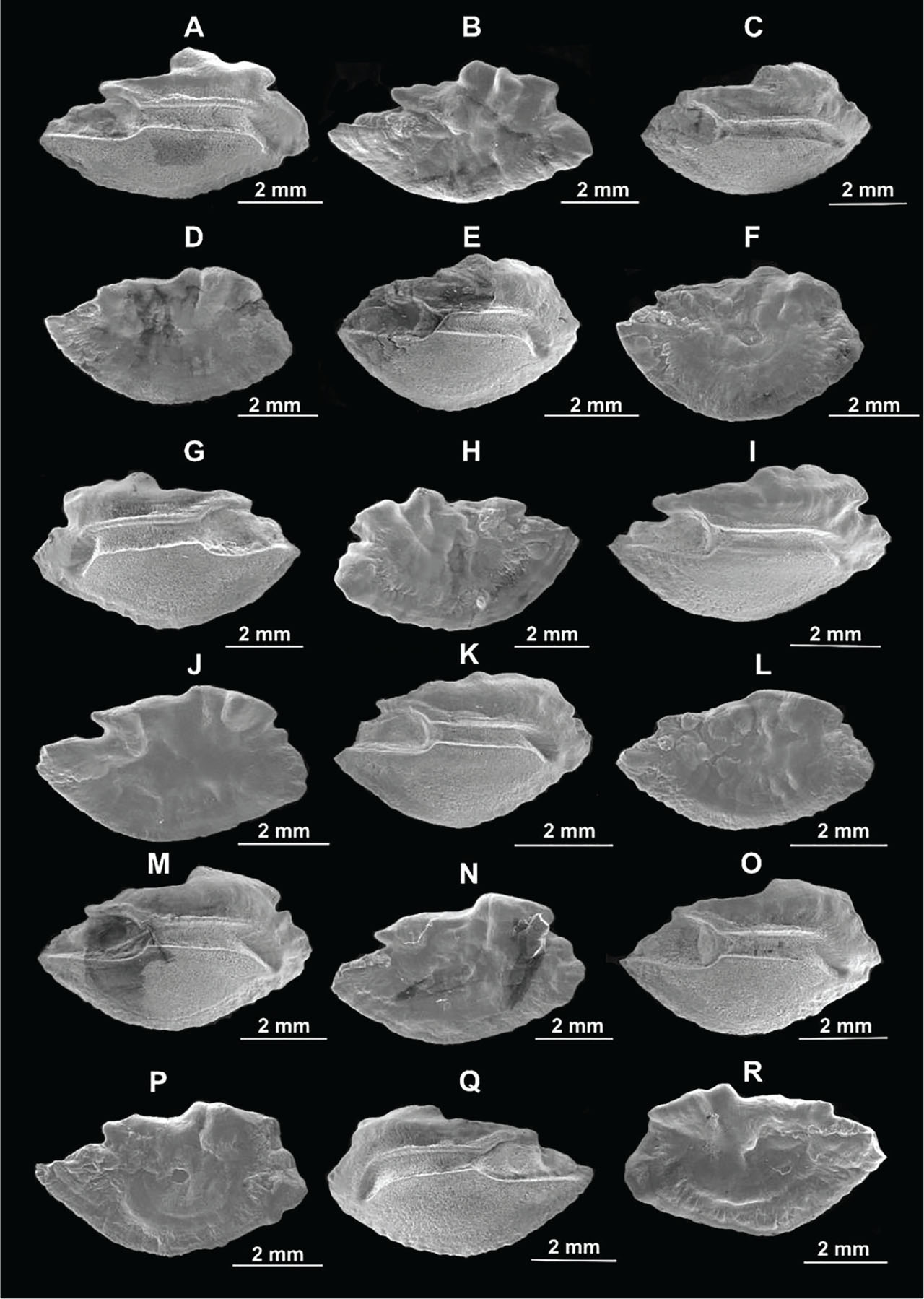
Figure 4
SEM images showing the lateral and the mesial sides of the otolith of B. boops from the Antalya Bay, Mediterranean coast of Türkiye. (A) Right otolith of a specimen with 138 mm TL male showing mesial side; (B) Left otolith of a specimen with 138 mm TL male showing lateral side; (C) Right otolith of a specimen with 140 mm TL male showing mesial side; (D) Left otolith of a specimen with 140 mm TL male showing lateral side; (E) Right otolith of a specimen with 140 mm TL female showing mesial side; (F) Left otolith of a specimen with 140 mm TL female showing lateral side; (G) Right otolith of a specimen with 142 mm TL male showing mesial side; (H) Left otolith of a specimen with 142 mm TL male showing lateral side; (I) Right otolith of a specimen with 145 mm TL female showing mesial side; (J) Left otolith of a specimen with 145 mm TL female showing lateral side; (K) Right otolith of a specimen with 149 mm TL male showing mesial side; (L) Left otolith of a specimen with 149 mm TL male showing lateral side; (M) Right otolith of a specimen with 150 mm TL male showing mesial side; (N) Left otolith of a specimen with 150 mm TL male showing lateral side; (O) Right otolith of a specimen with 155 mm TL male showing mesial side; (P) Left otolith of a specimen with 155 mm TL male showing lateral side; (Q) Left otolith of a specimen with 157 mm TL female showing mesial side; (R) Right otolith of a specimen with 157 mm TL female showing lateral side. SEM, scanning electron microscopy; TL, total length.
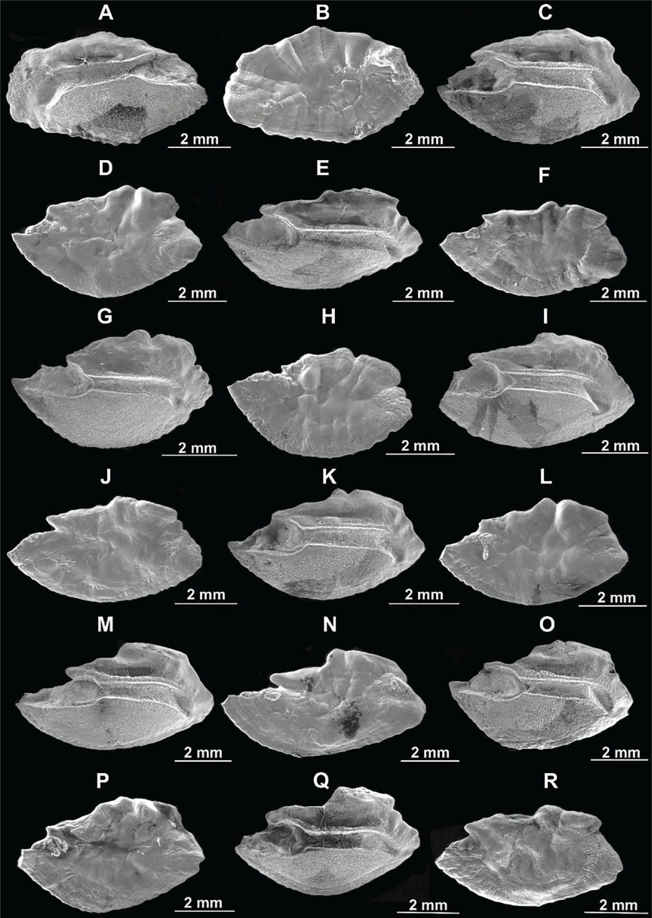
Figure 5
SEM images showing the lateral and the mesial sides of the otolith of B. boops from the Antalya Bay, Mediterranean coast of Türkiye. (A) Left otolith of a specimen with 162 mm TL male showing mesial side; (B) Right otolith of a specimen with 162 mm TL male showing lateral side; (C) Right otolith of a specimen with 163 mm TL female showing mesial side; (D) Left otolith of a specimen with 163 mm TL female showing lateral side; (E) Right otolith of a specimen with 165 mm TL male showing mesial side; (F) Left otolith of a specimen with 165 mm TL male showing lateral side; (G) Right otolith of a specimen with 165 mm TL female showing mesial side; (H) Left otolith of a specimen with 165 mm TL female showing lateral side; (I) Right otolith of a specimen with 166 mm TL female showing mesial side; (J) Left otolith of a specimen with 166 mm TL female showing lateral side; (K) Right otolith of a specimen with 166 mm TL female showing mesial side; (L) Left otolith of a specimen with 166 mm TL female showing lateral side; (M) Right otolith of a specimen with 166 mm TL male showing mesial side; (N) Left otolith of a specimen with 166 mm TL male showing lateral side; (O) Right otolith of a specimen with 167 mm TL male showing mesial side; (P) Left otolith of a specimen with 167 mm TL male showing lateral side; (Q) Right otolith of a specimen with 170 mm TL male showing mesial side; (R) Left otolith of a specimen with 170 mm TL male showing lateral side. SEM, scanning electron microscopy; TL, total length.
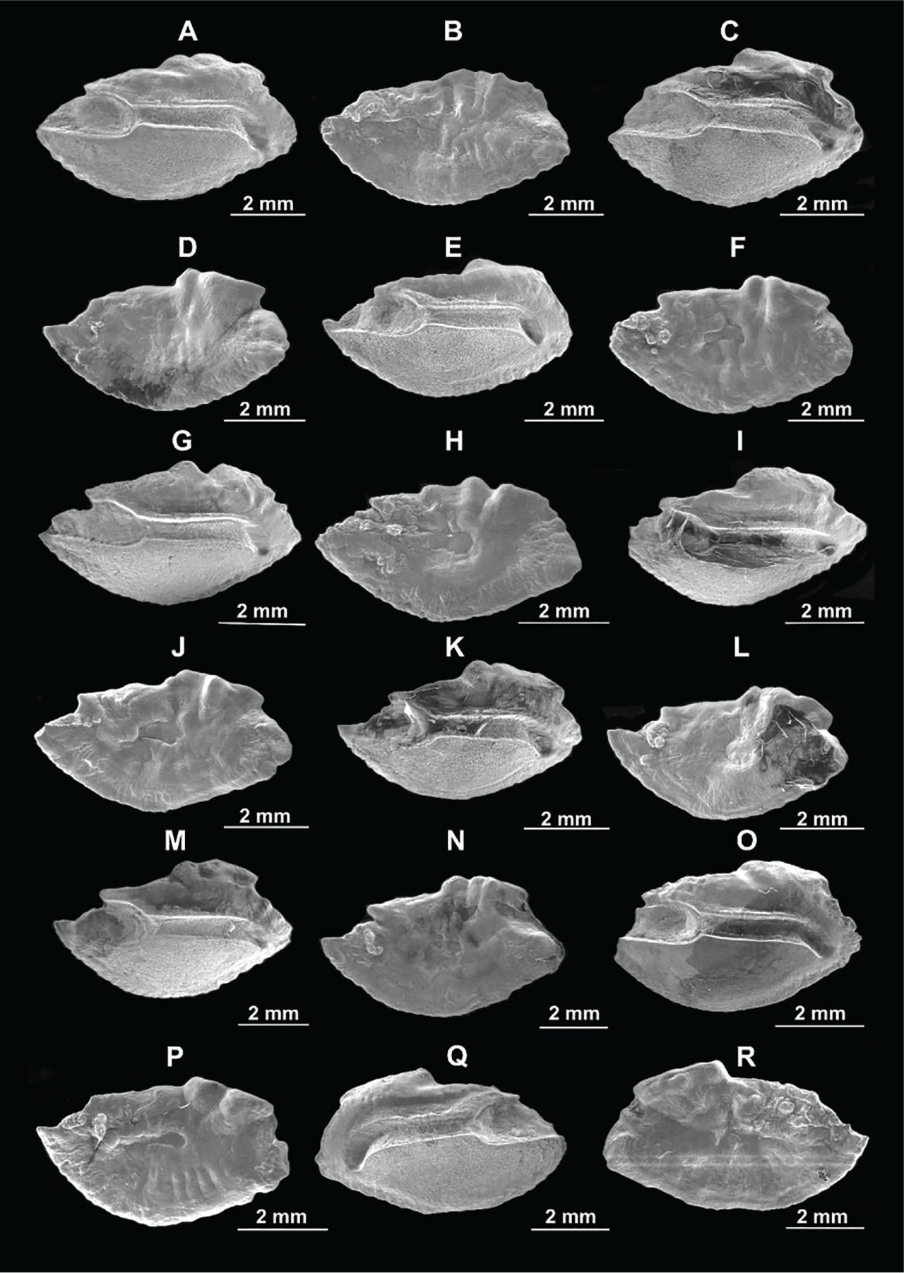
Figure 6
SEM images showing the lateral and the mesial sides of the otolith of B. boops from the Antalya Bay, Mediterranean coast of Türkiye. (A) Right otolith of a specimen with 172 mm TL female showing mesial side; (B) Left otolith of a specimen with 172 mm TL female showing lateral side; (C) Right otolith of a specimen with 172 mm TL male showing mesial side; (D) Left otolith of a specimen with 172 mm TL male showing lateral side; (E) Right otolith of a specimen with 175 mm TL female showing mesial side; (F) Left otolith of a specimen with 175 mm TL female showing lateral side; (G) Right otolith of a specimen with 175 mm TL male showing mesial side; (H) Left otolith of a specimen with 175 mm TL male showing lateral side; (I) Right otolith of a specimen with 175 mm TL male showing mesial side; (J) Left otolith of a specimen with 175 mm TL male showing lateral side; (K) Right otolith of a specimen with 178 mm TL female showing mesial side; (L) Left otolith of a specimen with 178 mm TL female showing lateral side; (M) Right otolith of a specimen with 180 mm TL male showing mesial side; (N) Right otolith of a specimen with 180 mm TL male showing lateral side; (O), Right otolith of a specimen with 180 mm TL male showing mesial side; (P) Left otolith of a specimen with 180 mm TL male showing lateral side; (Q) Left otolith of a specimen with 188 mm TL male showing mesial side; (R) Right otolith of a specimen with 188 mm TL male showing lateral side. SEM, scanning electron microscopy; TL, total length.
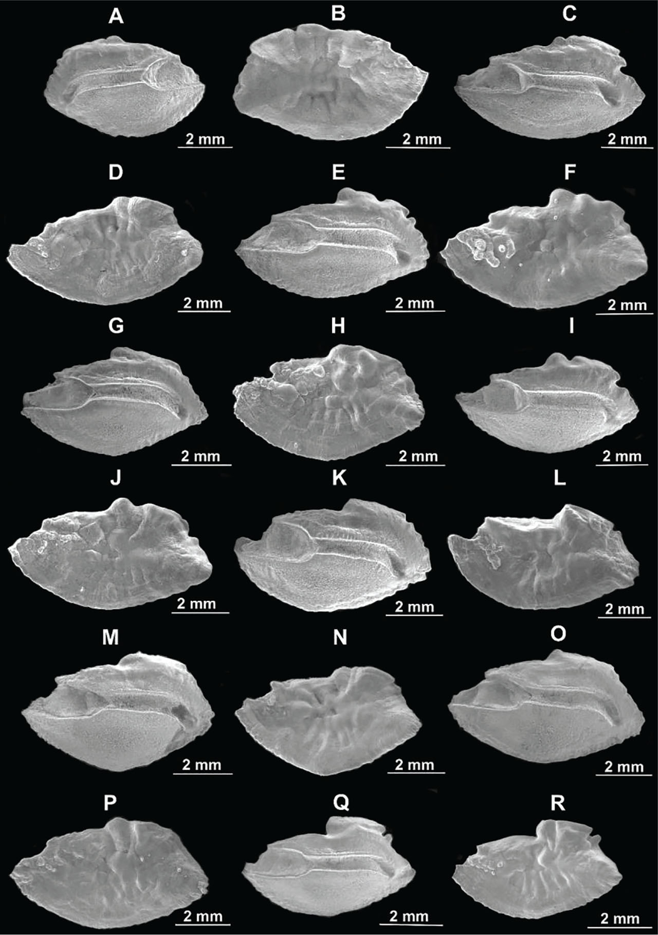
Figure 7
SEM images showing the lateral and the mesial sides of the otolith of B. boops from the Antalya Bay, Mediterranean coast of Türkiye. (A) Left otolith of a specimen with 191 mm TL male showing mesial side; (B) Right otolith of a specimen with 191 mm TL male showing lateral side; (C) Right otolith of a specimen with 195 mm TL male showing mesial side; (D) Left otolith of a specimen with 195 mm TL male showing lateral side; (E) Right otolith of a specimen with 195 mm TL female showing mesial side; (F) Left otolith of a specimen with 195 mm TL female showing lateral side; (G) Right otolith of a specimen with 197 mm TL female showing mesial side; (H) Left otolith of a specimen with 197 mm TL female showing lateral side; (I) Right otolith of a specimen with 202 mm TL male showing mesial side; (J) Left otolith of a specimen with 202 mm TL male showing lateral side; (K) Right otolith of a specimen with 203 mm TL female showing mesial side; (L) Left otolith of a specimen with 203 mm TL female showing lateral side; (M) Right otolith of a specimen with 203 mm TL male showing mesial side; (N) Left otolith of a specimen with 203 mm TL male showing lateral side; (O) Right otolith of a specimen with 204 mm TL male showing mesial side; (P) Left otolith of a specimen with 204 mm TL male showing lateral side; (Q) Right otolith of a specimen with 206 mm TL female showing mesial side; (R) Left otolith of a specimen with 206 mm TL female showing lateral side. SEM, scanning electron microscopy; TL, total length.
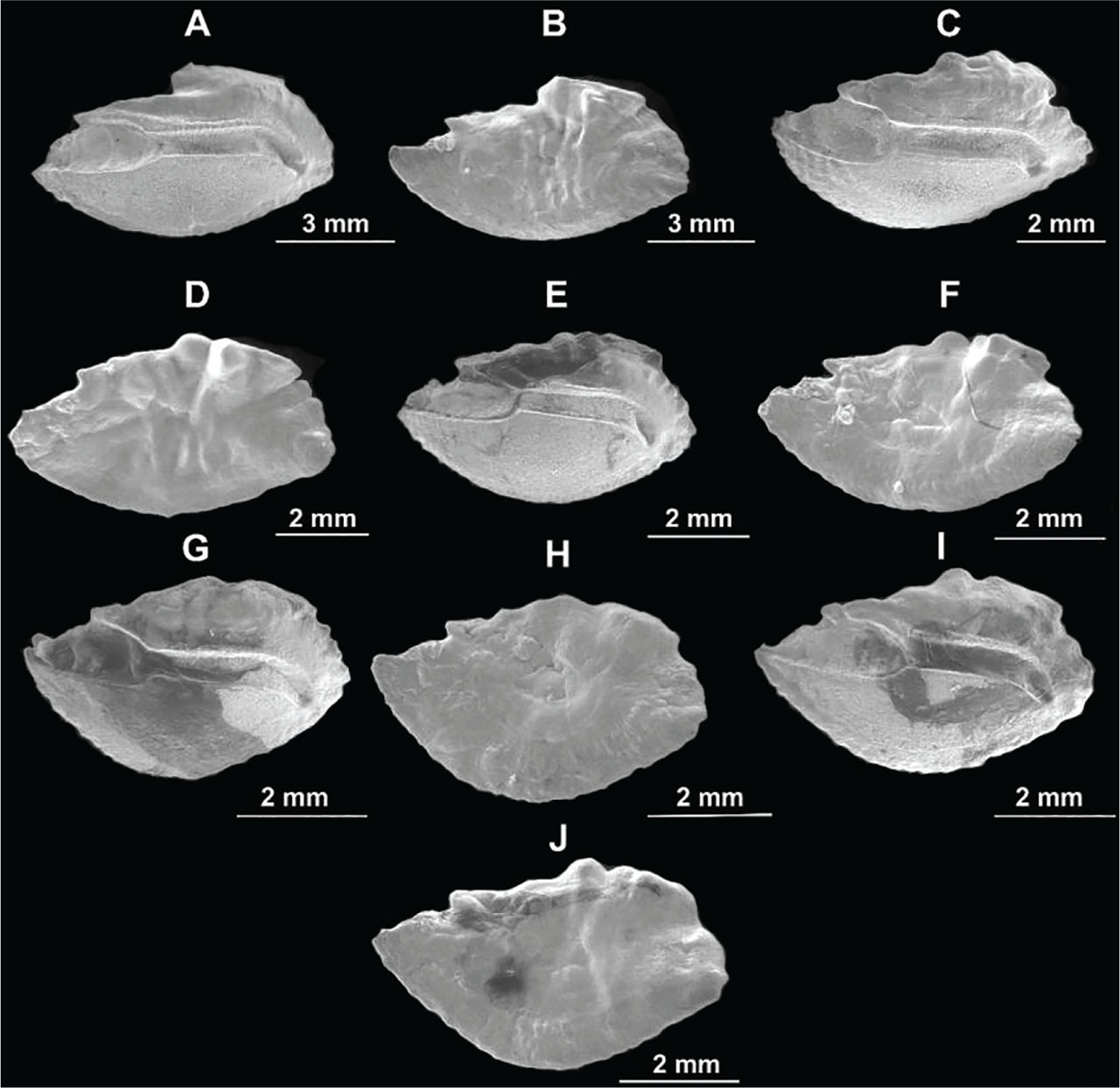
Figure 8
SEM images showing the lateral and the mesial sides of the otolith of B. boops from the Antalya Bay, Mediterranean coast of Türkiye. (A) Right otolith of a specimen with 215 mm TL female showing mesial side; (B) Left otolith of a specimen with 215 mm TL female showing lateral side; (C) Right otolith of a specimen with 215 mm TL female showing mesial side; (D) Left otolith of a specimen with 215 mm TL female showing lateral side; (E) Right otolith of a specimen with 217 mm TL female showing mesial side; (F) Left otolith of a specimen with 217 mm TL female showing lateral side; (G) Right otolith of a specimen with 223 mm TL female showing mesial side; (H) Left otolith of a specimen with 223 mm TL female showing lateral side; (I) Right otolith of a specimen with 225 mm TL female showing mesial side; (J) Left otolith of a specimen with 225 mm TL female showing lateral side. SEM, scanning electron microscopy; TL, total length.
In the otolith of N. randalli, coarse lobulations were observed on the lateral side mainly on the dorsal for the otolith of the four fish groups examined (Figs 9–12).
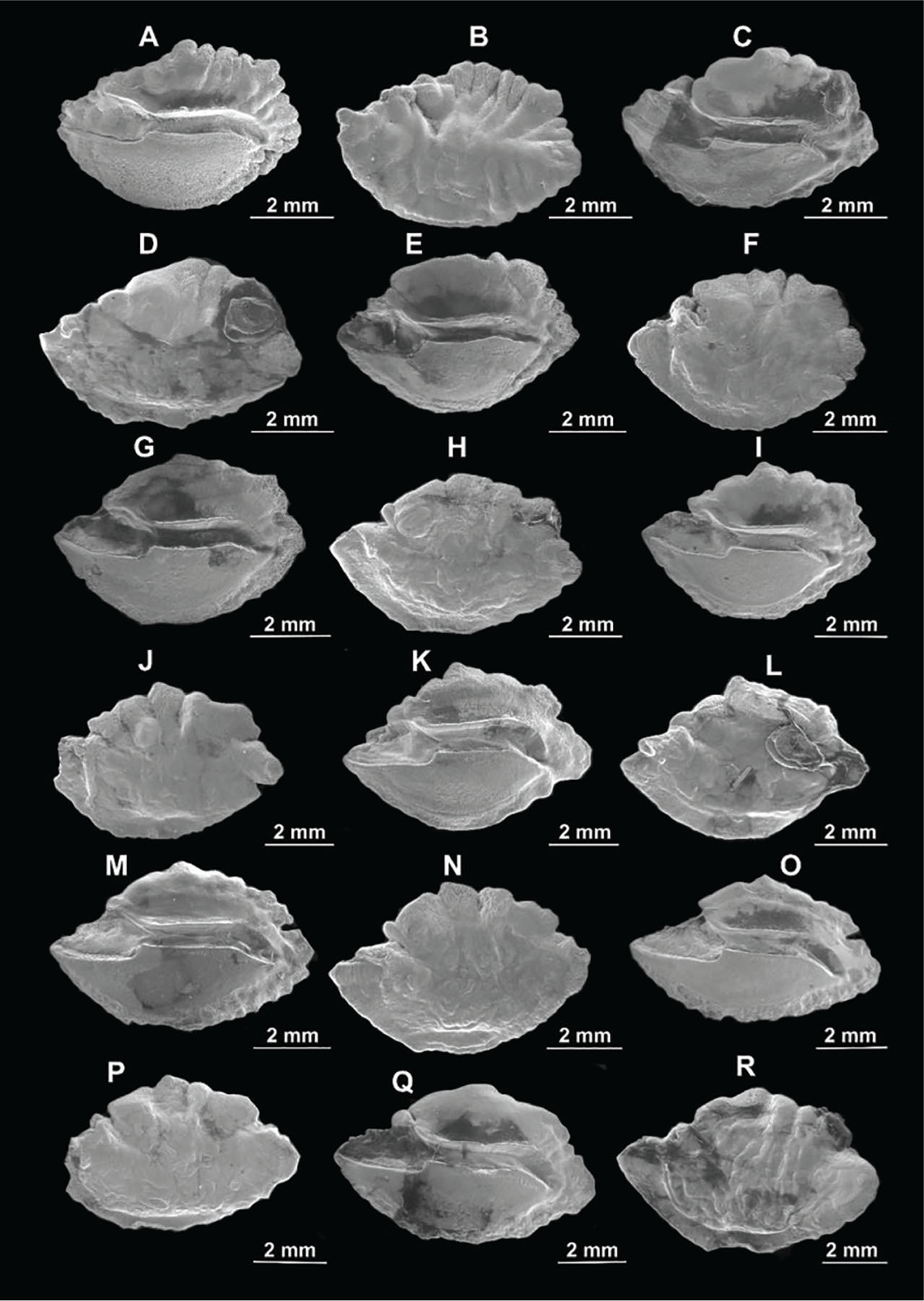
Figure 9
SEM images showing the lateral and the mesial sides of the otolith of N. randalli from the Antalya Bay, Mediterranean coast of Türkiye. (A) Right otolith of a specimen with 126 mm TL female showing mesial side; (B) Left otolith of a specimen with 126 mm TL female showing lateral side; (C) Right otolith of a specimen with 130 mm TL male showing mesial side; (D) Left otolith of a specimen with 130 mm TL male showing lateral side; (E) Right otolith of a specimen with 139 mm TL female showing mesial side; (F) Left otolith of a specimen with 139 mm TL female showing lateral side; (G) Right otolith of a specimen with 144 mm TL female showing mesial side; (H) Left otolith of a specimen with 144 mm TL female showing lateral side; (I) Right otolith of a specimen with 145 mm TL female showing mesial side; (J) Left otolith of a specimen with 145 mm TL female showing lateral side; (K) Right otolith of a specimen with 145 mm TL male showing mesial side; (L) Left otolith of a specimen with 145 mm TL male showing lateral side; (M) Right otolith of a specimen with 147 mm TL male showing mesial side; (N) Left otolith of a specimen with 147 mm TL male showing lateral side; (O) Right otolith of a specimen with 150 mm TL male showing mesial side; (P) Left otolith of a specimen with 155 mm TL male showing lateral side; (Q) Right otolith of a specimen with 155 mm TL male showing mesial side; (R) Left otolith of a specimen with 150 mm TL male showing lateral side. SEM, scanning electron microscopy; TL, total length.
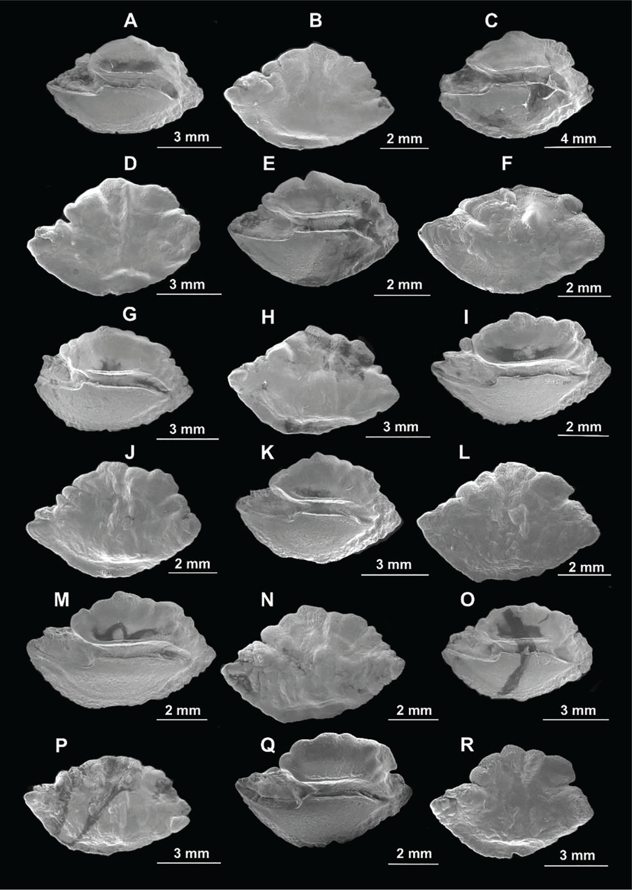
Figure 10
SEM images showing the lateral and the mesial sides of the otolith of N. randalli from the Antalya Bay, Mediterranean coast of Türkiye. (A) Right otolith of a specimen with 160 mm TL male showing mesial side; (B) Left otolith of a specimen with 160 mm TL male showing lateral side; (C) Right otolith of a specimen with 162 mm TL male showing mesial side; (D) Left otolith of a specimen with 162 mm TL male showing lateral side; (E) Right otolith of a specimen with 165 mm TL female showing mesial side; (F) Left otolith of a specimen with 165 mm TL female showing lateral side; (G) Right otolith of a specimen with 166 mm TL male showing mesial side; (H) Left otolith of a specimen with 166 mm TL male showing lateral side; (I) Right otolith of a specimen with 166 mm TL female showing mesial side; (J) Left otolith of a specimen with 166 mm TL female showing lateral side; (K) Right otolith of a specimen with 168 mm TL female showing mesial side; (L) Left otolith of a specimen with 168 mm TL female showing lateral side; (M) Right otolith of a specimen with 171 mm TL female showing mesial side; (N) Left otolith of a specimen with 171 mm TL female showing lateral side; (O) Right otolith of a specimen with 175 mm TL female showing mesial side; (P) Left otolith of a specimen with 175 mm TL female showing lateral side; (Q) Right otolith of a specimen with 175 mm TL female showing mesial side; (R) Left otolith of a specimen with 175 mm TL female showing lateral side. SEM, scanning electron microscopy; TL, total length.
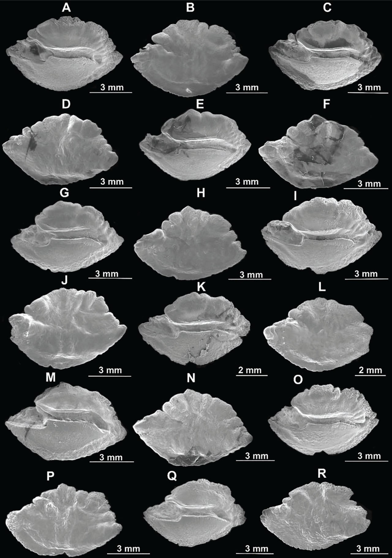
Figure 11
SEM images showing the lateral and the mesial sides of the otolith of N. randalli from the Antalya Bay, Mediterranean coast of Türkiye. (A) Right otolith of a specimen with 183 mm TL male showing mesial side; (B) Left otolith of a specimen with 183 mm TL male showing lateral side; (C) Right otolith of a specimen with 184 mm TL male showing mesial side; (D) Left otolith of a specimen with 184 mm TL male showing lateral side; (E) Right otolith of a specimen with 185 mm TL male showing mesial side; (F) Left otolith of a specimen with 185 mm TL male showing lateral side; (G) Right otolith of a specimen with 185 mm TL female showing mesial side; (H) Left otolith of a specimen with 185 mm TL female showing lateral side; (I) Right otolith of a specimen with 188 mm TL male showing mesial side; (J) Left otolith of a specimen with 188 mm TL male showing lateral side; (K) Right otolith of a specimen with 190 mm TL male showing mesial side; (L) Left otolith of a specimen with 190 mm TL male showing lateral side; (M) Right otolith of a specimen with 190 mm TL female showing mesial side; (N) Left otolith of a specimen with 190 mm TL female showing lateral side; (O) Right otolith of a specimen with 195 mm TL female showing mesial side; (P) Left otolith of a specimen with 195 mm TL female showing lateral side; (Q) Right otolith of a specimen with 197 mm TL female showing mesial side; (R) Left otolith of a specimen with 197 mm TL female showing lateral side. SEM, scanning electron microscopy; TL, total length.
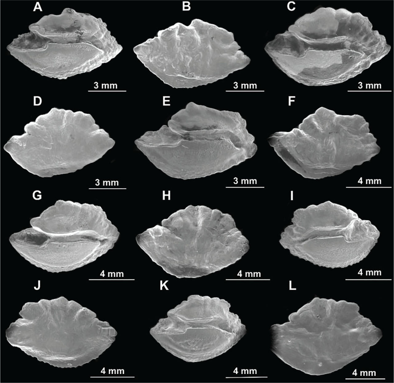
Figure 12
SEM images showing the lateral and the mesial sides of the otolith of N. randalli from the Antalya Bay, Mediterranean coast of Türkiye. (A) Right otolith of a specimen with 203 mm TL male showing mesial side; (B) Left otolith of a specimen with 203 mm TL male showing lateral side; (C) Right otolith of a specimen with 204 mm TL male showing mesial side; (D) Left otolith of a specimen with 204 mm TL male showing lateral side; (E) Right otolith of a specimen with 210 mm TL male showing mesial side; (F) Left otolith of a specimen with 210 mm TL male showing lateral side; (G) Right otolith of a specimen with 222 mm TL male showing mesial side; (H) Left otolith of a specimen with 222 mm TL male showing lateral side; (I) Left otolith of a specimen with 227 mm TL male showing mesial side; (J) Right otolith of a specimen with 227 mm TL male showing lateral side; (K) Right otolith of a specimen with 240 mm TL male showing mesial side; (L) Left otolith of a specimen with 240 mm TL male showing lateral side. SEM, scanning electron microscopy; TL, total length.
A distinct ontogenetic shift in otolith morphology was detected in the otoliths of B. boops and N. randalli. Eight otolith characteristics were observed to be unchanged in the otoliths of the small- and large-sized specimens of these two species. These are: otolith width and thickness; the contour of the mesial and lateral surfaces; the configuration of the sulcus acusticus; the ostio-caudal dif ferentiation; and the shapes of the ventral depression and the rostrum thickness (Tables 1 and 2, Figs 4–12).
The analysis of 13 key otolith features distinguishing small-sized specimens from large-sized specimens revealed significant variations in the otolith morphology of B. boops and N. randalli (Tables 1 and 2, Figs 4–12). In B. boops, the otoliths of juvenile individuals (Group I, 130–150 mm TL) exhibited elliptical, oblong, or rectangular shapes, which transformed into elongated, elliptical, oblong, and rectangular shapes in Group II (151–170 mm TL). This pattern continued in Group III (171–190 mm TL) with elliptical, hexagonal, oblong, and rectangular shapes, and in Group IV (191–210 mm TL) and Group V (211–230 mm TL) with a mix of elliptical, hexagonal, oblong, and rectangular shapes.
For N. randalli, juvenile otoliths (Group I, 120–150 mm TL) were elliptical or hexagonal, transitioning to elliptical, hexagonal, and rectangular shapes in Group II (151–180 mm TL), primarily elliptical or hexagonal in Group III (181–210 mm TL), and predominantly elliptical in Group IV (211–240 mm TL).
In B. boops, the dorsal margin of the otolith varied by developmental stage. In the small-sized specimens (Group I, 130–150 mm TL), it was either coarsely lobate or finely wavy, and high or low. In Group II (151–170 mm TL), the margin was raised or irregular posteriorly, and smooth, finely irregular, or coarsely irregular anteriorly. Similar trends were observed in Group III (171–190 mm TL), Group IV (191–210 mm TL), and Group V (211–230 mm TL) (Table 1, Figs 4–8).
N. randalli displayed similar otolith features to B. boops, with adult otoliths (Groups III and IV) showing a nearly identical, coarsely lobulated dorsal margin (Table 2, Figs 9–12).
The ventral margin of B. boops otoliths remained relatively consistent across all groups, being either finely serrated or smooth (Table 1, Figs 4–8). In N. randalli, the ventral margin in the small-sized specimens (Groups I and II) was either smooth, serrated, or finely irregular, while in larger fish (Groups III and IV), it was smooth or finely serrated (Table 2, Figs 9–12).
The posterior margin of B. boops otoliths varied significantly. In the small-sized specimens (Group I), it was either short or long and uniformly rounded or irregular, evolving into a long or short, irregularly rounded, or finely pointed shape in Group V (Table 1, Figs 4–8). Similar variability was noted in N. randalli, with the juvenile otoliths (Group I) showing irregularly rounded, finely pointed, or short, irregular shapes. The otoliths of larger groups (Group III and IV) displayed more consistency with broadly or finely pointed and irregular posterior margins (Table 2, Figs 9–12).
The shape of the ostium in B. boops showed limited variability across groups, generally being deep or shallow, long or short in Groups I through III, with slight variations in Groups IV and V (Table 1, Figs 4–8). Similar trends were noted in N. randalli across all groups (Table 2, Figs 9–12).
The cauda of B. boops otoliths exhibited variations, being either long or short, and deep or shallow posteriorly (Table 1, Figs 4–8). In N. randalli, the cauda showed some differences, particularly in Group III, where calcium carbonate secretions were observed filling the posterior part of the cauda (Table 2, Figs 9–12).
The shape of the dorsal depression in B. boops otoliths varied consistently, ranging from narrow, deep, or shallow in Groups I and II, to more complex shapes like elongated or curved in later groups (Table 1, Figs 4–8). In N. randalli, juvenile otoliths displayed broad or narrow, oval or elongated shapes, while larger fish (Groups III and IV) showed wide, oval, deep, or shallow characteristics (Table 2, Figs 9–12).
The rostrum shape and size also varied. In B. boops, the rostrum was broadly or finely pointed in Groups I and II, and broadly or finely pointed, straight, or curved upward in later groups. Rostrum size ranged from long to short across all groups (Table 1, Figs 4–8). Similarly, in N. randalli, the rostrum was broadly or finely pointed, straight or curved upward, and its size varied between long and short in all groups (Table 2, Figs 9–12).
The shape of the antirostrum in B. boops varied widely across all groups, ranging from absent to poorly developed, and from short to long, pointed, rounded, or broad (Table 1, Figs 4–8). Similar features were observed in N. randalli across all groups (Table 2, Figs 9–12).
Although the shape of the excisura and notch varied in B. boops, these variations were consistent across all five size groups (Table 1, Figs 4–8). The same pattern was observed in N. randalli (Table 2, Figs 9–12).
3.2. Sexual dimorphism
Variations in otolith surface morphology between female and male individuals were identified in both B. boops and N. randalli in this study (Tables 3 and 4, Figs 4–12).
Table 3
Comparison of the surface morphology of the otolith between female and male specimens of B. boops collected from the Antalya Bay, Mediterranean coast of Türkiye.
| Fish TL (mm) | Sex | Morphological characters | Fish size (mm) | Sex | Morphological characters |
|---|---|---|---|---|---|
| 140 | Female | Otolith with pentagonal shape, posterior margin slightly produced, posterior part of dorsal margin slightly raised, cauda curved vertically posteriorly, antirostrum absent, lateral side, with coarse lobes at posterior margin. | 140 | Male | Otolith with oblong shape, posterior margin broadly pointed, posterior part of dorsal margin with a pronounced broad projection, cauda straight posteriorly, antirostrum small, lateral side with five lobes at posterior dorsal margin. |
| 145 | Female | Posterior margin produced, dorsal depression broad, ostium shallow, cauda straight posteriorly, antirostrum long, excisura shallow and notch narrow, lateral side is mainly smooth. | 142 | Male | Posterior margin short and straight, dorsal depression narrow, ostium deep, cauda curved posteriorly, antirostrum poorly developed, excisura shallow and notch absent, lateral side is covered with coarse lobes. |
| 157 | Female | Posterior margin with no notch, dorsal depression narrow and deep, cauda deep, excisura shallow and notch narrow, lateral side waved, with no lobulations. | 155 | Male | Posterior margin with notch, dorsal depression broad and shallow, cauda shallow posteriorly, excisura absent, lateral side covered with coarse lobes. |
| 163 | Female | Otolith with elliptic shape, posterior margin broadly pointed, dorsal depression wide, ventral depression shallow, antirostrum small, but well developed, excisura shallow and notch narrow, the dorsal region of the lateral side covered with coarse lobulation. | 162 | Male | Otolith with oblong shape, posterior margin straight, dorsal depression narrow, ventral depression deep, antirostrum absent, excisura absent, the surface of the lateral side covered with fine wavy lobulation. |
| 165 | Female | Otolith with oblong shape, posterior margin finely lobulated, with irregular posterior end, dorsal depression wide, ostium and cauda shallow, rostrum with a pointed tip, antirostrum short. | 165 | Male | Otolith with elliptic shape, posterior margin broadly pointed, with coarsely lobulated posterior end, dorsal depression narrow, ostium and cauda deep, rostrum with a broadly pointed tip, antirostrum long. |
| 166 | Female | Otolith with elliptic shape, posterior margin pointed, dorsal depression shallow, ostium long and shallow, the surface of the lateral side is covered with coarse lobulation. | 166 | Male | Otolith with oblong shape, posterior margin broadly pointed, dorsal depression deep, ostium short and deep, the surface of the lateral side is covered with fine lobulation. |
| 172 | Female | Otolith with oblong shape, posterior margin produced, dorsal depression shallow, antirostrum small, lateral side is covered with fine lobulation mainly in the middle. | 172 | Male | Otolith with elliptical shape, posterior margin broadly pointed, dorsal depression deep, antirostrum absent, lateral side is covered with coarse lobulation mainly in the middle. |
| 175 | Female | Otolith with oblong shape, posterior margin rounded, dorsal depression narrow, rostrum with a pointed tip, antirostrum poorly developed, excisura absent. | 175 | Male | Otolith with elliptical shape, posterior margin broadly pointed, dorsal depression wide, rostrum with a broadly pointed, antirostrum long, excisura wide and notch deep. |
| 178 | Female | Otolith with elliptical shape, posterior margin nearly smooth, dorsal depression deep, ostium and cauda deep, antirostrum small, excisura absent, the surface of the lateral side is covered with coarsely lobulations near the posterior margin. | 180 | Male | Otolith with hexagonal shape, posterior margin wavy, dorsal depression narrow and deep, ostium and cauda shallow, antirostrum long, excisura wide and notch deep, the surface of the lateral side is entirely covered with fine lobulations. |
| 195 | Female | Dorsal side of the posterior margin irregular, dorsal depression narrow and shallow, ostium shallow, antirostrum absent, the surface of the lateral side is entirely covered with coarse lobulations mainly at the center. | 195 | Male | Dorsal side of the posterior margin with a notch, dorsal depression wide and deep, ostium deep, antirostrum developed but small, the surface of the lateral side is entirely covered with fine lobulations mainly at the centre. |
| 203 | Female | Otolith with oblong shape, dorsal and posterior margins with no notch, dorsal depression wide, rostrum with a broadly pointed tip and curved dorsally, ostium and cauda deep, antirostrum absent. | 202 | Male | Otolith with elliptical shape, dorsal and posterior margins with notch, dorsal depression narrow, rostrum with a pointed and straight tip, ostium and cauda shallow, antirostrum developed, but small. |
| 206 | Female | Otolith with spindle shape, dorsal margin with conspicuous projection, posterior margin finely pointed, rostrum with a pointed tip, antirostrum absent, excisura absent, cauda deep and straight, lateral side is covered evenly distributed lobulations. | 204 | Male | Otolith with elliptical shape, dorsal margin straight and raised, posterior margin broadly pointed, rostrum with a broad tip, antirostrum absent, excisura developed, but small, cauda shallow and slightly curved posteriorly, lateral side is covered with coarse lobes at the center. |
Table 4
Comparison of the surface morphology of the otolith between female and male specimens of N. randalli collected from the Antalya Bay, Mediterranean coast of Türkiye.
| Fish TL (mm) | Sex | Morphological characters | Fish size (mm) | Sex | Morphological characters |
|---|---|---|---|---|---|
| 126 | Female | Posterior part of the ventral margin coarsely lobulated, dorsal depression narrow and deep, ventral depression shallow, rostrum with straight tip, cauda curved posteriorly, ostium shallow, the surface of the lateral side is covered with small lobulations. | 130 | Male | Posterior part of the ventral margin irregular, dorsal depression wide and shallow, ventral depression deep, rostrum with broadly pointed tip, cauda straight posteriorly, ostium deep, the surface of the lateral side is waved and covered with small lobulations. |
| 145 | Female | Posterior margin with notch, antirostrum small, excisura wide and notch shallow, ostium and cauda narrow and shallow, cauda slightly curved posteriorly, in the lateral side, a few coarse lobes are present on the dorsal margin. | 145 | Male | Posterior margin with no notch and produced, antirostrum absent, excisura absent, ostium and cauda wide and deep, cauda strongly curved posteriorly, on the lateral side, the anterior and the posterior ends are covered with coarse lobes. |
| 165 | Female | Otolith with oval shape, dorsal margin with two prominent lobes anteriorly, ventral margin smooth, posterior margin slightly produced and rounded, dorsal depression deep, ostium and cauda are broad and deep, rostrum broad and well developed, the surface of the lateral side is evenly covered with coarsely lobes. | 166 | Male | Otolith with elliptical shape, dorsal margin finely emarginated, ventral margin finely serrated, posterior margin short and broadly pointed, dorsal depression shallow, ostium and cauda are narrow and shallow, rostrum short and poorly developed, the surface of the lateral side covered with coarsely lobes mainly on the dorsal margin. |
| 175 | Female | Otolith with broad rectangular shape, dorsal margin emarginated, ventral margin dentate. | 183 | Male | Otolith with elliptical shape, dorsal margin irregular, ventral margin smooth. |
| 185 | Female | Posterior margin produced and finely pointed, ventral margin slightly notched at center, posterior part of the dorsal margin with coarse lobes, dorsal depression shallow, ostium and cauda shallow, the surface of the lateral side is covered with coarsely lobes mainly on dorsal margin. | 185 | Male | Posterior margin short and finely pointed, ventral margin smooth, posterior part of the dorsal margin with fine lobes, dorsal depression deep, ostium and cauda deep, the surface of the lateral side is covered evenly with coarse lobes. |
| 190 | Female | Posterior margin broadly pointed, ventral margin smooth, rostrum with pointed tip, ostium and cauda deep, cauda sharply curved posteriorly, excisura narrow and notch deep, antirostrum well developed and broadly pointed, the surface of the lateral side is covered with irregular lobes mainly on dorsal margin. | 190 | Male | Posterior margin notched, ventral margin notched, rostrum with rounded tip, ostium and cauda shallow, cauda slightly curved posteriorly, excisura wide and notch shallow, antirostrum well developed, but small, the surface of the lateral side is covered with evenly distributed lobes. |
| 197 | Female | Posterior margin irregular, dorsal margin wavy and irregular posteriorly, dorsal depression shallow, antirostrum short and poorly developed. | 203 | Male | Posterior margin broadly pointed, dorsal margin coarsely wrinkelled, dorsal depression deep, antirostrum long and well developed. |
For B. boops, otolith morphology was compared between 12 pairs of females and males of similar or nearly equal TLs. Female TLs ranged from 149 mm to 206 mm, while males ranged from 140 mm to 204 mm.
In the smallest pair (140 mm female and 140 mm male), differences were observed in several otolith features, including overall shape, posterior margin, posterior section of the dorsal margin, cauda, presence or absence of the antirostrum, and the surface morphology of the lateral side. In the largest pair (206 mm female and 204 mm male), differences were noted in the otolith shape, dorsal and posterior margins, tip of the rostrum, presence or absence of the antirostrum, and the shapes of the excisura and notch, along with the lateral surface morphology.
The fewest differences between sexes were seen in the 157 mm female and 155 mm male pair, with four distinct traits. In contrast, the greatest number of differences—eight distinct characteristics—was found in two pairs: 163 mm female and 162 mm male.
For N. randalli, otolith morphology was compared between seven pairs of females and males of similar or nearly equal TLs. Females ranged from 126 mm to 197 mm, while males measured between 130 mm and 203 mm.
In the smallest pair (126 mm female and 130 mm male), differences were identified in seven otolith features, including the posterior section of the ventral margin, the shape and depth of both the dorsal and ventral depressions, the tip of the rostrum, the cauda, the shape and depth of the ostium, and the lateral surface morphology. In the largest pair (197 mm female and 203 mm male), variations were observed in four otolith features: the posterior and dorsal margins, the depth of the dorsal depression, and the shape of the antirostrum.
The fewest variations were seen in the 175 mm female and 183 mm male pair, with three distinct traits. The greatest number of differences—eight distinct characteristics—was noted in two pairs: 165 mm female and 166 mm male.
4. Discussion
4.1. General
The discussion on otolith morphology in B. boops (Sparidae) and N. randalli (Nemipteridae) can be significantly enhanced by incorporating SEM as a key analytical tool. SEM provides high-resolution, three-dimensional imaging of otolith surfaces and microstructures, enabling detailed morphological and microchemical analyses that are crucial for taxonomic, ontogenetic, and ecological studies.
The identification of species within the families Sparidae and Nemipteridae relies on a combination of morphological and meristic characters. In Sparidae, key identifying features include body shape, fin structure, and the arrangement of teeth, with many species exhibiting distinctive color patterns that aid in recognition (Froese & Pauly, 2024). The number and structure of dorsal and anal fins, as well as the presence of specific spines, are critical for differentiation among species. For Nemipteridae, characteristics such as elongated body form, prominent thread-like dorsal fin rays, and distinct lateral line patterns are essential for species identification (Matsuura, 2016). Additionally, the analysis of scale morphology and pigmentation patterns provides valuable information for taxonomists. Overall, the integration of these morphological traits, supported by molecular data, enhances the accuracy of species identification within these families.
Since the early 20th century, various features of the saccular otolith have been employed in taxonomic research (e.g., Chaine & Duvergier, 1934), despite significant variation among species (Jawad, 2007). This study focused on the otolith morphology of the sparid species B. boops and the nemipterid species N. randalli, exploring a broad array of characteristics. However, only a limited number of these traits were identified as taxonomically significant and useful for systematic classification.
The findings of this study identified two distinct categories of characteristics: (1) features that remain stable in otoliths across various length groups of fish, which are useful for species identification and (2) traits that change with ontogenetic development, yet still provide valuable information for determining developmental stages.
4.2. Ontogenetic variation
Several characteristics of the sagittal otolith can provide valuable insights for taxonomic research (see Fig. 3). Some of these features were first identified in the early 20th century (Nolf, 1985, 2013). Among teleost fish, the sagittal otolith is extensively utilized in comparative taxonomic studies due to its relatively large size and significant interspecies variation (Hoedemakers et al., 2024; Jawad, 2007; Jawad et al., 2018a, 2018b). In this study, we evaluate various otolith traits that are crucial for identifying species at the level of individual species. These traits encompass otolith width and thickness, the configuration of the mesial and lateral surfaces, the structure of the sulcus acusticus, the presence or absence of ostio-caudal differentiation, and the shapes of the crista superior and inferior in the otoliths of B. boops and N. randalli examined.
The crista superior and crista inferior in the otoliths of B. boops and N. randalli exhibit variations in shape, making them unreliable for distinguishing between otoliths from different length categories. The formation and positioning of the nucleus are influenced by the release of soluble Ca2+ on the proximal side (Ibsch et al., 2004). This calcium precipitates as CaCO3 crystals along an increasingly alkaline gradient from the sulcal area to the otolith’s periphery (Gauldie & Nelson, 1990). This process enhances the growth of the cristae, particularly on the sulcal side. In teleost fish, the macula is typically elongated and narrow, with the cristae superior and inferior playing a more critical role than the colliculum (Ladich & Popper, 2001). The macula’s orientation toward the column restricts growth in that region (Pannella, 1980; Popper & Hoxter, 1981). This pattern is evident in certain species where the column may be underdeveloped or absent. Research by Lombarte et al. (2003) found that the shape of the sagitta in Merluccius is influenced by genetic, developmental, and environmental factors. Studies on both fossil and contemporary fish otoliths indicate that sulcus morphology tends to remain consistent within species of the same genus (Nolf, 1985), suggesting a possible genetic basis for this characteristic (Gauldie, 1988).
The otoliths of juvenile individuals from the species B. boops and N. randalli differ from those of adults in 13 out of the 21 analyzed traits (Tables 1 and 2). These differences include variations in otolith shape and changes in the dorsal, ventral, and posterior margins, ostium and cauda, dorsal and ventral depressions, and the size, shape, and thickness of the rostrum. Additional differences were observed in the shape of the antirostrum and the structure of the excisura and notch. These changes reflect ontogenetic development. Similar findings have been documented by previous research, including Jawad (2007) on triplefin species, Jawad et al. (2008) on Scaridae species from the Solomon Islands, Jawad et al. (2023) on Lethrinidae species in Yemeni waters, and Reıs et al. (2023) on the mullet Chelon auratus from Köyceğiz Lagoon, Aegean Sea, Türkiye. Due to the variability observed across the nine size groups of the two species examined, these traits are not reliable for individual identification. The variations may be related to the organic matrix and the deposition process of CaCO3 during sagittal development (Volpedo & Echeverría, 2003). In the species examined, calcium deposition during otolith growth appears uneven, as indicated by the inconsistent shape across different stages of ontogenetic development. This observation is consistent with the findings of Smale et al. (1995) on Hipposcarus harid (325 and 424 mm), as well as with descriptions of other scarid species such as Scarus rivulatus (291 mm) by Rivaton and Bourret (1999), Hipposcarus longiceps (203 mm) by Lin and Chang (2012), and Scarus ghobban (191 mm) by Sadighzadeh et al. (2012).
In the early life stages, otoliths are small and exhibit a rapid rate of growth, making them highly susceptible to reshaping by environmental influences. The transition from a circular larval otolith to a more elongated form in adults is observed in many species (Hüssy, 2008). Research has shown that otolith shapes tend to remain stable in smaller individuals, with more pronounced changes occurring as the fish mature (Capoccioni et al., 2010; Vignon, 2012). These observations differ from findings that indicate significant habitat impacts on otolith growth in juvenile species compared to their adult counterparts (Lombarte et al., 2003).
The development of otolith shape is mainly governed by ontogeny beyond the early life stages, during which environmental factors have a limited impact on overall morphology. This initial stability may represent a biological constraint associated with the otolith’s function in sound perception (Gauldie & Nelson, 1990). Proteins present in the endolymphatic epithelium play a crucial role in regulating otolith shape, while the physiological factors within the endolymph that promote biomineralization are subject to genetic control (Borelli et al., 2003; Morales-Nin, 2000). This could explain why the early stages of otolith development adhere to a predetermined ontogenetic trajectory, with growth primarily driven by the deposition of calcium carbonate and proteins.
Members of both B. boops and N. randalli across different size categories display eight consistent otolith traits, irrespective of size (Tables 1 and 2). These reliable characteristics may provide valuable cues for identifying individuals of these species and can be regarded as key distinguishing features.
The surface morphology of the lateral side of the otolith of B. boops and N. randalli species exhibits ontogenetic variation. This variation is primarily reflected in the irregularities and lobulations around different edges of the otolith. These features are especially pronounced in both species investigated. The main features of the lateral side of the otolith of both species examined are the presence of coarse flocculation distributed unevenly on this surface and mainly on the dorsal side of the otolith. This characteristic could not hold taxonomic significance and may not be a useful diagnostic feature for the two species studied.
The results of this study indicated a consistent shape in the sulcus acusticus, specifically heterosulcoid and ostial, observed in the otoliths of both juvenile and adult specimens of B. boops and N. randalli. Previous investigations into fossil and modern otoliths have shown that this feature typically retains a uniform shape within species of the same genus (Jawad et al., 2023; Nolf, 1985), suggesting a potential genetic basis (Gauldie, 1988). In a similar vein, Reichenbacher et al. (2007) successfully classified species within the genus Aphanius (Cyprinodontiformes) into distinct groups based on the specific morphologies of the sulcus acusticus.
This study found that the otolith shapes of B. boops individuals varied, exhibiting elliptical, oblong, and rectangular forms. Young individuals primarily displayed elongated and elliptical shapes, while adults showed a mix of elliptical, hexagonal, oblong, and rectangular shapes. In N. randalli, there was considerable variation as well. Juvenile otoliths presented a range of forms, including elliptical and hexagonal, which transitioned to predominantly elliptical, hexagonal, and rectangular shapes in adults. The ontogenetic differences in otolith shape between the small and large-sized specimens are significant, as they may complicate the distinction between these age groups (Tables 1 and 2, Figs 4–12).
Differences in body coloration are also significant in brightly colored, sympatric species, as these variations play a crucial role in sexual selection. Among notothenioid species, there are notable correlations between otolith size, habitat, and behavior. The development of the anterodorsal region of the sagittal otolith may be associated with fish behavior. This relationship was identified by Sadighzadeh et al. (2014) in their examination of variations in snapper otoliths.
This study indicates significant variation in the shapes of the dorsal and posterior margins of the otoliths in B. boops and N. randalli, while the ventral margins showed minimal differences between the two species. These findings align with previous research on triplefin fish (Jawad, 2005), Saurida tumbil (Jawad et al., 2008), and parrotfish species Chlorurus sordidus and H. harid from the Red Sea coast of Egypt (Jawad et al., 2018b).
The shape of the ostium exhibited limited variation across the two species. It typically ranged from deep to shallow and long to short in Groups I through III, with only minor variations observed in Groups IV and V (Table 1, Figs 4–8). Consistent patterns were identified in N. randalli across all groups (Table 2, Figs 9–12). In contrast, the cauda demonstrated significant variation among the specimens of the two species studied, B. boops and N. randalli. The cauda of B. boops otoliths showed variations in length and depth, appearing either long or short, and deep or shallow posteriorly (Table 1, Figs 4–8). For N. randalli, notable differences were evident, especially in Group III, where calcium carbonate secretions were observed filling the posterior section of the cauda (Table 2, Figs 9–12). Previous research has similarly indicated that the cauda and ostium possess different textures, with the cauda curving toward the ventral margin and being significantly deeper than the ostium.
Additionally, the dorsal and ventral depressions, as well as the shape, size, and thickness of the rostrum, along with the configuration of the antirostrum and the presence or absence of the excisura and notch, displayed varying degrees of variation among the otoliths of individuals from different size groups within the two fish species examined. Environmental factors, including physical and chemical conditions, may play a role in the formation of calcareous structures in fish. Corrêa and Vianna (1992) noted differences in the textures of the cauda and ostium, observing that the cauda curves toward the ventral margin and is significantly deeper than the ostium. Similarly, Baldás et al. (1997) described a related calcareous structure and a dorsal depression on the inner face. Selective pressures may shape sagitta morphology to adapt to species-specific auditory needs.
The individuals of varying length groups for B. boops and N. randalli share eight consistent traits in their otoliths, ranging from smaller to larger specimens (Tables 1 and 2). These characteristics can serve as a means of identifying individuals of the studied species and may be indicative of their classification.
Beyond the morphology of the sulcus, no established correlation exists between specific otolith features, such as the proportions of the rostrum and antirostrum, and biological functions like swimming ability or feeding activities (Popper et al., 2005). Although some studies suggest a potential link between rostrum length and swimming performance in various teleost species (Nolf, 1985; Volpedo & Echeverría, 2003), this characteristic has not proven significant for distinguishing closely related species (Reichenbacher et al., 2007). In this study, the shape of the rostrum varied among the otoliths of the two species investigated, consistent with the findings of previous research (Reichenbacher et al., 2007).
Significant differences in the morphology of the otoliths of B. boops and N. randalli were observed in this study. These variations were found in specimens collected from nearby geographic areas.
4.3. Intra and inter specific variations
The otoliths of B. boops have been examined from three distinct regions: Turkish waters, the Mediterranean Sea, and African waters.
Turkish Waters: Çiçek et al. (2021) conducted a study on otoliths from B. boops in Iskenderun Bay, Turkey. Their findings indicated slight variations in otolith characteristics compared to the single specimen they described, likely attributed to the larger sample size. Nevertheless, the overall morphology of otoliths from both Iskenderun Bay and Antalya Bay is largely comparable. Given that these bays are situated only 400–450 km apart along Turkey’s southern coast, this distance is insufficient to produce significant differences in the surface morphology of B. boops otoliths.
Mediterranean Sea: Tuset et al. (2008) investigated otoliths of B. boops from the Western Mediterranean and the central and Northwest Atlantic Oceans. A comparison between the otoliths examined in this study and those analyzed by Tuset et al. (2008) revealed four primary features that exhibited variation between the Western Mediterranean and the central and northern Atlantic Ocean regions, as well as Antalya Bay. These features include the shapes of the dorsal and ventral margins, along with the size and shape of the rostrum.
African Waters: Veen and Hoedemakers (2005) explored otoliths of B. boops from West Africa. When comparing their findings with the current study, differences were noted in only three characteristics: the shape of the sulcus acusticus, the size of the rostrum, and the shape of the posterior margin.
The morphology of the otoliths of N. randalli has been analyzed from two distinct regions: the Turkish waters and the Western Arabian Gulf.
Turkish Waters: Yazici (2022) studied otolith specimens of N. randalli from Iskenderun Bay, Turkey. Due to the limited number of specimens described in this study, a comprehensive comparison was not possible. Although the fish specimens in both studies were collected from geographically proximate locations, variations in otolith characteristics were observed. These differences include the shape of the dorsal margin, the presence or absence of the antirostrum, as well as variations in the shape and depth of the ostium and cauda. Additional differences were noted in the shapes of the dorsal and ventral depressions, as well as in the crista superior and inferior.
Western Arabian Gulf: Lin and Al-Abdulkader (2019) investigated otoliths of N. randalli from the Western Arabian Gulf. When comparing the otoliths from this region with those from Antalya Bay in the current study, several differences were identified. Notable variations include the shapes of the dorsal and posterior margins, the sulcus acusticus, and the antirostrum, along with the presence or absence of the dorsal and ventral depressions, and both the crista superior and inferior.
Variations in specimens from nearby geographic areas have been observed. Research conducted in different regions of the Mediterranean Sea on B. boops and N. randalli has highlighted various aspects of otolith morphology without adhering to strict criteria. Changes in physical and chemical parameters can lead to differences in the formation of calcareous structures in fish (Kumar et al., 2012). Baldás et al. (1997) documented similar calcareous structures, noting a dorsal depression on the inner face of the otoliths. Selective pressures influence the morphology of sagittae to address specific auditory requirements (Gauldie, 1988; Platt & Popper, 1981; Popper & Coombs, 1982).
Differences in otolith size are associated with fish growth, although in some cases, larger species may have smaller otoliths than smaller species, and vice versa (Campana, 2004). Furthermore, growth rates can be affected by environmental factors, including water temperature, depth, and the availability of minerals and food (Aguirre & Lombarte, 1999; Lombarte & Fortuno, 1992; Lombarte & Lleonart, 1993). Physical constraints on the arrangement of sagittae within the skull have also been observed, particularly in closely related species with large sagittae (Gaemers, 1984; Smith, 1992). A limited comparison by Friedland and Reddin (1994) indicated that genetic factors may play a significant role in determining otolith shape.
The limited geographic variations observed in the otolith morphology of B. boops and N. randalli, as well as those from other regions of Turkish waters, may indicate intraspecific variation in otolith shape. This observation suggests a genetic exchange within populations of these species. The data collected in this study imply that variability in otolith shape can be considered an additional trait for characterizing each species. Bani et al. (2013) reached a similar conclusion in their investigation of gobiids from the Caspian Sea. Further research, including a comparative analysis of the shape and geometry of sagittal otoliths, is necessary to establish more taxonomic characteristics for the identification of these species.
According to the author, the genus Boops contains only one species, B. boops. Consequently, the otolith of N. randalli was compared with those of other species within the genus Nemipterus in this study. To highlight the similarities and differences in the morphology of the otolith of N. randalli compared to other Nemipterus species, a detailed comparison was conducted.
The otolith morphology of N. randalli was analyzed in conjunction with nine other species from various geographical regions: Nemipterus bathybius, located in the waters around Taiwan (Lin & Chang, 2012); Nemipterus bipunctatus, found in the Western Arabian Gulf (Lin & Al-Abdulkader, 2019); Nemipterus delagoae from South Africa (Hecht & Hecht, 1978); Nemipterus furcosus, also from Taiwan (Lin & Chang, 2012); Nemipterus japonicus, collected from the Western Arabian Gulf (Lin & Al-Abdulkader, 2019); and two species from the Iranian waters of the Arabian Gulf—Nemipterus peronii (Lin & Al-Abdulkader, 2019; Sadighzadeh & Tuset, 2012) and Nemipterus thosapornii. Additionally, Nemipterus virgatus and Nemipterus zysron were sampled from waters around Taiwan (Lin & Chang, 2012).
The otoliths of the nine Nemipterus species exhibit distinct morphological differences compared to those of N. randalli. These differences include variations in the shape of the dorsal and ventral margins, the size and shape of the rostrum and antirostrum, and the depth of the sulcus acusticus. Notably, the otolith of N. delagoae is markedly different in all 21 morphological features examined in this study.
4.4. Sexual dimorphism
The results of this study indicate sexual differences between pairs of females and males of varying sizes in the examined specimens of B. boops and N. randalli. These differences involve several otolith characteristics. Some features are shared between the two species, including the shape of the otolith, the configuration of the dorsal and ventral depressions, the shape and depth of the cauda, and the morphology of the lateral surface of the otolith.
Sexual dimorphism in the shape of the otoliths of B. boops and N. randalli has been documented, revealing distinct morphological differences between male and female specimens. In B. boops, males typically exhibit otoliths with more pronounced dorsal and ventral margins, contributing to a unique shape that aids in species identification (Srihari et al., 2021). Similarly, N. randalli displays variations in otolith morphology between sexes, with females often showing a deeper sulcus acusticus and a broader rostrum compared to males (Sadighzadeh et al., 2012). These sexual differences not only highlight the adaptability of these species to their environments but also provide valuable insights into their reproductive strategies and ecological roles (Hecht & Hecht, 1978). Understanding these dimorphic traits is crucial for effective management and conservation efforts in marine ecosystems.
Different sexual behaviors during courtship may contribute to the evolution of observable sexual dimorphism in otolith shape. This phenomenon has been proposed for Micropogonias americanus (Teleostei; Sciaenidae) in research conducted by Carvalho et al. (2020). Additionally, factors such as distinct habitat utilization by male and female fish, differing growth rates, and sex-specific hormone levels are known to influence the resulting otolith shape, potentially leading to significant sexual dimorphism (Parmentier et al., 2018; Tuset et al., 2015, 2016; Vaux et al., 2019). However, it is important to note that several fish species do not display any sexual dimorphism in their otoliths, including the red scorpionfish (Scorpaena scrofa) (Jaramillo et al., 2014), swordfish (Xiphias gladius) (Mahé et al., 2016), polar cod (Boreogadus saida) (Fey & Węsławski, 2017), as well as the coastal trevally (Carangoides caeruleopinnatus), longnose trevally (Carangoides chrysophrys), and torpedo scad (Megalaspis cordyla) (Fashandi et al., 2019).
Research has shown that in species like Scorpaenopsis papuensis and Dendrochirus zebra, males and females exhibit distinct otolith shapes. These differences are likely driven by the varying acoustic and navigational needs of each sex (Pavlov, 2021). For instance, males may develop more robust otoliths to enhance their ability to communicate acoustically during mating rituals, while females might have otoliths shaped to optimize their sensory capabilities for foraging and navigation. Advanced morphometric techniques, such as Elliptical Fourier Analysis, are often employed to detect these subtle variations in otolith shape (Pavlov, 2021).
Overall, the study of sexual dimorphism in otolith shape among the individuals of the two species in question provides valuable insights into the adaptive strategies of these fish. It highlights the importance of considering both environmental and biological factors when studying fish morphology and behavior. Understanding these differences can also have practical implications for fisheries management and conservation, as it allows for more targeted and effective strategies that consider the distinct needs and roles of each sex (Pavlov, 2021).
5. Conclusion
Integrating SEM into otolith studies of B. boops and N. randalli would provide unprecedented resolution for: (1) taxonomic discrimination (e.g., sulcus morphology, cristae structure); (2) ontogenetic tracking (growth patterns, environmental adaptations); (3) sexual dimorphism analysis (microstructural differences between males and females); and (4) geographic variation assessment (population-specific otolith signatures). This approach aligns with modern ichthyological research, where high-resolution imaging complements genetic and ecological data, offering a more comprehensive understanding of fish biology and evolution. The diagnostic features of the otoliths obtained in the present study can be added to the specific characteristics of the fish species studied. Comparable results were obtained by Jawad (2007); Reichenbacher et al. (2007); Jawad et al. (2018a, 2018b, 2023); Echreshavi et al. (2021), and Mehraban et al. (2023) on different fish species. Further studies are fundamental, comprising a comparative study of the shape and geometry of the sagittal otolith, to aid further taxonomic characteristics for the distinction of these species. Preferably, other populations of the species studied should be sampled to compare the otoliths through morphological descriptions and landmarking.
Acknowledgements
The authors would like to thank the following individuals for their assistance in stitching the SEM images of the otoliths: Samroz Majeed and Joo Myun Park from the Korea Institute of Ocean Science and Technology, Republic of Korea; Jeffrey Low from The Photon Factory, University of Auckland, New Zealand; and Joacim Näslund from the Department of Aquatic Resources, Institute of Freshwater Research, Swedish University of Agricultural Sciences, Sweden. We would especially like to thank Dr Salih Akyürekli (Isparta), SEM responsible of Süleyman Demirel University YETEM-Innovative Technologies Application and Research Centre.
Notes
[3] Credit authorship contribution statement
Laith Jawad: Conceptualization; formal analysis; investigation; methodology; project administration; supervision; validation; visualization; writing original draft; writing review and editing.
Habil Uğur Koca: Conceptualization; data curation; investigation; methodology; project administration; resources.
Seval Bahadir Koca: Data curation; investigation; methodology; resources; validation; writing review and editing.
[5] Ethics statement
This work is based on commercial fish species, and the specimens were collected from a commercial catch. Therefore, ethical aspects are not applicable.