
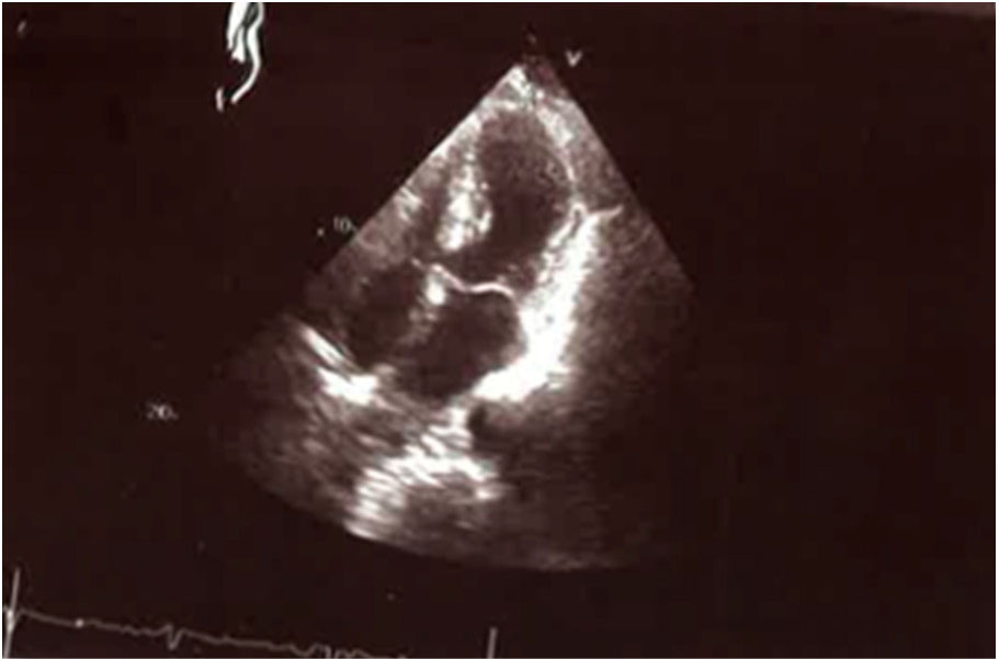
Figure 1.
Transthoracic echocardiographic apical four-chamber view in systole showing ballooning of the apical segments.
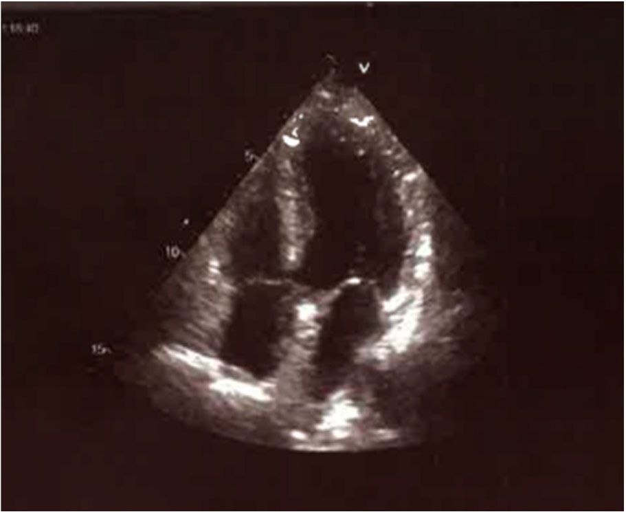
Figure 2.
Transthoracic echocardiographic apical four-chamber view in systole showing ballooning of the apical segments 3 days later