
INTRODUCTION
Giant cell arteritis (GCA), a systemic granulomatous vasculitis that primarily affects large and medium arteries, represents one of the most important medical emergencies in ophthalmology, due to its profound effect on visual function[1]. Waldman et.al. emphasized that GCA is a vision-threatening vasculitis in which prompt recognition and aggressive treatment are essential to prevent permanent blindness[2].
Cotton wool spots (CWS) are localized accumulations of axoplasmic debris due to terminal arteriolar occlusion leading to inner retinal ischemia[3]. CWS have been described in GCA predominantly in association with optic disc edema or a pallid optic disc, consistent with anterior ischemic optic neuropathy (AION). Ocular presentation of GCA with isolated CWS on retinal examination has rarely been reported, [4],[5] making this condition difficult to recognize.
GCA is a well-known masquerader of many systemic conditions, and patients may present with nonspecific symptoms like headache, neck pain, fever, and weight loss. Other cited systemic manifestations are myocardial infarction, stroke, aortic aneurysm or dissection, tongue necrosis, and limb ischemia[6].
Occult GCA (or “silent” or “atypical” GCA) is a well-established entity described as the development of visual symptoms as the first or the only sign of GCA[7]. This ocular variant has an incidence ranging between 14.3%[8] – 21.2%[9] from the total of GCA cases. These patients lack systemic symptoms of GCA despite a positive temporal artery biopsy (TAB) for GCA, thereby challenging the diagnosis. Furthermore, the association between the occult presentation of GCA and CWS as the sole fundoscopic finding can be extremely difficult to recognize.
GCA preferentially affects people of Northern European ancestry[10], peaking in the eighth decade of life, but the risk of visual loss from GCA is also critical in Asian populations [11],[12]. Visual loss is among the most feared complications, attributable to preferential involvement of the short and long posterior ciliary arteries supplying the retina, choroid, optic nerve head, and extraocular muscles.[8] Their involvement can cause arteritic anterior ischemic optic neuropathy (AAION), central retinal artery obstruction (CRAO), choroidal infarction, or posterior ischemic optic neuropathy[13]. The first two ocular syndromes can cause permanent visual loss. Visual loss usually occurs in only one eye, but if unrecognized and left untreated, it may affect the other eye simultaneously or sequentially over a short period of time[14].
This study aimed to search the literature for peer-reviewed articles, randomized controlled trials, case-control studies, cohort studies (prospective and retrospective), cross-sectional studies, case series, and case reports, reported on GCA, and to include a descriptive analysis of the cases, focusing on those subjects presenting with unilateral or bilateral CWS as the only manifestation (on retinal examination) of GCA. Given the rarity of this condition, the available literature is limited to case reports and case series.
We sought to describe and compare the demographic and clinical characteristics of patients with occult and systemic GCA presenting with isolated CWS. We also sought to assess the frequency of occult GCA presenting with CWS and to determine the rate of ophthalmic and systemic manifestations in this subgroup of patients. To our knowledge, there is currently no published data on this topic.
MATERIALS AND METHODS
This scoping review was conducted in accordance with the Preferred Reporting Items for Systematic Reviews and Meta-Analyses (PRISMA) extension for scoping reviews.[15][16] The literature search was conducted in PubMed, Scopus, and Web of Science using database-specific search strategies combining terms related to GCA and CWS. A literature search was conducted using the following Boolean query, searching all fields:
(ALL("cotton wool spots") OR ALL("soft exudate") OR ALL("nerve fibre layer infarct")) AND (ALL("giant cell arteritis") OR ALL("temporal arteritis") OR ALL(Horton disease)).
Peer-reviewed articles published between 1960 and June 2026 were eligible for inclusion if they described patients with a diagnosis of GCA, established by temporal artery biopsy (TAB), vascular imaging, or compatible clinical criteria, when sufficient diagnostic justification was provided by the source author, who presented with unilateral or bilateral CWS as the sole finding on fundoscopic examination. TAB-negative, but imaging-confirmed or clinically supported cases were therefore included and considered equivalent for eligibility assessment. The reference lists of included articles were manually screened to identify additional relevant studies. Non-English articles were considered eligible when sufficient data could be extracted for analysis. The exclusion criteria included cases with concurrent papilledema and review articles. The screening process was conducted using the Rayyan app (version 1.4.3). No review protocol was prospectively registered.
Figure 1 presents a PRISMA flow diagram summarizing the identification, screening, eligibility, and inclusion/exclusion processes for the studies included. Data were extracted independently by two reviewers (IV and RI) using a pre-defined Microsoft Excel form. Discrepancies were resolved through discussion and consensus with a third reviewer available if needed.
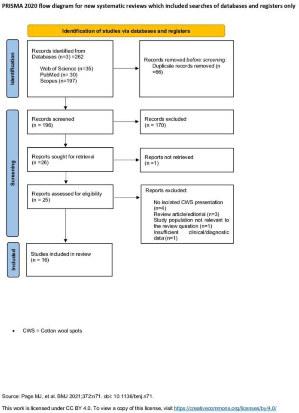
Figure 1:
PRISMA 2020 flow diagram for new systematic reviews which included searches of databases and registers only
Data collected included patient demographics (age, gender, ethnicity), inflammatory markers at presentation - such as C-reactive protein (CRP), erythrocyte sedimentation rate (ESR), Platelet count - visual acuity, details of systemic and ophthalmic symptoms, and treatment initiated at presentation. Patients with solely ocular signs (diplopia, amaurosis fugax, blurred vision) and without systemic manifestations (jaw claudication, scalp tenderness, fatigue, weight loss, etc.) were classified as occult GCA.
For data analysis, patients were divided into two groups: those with systemic symptoms and signs of GCA and those without. Groups were compared descriptively using proportions, medians, and ranges.
RESULTS
The literature search identified 262 records across the three databases. 66 duplicates were removed. After screening of 196 records, 170 were excluded for lack of relevance to our study. 25 papers underwent full-text review for study inclusion and eligibility criteria. Review articles and papers failing to meet the inclusion criteria were excluded. As a result, 16 articles (15 case reports and one case series) were included in the scoping review.
We identified 23 patients with GCA and CWS between 1960 and 2026. One case reported by Sánchez et al.19 was excluded because of concurrent papilledema, and another one was excluded because the diagnosis was limited to polymyalgia rheumatica without confirmed GCA. Consequently, 21 patients were included in the final analysis. The demographic and clinical characteristics of patients with and without systemic symptoms of GCA are detailed as follows. A biopsy-proven diagnosis (TAB+) was present in 19 of 21 patients (90,5%). In one patient, the diagnosis was supported by a positive “halo sign” on TA ultrasonography. In one patient described by Daudin et.al.,[20], the diagnosis was made solely on clinical criteria, as TAB was negative. The mean age at presentation was 69.52 ±7.61 and 57.14% (12/21) were women. Occult GCA was reported in five patients (23.8%), while 16 patients (76.19%) presented with systemic symptoms.
Patients with occult GCA tended to be younger at presentation than those with systemic disease (occult GCA: 62.2 ± 6.05 years, systemic GCA: 71.81 ± 6.63 years. Differences in sex distribution were also observed between groups, with men representing 80% of occult GCA cases compared with 31% of systemic GCA cases. Furthermore, CWS involvement was unilateral or bilateral in both groups. Unilateral involvement was reported in three of five patients (60.0%) with occult GCA and in seven of 16 patients (43.75%) with systemic GCA. Presenting visual acuity in the affected eye(s) ranged from 0.12 ± 0.21 LogMAR in occult GCA to 0.51± 0.48 LogMAR in systemic GCA.
Table 1:
The main characteristics of the sixteen included articles
| Author, year, country | No. of patients | Diagnosis |
|---|---|---|
| 1. MacLeod JDA[17], 1993, UK | 1 | TAB+ |
| 2. Melberg NS[18], 1995, US | 7 (6a) | TAB+ |
| 3. Asensio Sánchez VM[19], 2004, Spain | 2 (1a) | TAB+ |
| 4. Velusami P[4], 2005, UK | 1 | TAB+ |
| 5. Daudin JB[20], 2006, France | 1 | TAB− |
| 6. Jaggi[5], 2007, Switzerland | 2(2a) | TAB+ |
| 7. Johnson MC[21], 2008, US | 1 | TAB+ |
| 8. Levin[22], 2011, US | 1 | TAB+ |
| 9. De Smit E[23], 2013, UK | 1 | TAB+ |
| 10. Thanos A[7], 2015, USA | 1 | TAB+ |
| 11. Gospe[24], 2017, US | 1 | TAB+ |
| 12. Virdee[25], 2019, UK | 1 | TAB+ |
| 13. Rai AS[26], 2020, Canada | 1 | TAB+ |
| 14. Fu L[27], 2022, UK | 1 | “Halo sign”b |
| 15. Yagci BA[28], 2023, Turkey | 1 | TAB+ |
| 16. Pellegrini[29], 2023, Italy | 1 | TAB+ |
Descriptive comparison of ESR and CRP values showed similar medians and ranges across the two cohorts. Notably, one patient with systemic presentation of GCA, reported by Gospe et al.[24], had normal CRP (=4.9mg/L) and ESR (=2mm/h) levels.
Among the 21 patients included in the study, hemoglobin levels were reported for three patients with systemic GCA, whereas platelet counts were reported for six patients. In this subgroup, all three patients (100%) had low hemoglobin levels (Mean±SD:11.2g/dl±1.31), while three (3/6;50%) showed thrombocytosis (Mean±SD: 554.666 platelets per microliter ±83679).
The ocular, cranial, and systemic manifestations of patients with both occult and systemic GCA can be summarized as follows. The most frequently described ocular symptom was blurred vision reported in two of five (40%) patients with occult GCA and in 14 of 16 patients (87.5%) with systemic GCA. Descriptively, blurred vision appeared to be more frequently reported among patients with systemic manifestations of GCA. Amaurosis fugax was also described in two of five patients (40.0%) with occult GCA and in four of 16 (25.0%) patients with systemic disease, whereas diplopia was described in one patient (6.25%) with systemic GCA and was not reported in the occult presentation.
Jaw claudication was the predominant cranial manifestation (6/16, 37.5%), followed by temporal headache and scalp tenderness (5/16, 31.25%). Furthermore, general headache was reported by 4/16 patients (25%), while neck pain was noted in one of 16 patients (6.25%). Four patients (4/16, 25%) had a prominent TA, while one of 16 (6.25%) had a non-pulsatile TA. The most common systemic symptom noted was weight loss (3/16;18.75%), followed by fatigue (2/16, 12.5%). Joint pain and tongue fatigue while eating were each reported by one patient (6.25%).
A positive ANA result was identified in one patient in the systemic group, without fulfillment of criteria for systemic lupus erythematosus.
One patient with an occult presentation was concurrently diagnosed with occipital stroke.
Among the 16 patients for whom treatment data were available, half received high-dose oral steroids, and half were treated with high-dose intravenous steroids. Post-treatment resolution of CWS was documented in nine patients. Due to the small sample size, no further conclusions could be drawn.
DISCUSSION
This study provides a scoping review of the literature on CWS in GCA and is the first to clarify the detailed clinical presentation of this rare entity. Historically, experimental studies demonstrated that platelet-fibrin microemboli could induce cotton-wool spots by causing precapillary arteriolar occlusion and retinal ischemia[30]. McLeod et al.[31] described CWS as sentinels of oncotic inner retinal infarction after occlusions of branch arterioles. GCA affects medium-sized and larger arteries, therefore not causing CWS directly. CWS may be seen in a range of systemic conditions, including diabetes, hypertension, retinal vein occlusion, neoplastic disorders (e.g., leukemia and lymphoma), HIV infection, and inflammatory diseases such as systemic lupus erythematosus.[3] Accordingly, an extensive diagnostic evaluation is required to rule out the above-mentioned diagnoses.
At present, evidence regarding the incidence and prevalence of CWS in GCA remains uncertain. Melberg et al. [18] recognized CWS as an early fundoscopic finding in GCA. They suggested that their presence in older patients may serve as a useful clinical clue to the diagnosis, potentially preceding AION or other ischemic lesions. Furthermore, our results suggested that the occult group presenting with CWS tends to be younger than the group with systemic symptoms. One proposed explanation is that CWS may develop when partial ischemia affects retinal arterioles before systemic symptoms manifest and before complete posterior ciliary occlusion causes optic nerve infarction.
The observed prevalence of occult GCA presenting with CWS (23.8%) is comparable to previously reported data on occult GCA (21.2% [9]). Occult GCA has previously been thought to present with lower ESR values[9]. However, in this scoping review, ESR and CRP values showed similar medians and ranges among patients with systemic and occult presentation, consistent with the results reported by Issa et al.[8] In rare cases (<1%), both ESR and CRP may be normal in patients with GCA[32], as seen in the case reported by Gospe et.al.[24]. CWS are, therefore, nonspecific findings with a broad differential diagnosis, among which GCA should also be considered.
Our study has several limitations, one of which is the retrospective nature of the review. The sample size was also limited, and the occult presentation of GCA may not be fully representative. We included only peer-reviewed articles, but two TAB-negative cases as well, leading to the uncertainty in the evidence level discussed. However, one case had a positive ultrasound of the temporal arteries, confirming the diagnosis, according to the 2022 EULAR recommendations[33]. Nevertheless, the other case reported by Daudin et.al.[20], presented with inflammatory syndrome, clinical signs and symptoms suggestive of GCA, and a positive response to corticosteroid therapy, therefore fulfilling the EULAR[33] criteria for GCA.
Moreover, incomplete reporting in some case reports and series limited the depth of analysis. Critical appraisal of included sources was conducted using the JBI Checklist for Case Reports and Case Series and is presented in the Supplementary Appendix (Table S1 and Table S2). Most reports adequately described patient characteristics, clinical presentation, diagnostic procedures, and outcomes; however, incomplete reporting of adverse events was noted in several studies. Additionally, the findings of this review should be interpreted considering the limitations associated with the exclusively case report – and case series–based evidence. Nevertheless, to our knowledge, this represents the first scoping review investigating CWS in GCA.
CONCLUSION
In conclusion, this review summarizes the current evidence and characteristics of CWS as an isolated ocular manifestation on retinal examination of GCA. CWS may occur even in the absence of systemic symptoms, further complicating the diagnostic process. Recognizing this atypical presentation in older patients with unexplained CWS is critical, as delayed diagnosis may result in irreversible visual loss. Increased awareness of this presentation among ophthalmologists and clinicians may facilitate earlier diagnosis. Further prospective studies are required for a detailed analysis of treatment response and therapeutic strategies in this uncommon presentation.
In patients over 50 years with unexplained, new, or bilateral CWS — particularly when common causes (diabetes, hypertension, retinal vascular occlusion) have been excluded — GCA should be included in the differential diagnosis, and inflammatory markers should be measured urgently.