
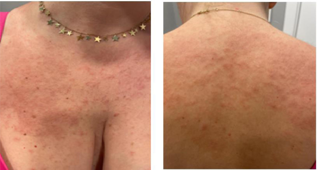
Figure 1.
Clinical aspect of cutaneous lesions on admission
Table 1.
Diagnostic workup
| Diagnostic domain | Specific tests performed | Clinical interpretation |
|---|---|---|
| Infectious Disease | Toxocara canis IgG: positive; Trichinella, Ascaris, and Toxoplasma IgG serology: Negative; Strongyloides stercoralis by agar plate culture and repeated stool microscopy: Negative | Toxocara exposure noted, however lack of response to a 6-week course of albendazole argues against active infection. Strongyloides infection was strictly ruled out prior to corticosteroid initiation to eliminate the risk of hyperinfection. |
| Allergy | ALEX Multiplex Test: Class 3 sensitization to ragweed pollen (r Amb a4); Elevated total serum IgE: 3,603 IU/mL | Hypersensitisation to Ragweed pollen confirmed, without clinical allergy symptoms. |
| Autoimmune and inflammation | ANA, ANCA, C3, C4, anti-dsDNA, Rheumatoid Factor: Negative; ESR, CRP, fibrinogen – normal, Anti-TPO, Anti-tireoglobulin antibodies: Negative | No evidence of systemic autoimmune disease, active vasculitis (such as EGPA), or autoimmune thyroiditis. |
| Hematological and molecular | LDH, B12 vitamin, Beta-2 microglobulin, immunoelectrophoresis. Serum Tryptase: Normal (Repeated); FISH: Negative for FIP1L1-PDGFRA, PDGFRB, JAK2, c-KIT mutations. ECP: Persistently elevated (>200 ng/mL) | Excludes primary clonal myeloid neoplasms. High ECP confirms systemic eosinophil activation. |
| Peripheral blood pheno-typing (flow cytometry): | The expression of the following antigens: CD3, CD4, CD5, CD8, CD10, CD19, CD20, CD23, CD33, CD38, CD45, CD56, CD57, kappa and lambda light chains, and T cell gamma/delta receptor Number of lymphocytes with mildly increased B cells, no evidence of clonal B cell population, normal CD4/CD8 ratio, expression of the T cell antigens CD3 and CD5, kappa/lambda light chain ratio | No aberrant T-cell phenotype or clonal B-cell population detected; findings argue against lymphocytic-variant HES, although this cannot be definitively excluded without lymph-node biopsy and/or molecular clonality assessment. |
| Bone marrow examination | Hypercellular bone marrow (85%) with granulocytic hyperplasia; marked granulocytic hyperplasia G/E ratio 7:1, mild left shift, maturation preserved, mild eosinophilia (<10%); normoblastic maturation; normal size megakaryocytes. Gomori sylver impregnation: MF-1 (>30%). Immunohistochemistry: CD20 positive in isolated reactive B cells, CD14 positive in rare monocytes (10%), CD 34 ~ 3%. | No morphology of evident leukemia or lymphoma. Mild reticulin fibrosis requires close monitoring as a potential indicator of early clonal hematopoiesis |
| Imaging | Whole-body CT: Multiple small lymph nodes ≤11 mm (cervical, thoracic, abdominal, pelvic); Echocardiography: Normal EF (60%), no fibrosis/thrombi; Spirometry: Normal (FEV1 90%, FVC 95%); | Generalized small reactive lymphadenopathy documented. Absence of definitive eosinophil-mediated end-organ damage |
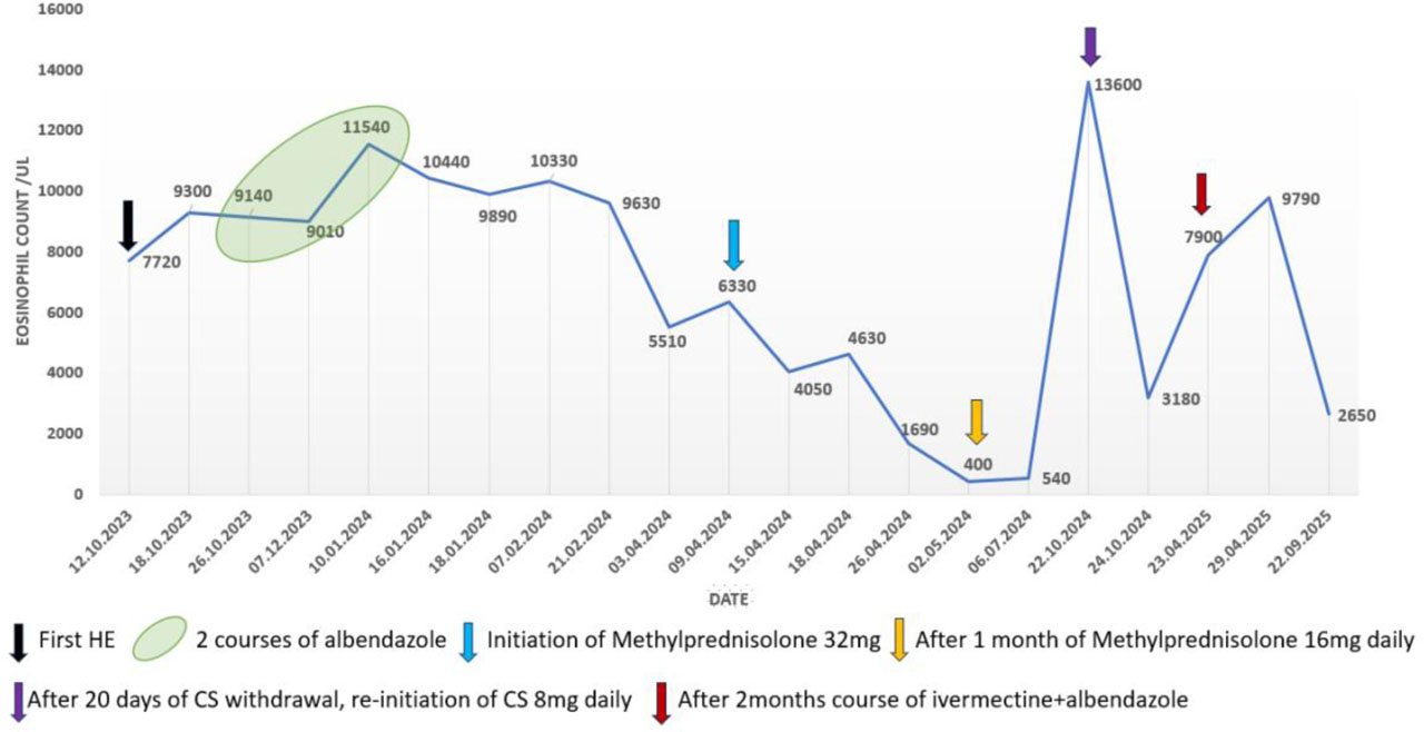
Figure 2.
Blood eosinophilia and therapeutic response between 2023–2025