
What is New? What is Important?
- Illustrates the clinical challenge of managing severe hypereosinophilia diagnosed as HE-US with no histopathologic confirmation of HES
- Highlights the need for rigorous long-term follow-up to identify early transition into lymphocytic-variant HES or occult malignancies
Emphasize how clinicians must carefully weigh therapeutic options in HE-US, including beneficial and side effects of corticosteroids versus patient preference and tolerance
1. Introduction
Hypereosinophilia (HE) is a laboratory finding encountered across multiple medical specialties, with etiologies ranging from benign allergic conditions to severe hematologic malignancies. An increasing interest was proved during the last decades in studying eosinophils, considered “still enigmatic cells “after 140 years from their discovery by Paul Ehrlich [1]. Significant progress was done in understanding eosinophils physiology and functions, as well as in defining the main clinical entities, improve diagnosis, classification and treatment of these extremely versatile diseases [2,3]. Blood eosinophilia between 500/uL and 1500/ul is relatively common, with reported incidence between 1–2%, while HE (> 1500/uL) is rare, with an estimated incidence between 0,3–6,3/100.000 [4,5].
The initial classification algorithm for HES was proposed by the International Co-operative Working Group on Eosinophil Disorders (ICOG-EO) in 2006, based on clinical phenotypes [6] and updated in 2011 [3]. This classification was recently revised, making a clear distinction between blood eosinophilia, tissue eosinophilia, and hypereosinophilic syndromes (HES) 7,8,9]. According to current guidelines, the difference between asymptomatic HE and HES is critical, since HES requires clear evidence that clinical organ failure is caused by eosinophil toxicity. This distinction has major therapeutic implications, as organ dysfunction can develop independently of the source of HE, occasionally justifying the initiation of therapy even during the etiological workup based on clinical severity. [10].
Evaluation of HE requires a structured diagnostic approach to differentiate secondary, clonal, hereditary and idiopathic causes. Although most cases are reactive, a subset of patients present with persistent eosinophilia and no identifiable cause despite comprehensive evaluation [11,12]. In some cases, hypereosinophilia with initial severe organ damage may progress to systemic disease such as eosinophilic granulomatosis with polyangiitis (EGPA) [13].
A considerable fraction of HE/HES cases remain unresolved and are designated as idiopathic hypereosinophilic syndrome (I-HES). This term refers to cases of HE with AEC >1500/uL, accompanied by tissue and end organ damage, with unknown etiology despite thorough examination [14]. Conversely, HE of undetermined significance (HE-US) is defined as a persistent idiopathic eosinophilia >1500/uL, with no organ damage or clinical symptoms attributed to eosinophils. Both the etiological and organ impact assessments are negative, but maintained potentially severe unexpected evolution. HE-US remains a provisional diagnosis of exclusion based on a thorough workup to rule out reactive, familial and clonal HE [10]. Because these patients retain the potential to evolve into specific HES subtypes, close clinical monitoring is mandatory [7,15].
Both “undetermined” and “uncertain” terms are used in HE-US. While “undetermined” refers more to unknown mechanisms, “uncertain” may better reflect the unpredictable outcome, as it remains unclear why some patients tolerate severe HE for years without organ damage and others develop irreversible lesions rapidly. No correlation between peripheral and tissue eosinophilia could be demonstrated so far [16,17].
The aim of our paper is to report the evaluation and two-year follow-up of a patient provisionally diagnosed with HE-US and to evaluate the practical applicability of current guidelines and experts’ recommendations for diagnosis and management of eosinophilic diseases, particularly of HE-US.
2. Case Presentation
We report the case of a 47-year-old woman with no personal history of food or drug allergies, referred to our hospital Department of Allergology from Bucharest in February 2024, for severe, persistent blood hypereosinophilia, ranging between 7.220 – 13.600 /uL. This was discovered five months before presentation during dermatologic evaluation for fixed, slightly pruritic eczematous skin lesions of the face, progressively extended to the limbs, thorax, and abdomen over four months. She denied systemic symptoms, including fever, weight loss, respiratory, cardiac or gastrointestinal complaints.
Initial ambulatory medical evaluation revealed positive IgG serology for Toxocara canis, for which albendazole was administered during six weeks, without significant reduction of blood eosinophilia. Screening for autoimmune diseases and hematologic evaluation for mastocytosis-serum tryptase and c-KIT mutation were negative. Skin biopsy showed chronic vascular inflammatory reaction with eosinophilic infiltration, without evidence of vasculitis or malignancy.
Physical examination on admission revealed multiple fixed erythematous–violaceous lesions on the neck and thorax, accompanied by soft, painless laterocervical lymphadenopathy with diameter max.10 mm (Figure 1).
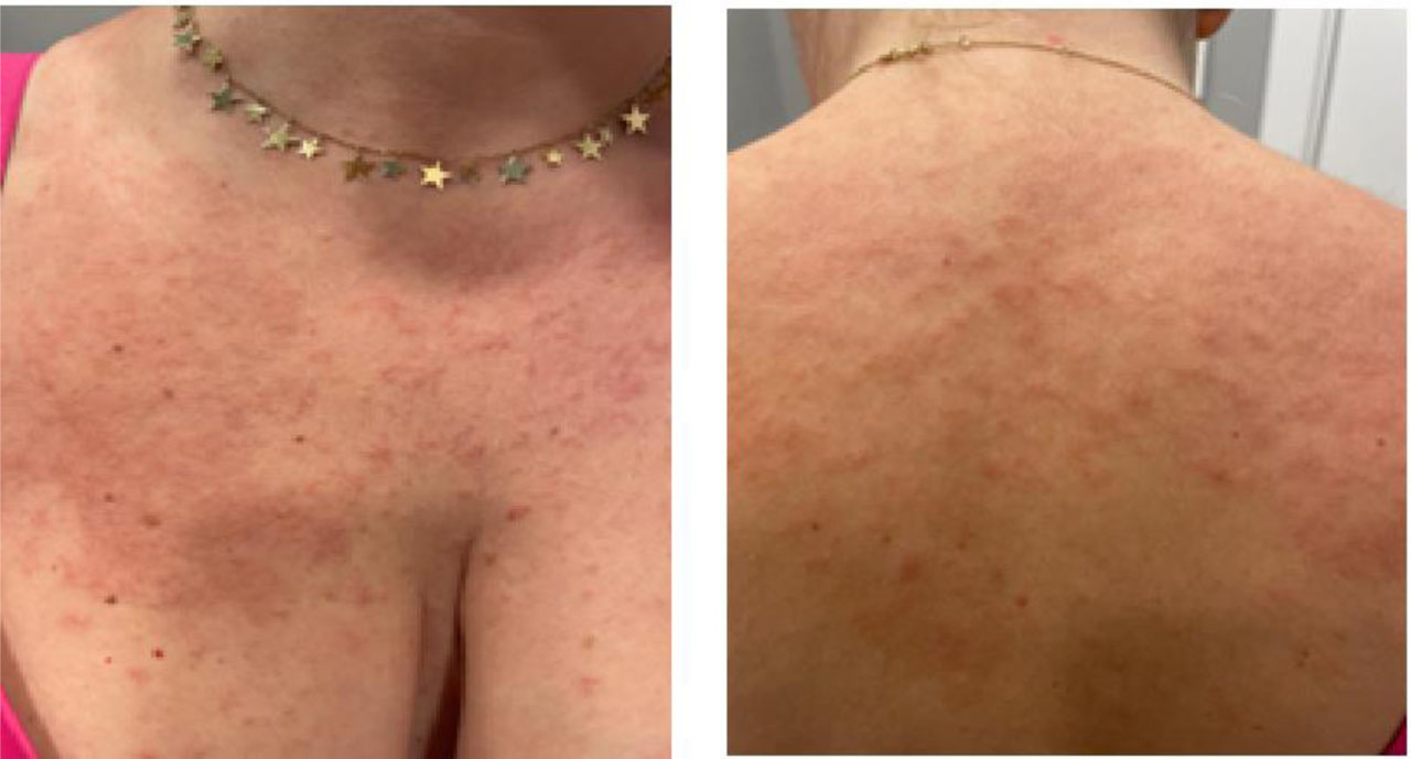
Figure 1.
Clinical aspect of cutaneous lesions on admission
Laboratory tests confirmed severe hypereosinophilia (9.630/uL), leukocytosis (15.610/uL), and markedly elevated total serum IgE (3.603 IU/mL). Other leukocyte subsets were within normal limits, including absolute neutrophils (4180/uL) and lymphocytes (1000/uL). Hemoglobin (12.10g/dL) and platelet counts (222000/uL) were normal, and peripheral blood smear confirmed mature eosinophils without blasts or dysplastic features. Inflammatory markers were unremarkable, with erythrocyte sedimentation rate (ESR) of 20 mm/h, C-reactive protein (CRP) of 5 mg/L and fibrinogen of 365 mg/dL. Biochemical screening, including liver enzymes (AST – 28UI/L, ALT -33 UI/L), renal function tests (creatinine – 0.69 mg/dL, urea – 12.6 mg/dL, eGFR - 108.32mL/min/1.73m2), serum vitamin B12 (197 pg/mL) and lactate dehydrogenase (LDH – 148 UI/L) revealed no abnormalities. Vitamin D deficiency was noted (12.8 ng/mL). Allergy Explorer ELISA multiplex test (ALEX) identified class 3 sensitization to ragweed pollen (rAmb a4), with no symptoms of clinical allergy. Extended parasitological screening was performed and Strongyloides stercoralis infection was assessed before corticosteroid initiation by agar plate culture with negative results. Serologies for Toxocara canis, Toxoplasma gondii, Trichinella spiralis, Ascaris lumbricoides and stool microscopy also returned negative. Autoimmune evaluation was negative for antinuclear antibodies (ANA – 12ng/mL), anti-neutrophil cytoplasmatic antibodies (ANCA), C3 and C4 complement proteins. Total immunoglobulins IgA, IgM, and IgG (940 mg/dL) were within normal limits, however, serum IgG subclasses, including IgG4, were not measured and the absence of typical clinical or radiological features made IgG4-related disease unlikely. Comprehensive cardiac and respiratory assessment excluded occult end-organ damage: transthoracic echocardiography was normal (60% ejection fraction, no valvular or endomyocardial thickening); cardiac biomarkers were normal (hs T Troponin 4.5 pg/mL, NT-proBNP 42.5 pg/mL), and baseline spirometry was within normal limits (FEV1 90 %, FVC 95 %). Whole-body computed tomography (CT) revealed multiple lymph-nodes up to 11 mm in the thorax, abdominal and pelvic regions, with no evidence of hepatosplenomegaly or parenchymal organ infiltration.
Fluorescence in situ hybridization (FISH) analysis confirmed the absence of the FIP1L1-PDGFRA fusion gene, also the other specific PDGFRB and JAK2 mutations were negative. Lymph-node biopsy was proposed, but the patient constantly declined the procedure. Bone marrow biopsy revealed granulocytic hyperplasia with eosinophilia <10% and no evidence of clonal hematologic proliferation. Peripheral blood immunophenotyping showed no evidence of lymphoproliferative syndrome (Table 1). Additionally, T-cell receptor (TCR) gene rearrangement testing was not performed. During the next year, extended clinical and laboratory monitoring was performed. Serum tryptase was within normal range on repeated measurements and molecular testing for myeloproliferative neoplasms remained negative. Serial electrocardiograms, cardiac biomarkers (troponin, NT-proBNP), liver and renal function tests showed no abnormalities. Eosinophil cationic protein (ECP) as a marker of eosinophil activation was persistently elevated (>200ng/ml).
Table 1.
Diagnostic workup
| Diagnostic domain | Specific tests performed | Clinical interpretation |
|---|---|---|
| Infectious Disease | Toxocara canis IgG: positive; Trichinella, Ascaris, and Toxoplasma IgG serology: Negative; Strongyloides stercoralis by agar plate culture and repeated stool microscopy: Negative | Toxocara exposure noted, however lack of response to a 6-week course of albendazole argues against active infection. Strongyloides infection was strictly ruled out prior to corticosteroid initiation to eliminate the risk of hyperinfection. |
| Allergy | ALEX Multiplex Test: Class 3 sensitization to ragweed pollen (r Amb a4); Elevated total serum IgE: 3,603 IU/mL | Hypersensitisation to Ragweed pollen confirmed, without clinical allergy symptoms. |
| Autoimmune and inflammation | ANA, ANCA, C3, C4, anti-dsDNA, Rheumatoid Factor: Negative; ESR, CRP, fibrinogen – normal, Anti-TPO, Anti-tireoglobulin antibodies: Negative | No evidence of systemic autoimmune disease, active vasculitis (such as EGPA), or autoimmune thyroiditis. |
| Hematological and molecular | LDH, B12 vitamin, Beta-2 microglobulin, immunoelectrophoresis. Serum Tryptase: Normal (Repeated); FISH: Negative for FIP1L1-PDGFRA, PDGFRB, JAK2, c-KIT mutations. ECP: Persistently elevated (>200 ng/mL) | Excludes primary clonal myeloid neoplasms. High ECP confirms systemic eosinophil activation. |
| Peripheral blood pheno-typing (flow cytometry): | The expression of the following antigens: CD3, CD4, CD5, CD8, CD10, CD19, CD20, CD23, CD33, CD38, CD45, CD56, CD57, kappa and lambda light chains, and T cell gamma/delta receptor Number of lymphocytes with mildly increased B cells, no evidence of clonal B cell population, normal CD4/CD8 ratio, expression of the T cell antigens CD3 and CD5, kappa/lambda light chain ratio | No aberrant T-cell phenotype or clonal B-cell population detected; findings argue against lymphocytic-variant HES, although this cannot be definitively excluded without lymph-node biopsy and/or molecular clonality assessment. |
| Bone marrow examination | Hypercellular bone marrow (85%) with granulocytic hyperplasia; marked granulocytic hyperplasia G/E ratio 7:1, mild left shift, maturation preserved, mild eosinophilia (<10%); normoblastic maturation; normal size megakaryocytes. Gomori sylver impregnation: MF-1 (>30%). Immunohistochemistry: CD20 positive in isolated reactive B cells, CD14 positive in rare monocytes (10%), CD 34 ~ 3%. | No morphology of evident leukemia or lymphoma. Mild reticulin fibrosis requires close monitoring as a potential indicator of early clonal hematopoiesis |
| Imaging | Whole-body CT: Multiple small lymph nodes ≤11 mm (cervical, thoracic, abdominal, pelvic); Echocardiography: Normal EF (60%), no fibrosis/thrombi; Spirometry: Normal (FEV1 90%, FVC 95%); | Generalized small reactive lymphadenopathy documented. Absence of definitive eosinophil-mediated end-organ damage |
The provisional diagnosis of hypereosinophilia of undetermined significance was established. After considering the patient acceptance, we recommended a trial of oral corticosteroids in accordance with expert recommendations for HE-US. Methylprednisolone 32 mg daily for one week and 16 mg daily afterwards resulted in normalization of eosinophil counts within one month. After 20 days of corticosteroid withdrawal due to treatment-related side effects (arthralgia and myalgia), in October 2024, severe eosinophilia recurred (13.600/uL favouring reinitiation of methylprednisolone, with lower dose 8 mg daily, which was not enough to reduce eosinophilia) (Figure 2). After 10 months from the initial evaluation and start of corticosteroids, the patient self-administered oral ivermectin (12mg) and albendazole (444mg) daily in addition to corticosteroids, without clinical or laboratory improvement. Repeated CT in April 2025 revealed unchanged lymph-nodes aspect and size.
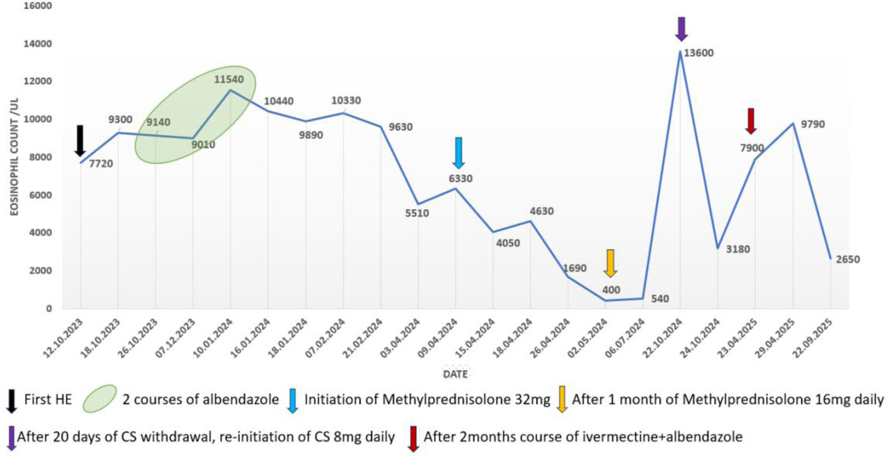
Figure 2.
Blood eosinophilia and therapeutic response between 2023–2025
Since the minimum dose of Methylprednisolone 16 mg was required to maintain blood eosinophils below 1500/uL and the patient complained of side effects, we discussed possible cytoreductive therapy with hydroxyurea as a corticosteroid-sparing option, but the patient declined this therapy. Anti-IL-5/IL-5R biologics may become relevant if the patient develops steroid-dependent HES or definite eosinophil-mediated organ involvement, but their role in HE-US without organ damage remains undefined. An ongoing plan for structured clinical and laboratory monitoring and close contact with the patient are performed in order to facilitate early detection of potential organ involvement.
3. Discussion
HE can be encountered in the clinical practice of many physicians, either incidentally during a routine medical workup or due to symptoms and signs of HE-related organ involvement, because of various manifestations of HE. Mild and transient cases can be managed by many specialists, while evaluation and follow-up of unclear or more severe cases need a multidisciplinary approach and experienced medical staff, depending on the predominant clinical manifestations [17]. Within this spectrum, the clinical relevance and natural history of HE-US remain incompletely understood. Nevertheless, the potential for delayed organ involvement or evolution toward clonal disease requires ongoing clinical and laboratory monitoring, as contemporary consensus statements emphasize the importance of multidisciplinary evaluation, repeated molecular testing, and periodic reassessment of organ function in patients with persistent eosinophilia [8,18,19]. Representing one of the most challenging diagnoses within eosinophil-associated disorders, the presented case illustrates the complexity of evaluating and diagnosing severe, persistent eosinophilia in the absence of identifiable secondary causes, clonal hematologic disease or eosinophil-mediated organ damage. This aligns with contemporary literature, which increasingly recognizes HE-US as a dynamic condition requiring structured evaluation and long-term surveillance rather than a static diagnosis [20].
The reported patient underwent extensive investigations consistent with current diagnostic algorithms, including repeated parasitological testing, autoimmune panels, serum immunoglobulins, vitamin B12, tryptase measurements, immunophenotyping, and cross-sectional imaging. [8]. Indication for empirical anti-parasitic therapy in unexplained chronic HE remains controversial. However, it may be considered due to the variable sensitivity of serological and stool examinations and the critical risk of severe Strongyloides hyperinfection during systemic corticosteroid use [10]. In our patient, although parasitological serology for Toxocara canis was initially positive, the complete lack of clinical or hematologic response to a prolonged course of albendazole argued against an active infection, confirming that serology alone is insufficient to establish causality. The patient's subsequent self-administration of ivermectin and albendazole yielded no improvement of eosinophilia, indicating no parasitological involvement.
Cutaneous manifestations are frequently encountered in various HE. In the reported patient, skin involvement presented as non-specific, variable, fixed, non-pruritic erythematous–violaceous lesions. The classification of these cutaneous lesions represented a key diagnostic challenge, as the skin biopsy revealed eosinophilic infiltration. However, the absence of focal degranulation, leukocytoclastic vasculitis, or tissue necrosis indicated non-specific reactive pattern. To accurately define HE-US, eosinophil mediated toxicity must be completely ruled out. We resolved this ambiguity by concluding that the skin infiltration was reactive and non-toxic, which supported the provisional HE-US diagnosis over suggesting the clinical picture of HES. Furthermore, while the co-existing lymphadenopathy raised concern for L-HES, peripheral blood immunophenotyping studies by flow cytometry were normal, inconsistent with this diagnosis. Data from the literature mention that in cases of lymph-node involvement the diagnosis of Hodgkin’s lymphoma, angioimmunoblastic or T-cell lymphoma should be considered. In cases with chronic presentation and small lymphadenopathy, HE may suggest lymphocytic HES or IgG4 related disease and the diagnosis can be confirmed by peripheral T-cell phenotyping, CT scan of the chest, abdomen and pelvis and lymph-node biopsy [21, 22].
Exclusion of clonal myeloid disease is critical in persistent HE. Prior studies have shown that a small but clinically significant proportion of patients initially classified as HE-US harbour specific mutations such as FIP1L1–PDGFRA, KIT D816V, or JAK2 V617F [23]. In the present case, repeated molecular testing and consistently normal serum tryptase levels, supported the provisional diagnosis of HE-US rather than classic myeloid HES or systemic mastocytosis. However, the patient’s bone marrow examination did reveal a hypercellular marrow with mild reticulin fibrosis (grade MF-1) and in the setting of severe, persistent blood HE, mild reticulin fibrosis cannot be deemed clinically negligible. Although the bone marrow lacked overt clonal features and specific mutations, this finding may represent an early morphological reflection of occult hematopoiesis or subclinical myeloproliferation that lacks typical genetic drivers, further emphasizing the necessity of cautious and long-term monitoring.
The clinical profile of this patient resembles that of the patients included in HE-US cohorts described in the literature including persistent high eosinophil counts, elevated IgE levels, bone marrow eosinophilia, variable corticosteroid responsiveness, and relapse upon steroid tapering. Importantly, progression to overt HES was rare, supporting a generally favourable prognosis, but underscoring the necessity of long-term monitoring [24,25]. Helbig’s study also highlighted the controversy surrounding corticosteroid use in asymptomatic HE-US, noting that treatment should be individualized and that no evidence supports early intervention to prevent progression. This aligns with our therapeutic dilemma: while the patient was asymptomatic regarding organ failure, the severe level of eosinophilia (AEC>5000/uL), elevated ECP levels (>200ng/mL) and relapsing skin lesions drove the decision to initiate steroids. Methylprednisolone was selected over prednisone as a first-line agent due to its lower mineralocorticoid activity compared to prednisone, thereby minimizing the risk of fluid retention, edema and steroid-induced hypertension.
Because management of HE-US is not strictly standardized and data favoring cytoreductive therapies are insufficient, the therapeutic approach must be highly individualized, depending on patient preferences, comorbidities, tolerance, and adherence to follow-up [17]. This aligns with current consensus guidelines, which recommend prioritizing strict long-term monitoring over systemic interventions in asymptomatic patients with HE counts below 5000/uL [26,27].
Alternative steroid-sparing options were considered due to the patient’s notable corticosteroid-related side effects (arthralgia and myalgia). Hydroxyurea was proposed as a cytoreductive option to reduce steroid use. However, its rationale remains controversial in a patient without confirmed clonal myeloid disease and it was subsequently declined by the patient [28]. Additionally, targeted biologic therapies could be considered as potential corticosteroid-sparing agents in the future. Anti-IL-5/IL-5R biologics may become relevant if the patient develops steroid-dependent HES or definite eosinophil-mediated organ involvement, but their role in HE-US without organ damage remains currently undefined. [29,30,31,32].
This report emphasizes the clinical challenges associated with the diagnosis and management of the hypereosinophilia spectrum, particularly in medical settings without dedicated reference centers or specialist multidisciplinary teams for eosinophilic disorders. When patients are initially sent to multiple distinct medical specialties, the diagnosis confirmation can be delayed and potentially increase the risk of severe complications.
The main limitation of our study is the missing lymph node biopsy. Despite extensive counselling regarding potential more severe outcome, the patient consistently declined the recommended lymph-node biopsy and blood-based examinations were unable to demonstrate an active L-HES variant. The transition to lymphocytic-variant HES (L-HES) remains a serious concern, given the context of relapsing skin involvement, diffuse small lymphadenopathy and persistently elevated ECP. Furthermore, while the two-year clinical follow-up provides valuable longitudinal insights, it is still insufficient to accurately predict future progression to more severe clonal or idiopathic conditions, highlighting the critical need for continuous, long-term monitoring.
4. Conclusions
The reported case underscores the diagnostic complexity and difficulties in managing hypereosinophilia of undetermined significance in clinical practice. While current evidence suggests a generally benign course, rare progression to hypereosinophilic syndrome or other malignancies cannot be ignored and requires structured long-term monitoring and individualized therapeutic decision-making. The practical lesson from this case is that a provisional HE-US classification must be dynamically reassessed, considering the potential transformation into a more severe disease such as L-HES or other hematologic malignancies. Clear recommendations for monitoring and therapeutic options of idiopathic and undetermined HE are needed, considering extended indications of new anti-eosinophilic biologic therapies.