
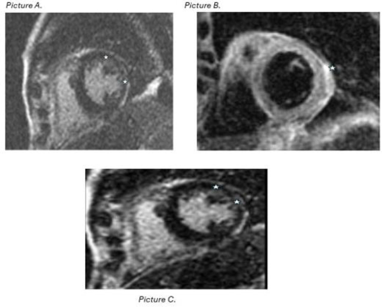
Figure 1:
Cardiac magnetic resonance (CMR) with gadolinium showing Myocarditis from patient in case #4. Picture A. CMR with gadolinium contrast reveals no evidence of first-pass perfusion abnormalities. Picture B. Mid-myocardial to subepicardial linear late gadolinium enhancement (LEG) is present in the lateral and inferior walls at the mid-apical level. Picture C. There is associated lateral wall myocardial edema. Findings are most consistent with a nonischemic myocarditis
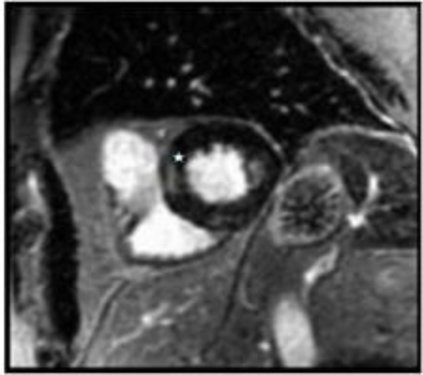
Figure 2:
Myocardial imaging (MR) from patient in case #5 showing patchy focal late gadolinium enhancement.(LGE) with subtle edema in the basal and mid-inferior segments of the left ventricle as depicted by the star in the image.
Table 1.
Summary of Clinical Cases
| Case | Age/Sex | Malignancy | ICI & Timing | Presentation | Peak troponin normal ≤15 ng/L | ±ECG | Echo (LVEF) | CMR/Biopsy | Therapy | Classification * | Outcome |
|---|---|---|---|---|---|---|---|---|---|---|---|
| 1 | 74/M | RCC | Nivolumab + cabozantinib; approx 30 d | Dyspnea; MMM overlap (myasthenia) | 1,871 | Sinus brady; NSVT | Preserved; LVH | CMR deferred | High dose steroids; IVIG; plasmapheresis (for myasthenia) | Possible myocarditis | Recovery |
| 2 | 82/M | Urothelial/RCC | Nivolumab; 28 d | Dyspnea | 1,155 | RBBB; LAFB; 1° AV block | Preserved | Biopsy: hypertrophic changes; no active inflammation | High dose steroids; abatacept (stopped) | Possible myocarditis | Recovery |
| 3 | 62/M | Hepatocellular carcinoma | Nivolumab; 23 d | Chest pain; ST elevation | 408 | RBBB; QT prolongation | EF 18%; apical ballooning | CMR: non-ischemic myopericarditis | High dose steroids | Probable myocarditis | Death (6 months) |
| 4 | 82/F | Colon adenocarcinoma | Pembrolizumab; recent | Respiratory failure; VT | 6,019 | Wide QRS; LBBB; PVCs | EF 64% | CMR not done; angiography declined | High dose steroids | Possible myocarditis | Death (comfort care) |
| 5 | 73/M | Lung mucinous adenocarcinoma | Pembrolizumab; after 3 cycles | Asymptomatic biomarker rise | 292 | PACs; RBBB | Normal | CMR: focal LGE & edema | High-dose steroids (escalated from oral to IV) | Definitive myocarditis | Recovery |
* Classification: Definite = CMR meeting revised 2018 Lake Louise criteria or diagnostic EMB; Probable = elevated troponin with supportive but non-diagnostic imaging; Possible = elevated troponin with compatible clinical features but no confirmatory imaging or biopsy.
±Baseline ECG comparison: Case 1 and Case 5 had no prior ECG available. Case 2 had ECG findings similar to baseline. Case 3 had baseline right bundle branch block and right axis deviation with new precordial ST elevations. Case 4 had baseline LBBB with premature ventricular complexes, with new wide-complex tachycardia (approximative 140 bpm) on presentation.
Table 2.
ASCO Cardiovascular irAE (Myocarditis) Grading and Management.
| Grade | Clinical description (concise) | Recommended management (ASCO) |
|---|---|---|
| 1 | Asymptomatic biomarker elevation; normal ECG/Echo | Hold ICI; oral prednisone 1-2 mg/kg/day; close monitoring; escalate if no response [2]. |
| 2 | Mild symptoms or new ECG/Imaging changes | Hold ICI; oral prednisone 1-2 mg/kg/day; cardiology involvement; consider admission; gradual taper [2]. |
| 3 | Severe symptoms, arrhythmias, or LV dysfunction | Permanently discontinue ICI; pulse IV methylprednisolone 500–1,000 mg/day for 3–5 days; transition to taper; add second-line immunosuppression if refractory [2]. |
| 4 | Life-threatening; hemodynamic compromise | ICU care; pulse IV steroids; early second-line therapy (mycophenolate, IVIG, ATG, plasmapheresis); consider abatacept as rescue in refractory cases [2,9,10]. |
Table 3.
Proposed Clinical Risk Score for ICI-Myocarditis (adapted from Power JR et al., Eur Heart J 2025)
| Parameter | Points |
|---|---|
| Underlying thymoma | 2 |
| Coexisting skeletal muscle or neuromuscular involvement | 1 |
| Low QRS voltage on ECG (≤0.5 mV, Sokolow–Lyon) | 1 |
| Depressed left ventricular systolic function (LVEF <50%) | 1 |
| Mild–moderate troponin elevation ( approximative 20–200 × ULN) | 1 |
| Marked troponin elevation ( approximative \200–2000 × ULN) | 2 |
| Extreme troponin elevation (>2000 × ULN) | 3 |