
What is new/important
This manuscript explores a rare but serious complication of immune checkpoint inhibitor (ICI) therapy: myocarditis. We highlight the critical need for early recognition, thorough risk evaluation, and prompt treatment. In one notable case, myocarditis occurred alongside myasthenia gravis, adding complexity to both diagnosis and management. Our findings emphasize the value of a multidisciplinary approach. We also discuss how ASCO grading and tailored immunosuppressive strategies based on individual risk can support clinical decision-making and potentially improve outcomes for patients receiving ICI treatment.
INTRODUCTION
Cancer remains the second most common cause of illness and death in the United States, with over two million new cases and more than 600,000 cancer-related deaths expected in 2024 [1]. In recent years, immune checkpoint inhibitors (ICIs) have significantly changed cancer treatment, improving survival rates across a range of cancers. Despite these advances, ICIs carry certain risks. By interfering with the body’s natural immune regulation, they can lead to immune-related adverse events (irAEs). These events occur when activated T-cells mistakenly target healthy tissues, potentially triggering autoantibody production and the release of inflammatory cytokines [2].
Although irAEs most commonly affect the skin, gastrointestinal tract, endocrine organs, lungs, liver, and musculoskeletal system [3], cardiovascular complications, though less frequent can be particularly severe. Among them, myocarditis is one of the most dangerous, with an estimated incidence of 0.04% to 1.14% and a mortality rate approaching 60% in some cases [4,5]. An especially concerning manifestation involves an overlap syndrome that includes myocarditis, myositis, and myasthenia gravis (MMM). Though rare, this condition has been associated with rapid clinical deterioration and early mortality [6].
We present five cases of suspected ICI-associated myocarditis encountered at our institution and explore current literature to better understand its diagnosis, management, and implications for clinical practice.
MATERIALS AND METHODS
We conducted a retrospective case series at Mayo Clinic Florida, reviewing patients treated between January 2019 and December 2024. Eligible participants had received at least one FDA-approved immune checkpoint inhibitor (ICI) and were clinically suspected of myocarditis. Cases were categorized based on degree of diagnostic confirmation. “Definite myocarditis” required fulfillment of revised 2018 Lake Louise CMR criteria [7] or histopathologic confirmation by endomyocardial biopsy. “Probable myocarditis” was defined as elevated cardiac biomarkers with supportive but non-diagnostic imaging and strong clinical context. “Possible myocarditis” was defined as biomarker elevation with compatible clinical features but limited or absent confirmatory imaging.
CMR scans were assessed according to the revised 2018 Lake Louise criteria, which require at least one marker of edema (T2-based) and one marker of tissue injury (T1-based) to support the diagnosis [7]. For cases where endomyocardial biopsy (EMB) was performed, interpretations adhered to established consensus criteria for myocarditis, while acknowledging the known limitations of myocardial tissue sampling [8]. Data related to clinical progression, immunosuppressive treatment strategies, and patient outcomes were extracted from electronic health records and reviewed manually by the study team.
RESULTS
A total of five patients had potentially developed myocarditis following treatment with anti–PD-1 immune checkpoint inhibitors (nivolumab or pembrolizumab). The median age was 74 years, and four of the five were male. On average, myocarditis onset occurred 23 days after the first ICI dose, with a range of 5 to 30 days. The patients had a variety of underlying malignancies: two had renal cell carcinoma, while the others had hepatocellular carcinoma, colon adenocarcinoma, and mucinous lung adenocarcinoma. Clinical presentation varied: three patients reported dyspnea, one had chest pain, and one was entirely asymptomatic, with elevated troponin detected during routine monitoring.
Troponin T levels ranged from 292 to 6,019 ng/L (normal ≤15 ng/L), and ECG findings included conduction abnormalities or ventricular ectopy in all cases. Baseline ECGs were not consistently available; when present, comparison suggested that some abnormalities were pre-existing, while others represented new changes during presentation. Echocardiograms revealed preserved ejection fraction in four patients, while one patient had a markedly reduced ejection fraction of 18%.
Cardiac MRI supported the diagnosis of myocarditis in two cases. Endomyocardial biopsy was performed in one patient but did not yield diagnostic findings.
All patients in the series were treated with corticosteroids. Four began therapy with intravenous methylprednisolone at a dose of 1,000 mg per day, while one patient initially received high-dose oral prednisone, which was later switched to an intravenous regimen. Two patients required additional immunosuppressive treatments; one was given intravenous immunoglobulin (IVIG) along with plasmapheresis, and the other received abatacept.
In Case 1, there was concern for myocarditis–myositis–myasthenia gravis overlap. Plasmapheresis was initiated primarily due to concern for progression to myasthenic crisis after the patient developed respiratory failure requiring intubation. The patient underwent four sessions with subsequent clinical improvement. Creatine kinase was mildly elevated at 505 U/L (reference range 39–308 U/L), supporting concurrent skeletal muscle involvement.
Outcomes differed among the group: three patients recovered, while two unfortunately died. In one case, death occurred months later in the setting of progressive immune-related complications. In the other, respiratory failure and systemic irAEs predominated. While myocarditis may have contributed, definitive attribution was not possible.
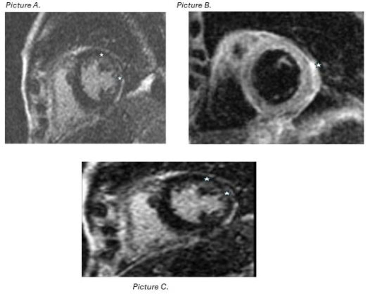
Figure 1:
Cardiac magnetic resonance (CMR) with gadolinium showing Myocarditis from patient in case #4. Picture A. CMR with gadolinium contrast reveals no evidence of first-pass perfusion abnormalities. Picture B. Mid-myocardial to subepicardial linear late gadolinium enhancement (LEG) is present in the lateral and inferior walls at the mid-apical level. Picture C. There is associated lateral wall myocardial edema. Findings are most consistent with a nonischemic myocarditis
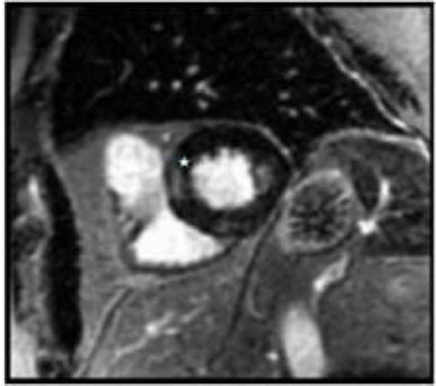
Figure 2:
Myocardial imaging (MR) from patient in case #5 showing patchy focal late gadolinium enhancement.(LGE) with subtle edema in the basal and mid-inferior segments of the left ventricle as depicted by the star in the image.
DISCUSSION
ICI myocarditis is diagnostically challenging, as no single test is definitive. Clinical features range from asymptomatic biomarker elevation to fulminant heart failure, malignant arrhythmias, and sudden death [4,5]. Troponin I is preferred over troponin T due to confounding by myositis; higher levels predict major adverse cardiovascular events [9]. In our cohort, only troponin T was routinely available, as troponin I required send-out testing with an approximately one-week turnaround. This may have limited biomarker specificity, particularly in overlap syndromes.
ECG frequently demonstrates new conduction abnormalities in ICI-associated myocarditis. In our series, conduction disturbances or ventricular ectopy were observed in all patients; however, documentation confirming that these changes were newly developed was not consistently available.
TTE may reveal preserved or impaired systolic ejection function. CMR is the most informative noninvasive modality, with the updated 2018 Lake Louise criteria requiring both T2 and T1 based abnormalities supporting robust diagnostic confidence [10].
In Case 3, the presence of severely reduced ejection fraction with apical ballooning raised consideration of Takotsubo syndrome. However, CMR demonstrated mid-myocardial to subepicardial delayed enhancement with associated myocardial edema and mild pericardial enhancement, findings more consistent with nonischemic myopericarditis in the setting of ICI therapy. While Takotsubo cardiomyopathy is a diagnosis of exclusion, overlap cannot be entirely excluded, especially given recovery of ventricular function about two months after the initial admission.
EMB remains the reference standard but is limited by invasiveness and sampling error [10]. The presence of inflammatory cell infiltrates, myocyte necrosis or damage without ischemia, with the use of hematoxylin and eosin (H&E) and immunohistochemistry (IHC) can confirm the diagnosis. Specific criteria include ≥14 leucocytes/mm2 including up to 4 monocytes/mm2 with the presence of CD-3 positive T-lymphocytes ≥7 cells/mm2 [11].
Contemporary cohorts have developed ICI myocarditis risk predictors score for prognosis [11]. These scores are prognostic tools and have not been validated to guide treatment intensity (Table 3). Therefore, they should not be interpreted as directing immunosuppressive escalation or de-escalation strategies.
Table 1.
Summary of Clinical Cases
| Case | Age/Sex | Malignancy | ICI & Timing | Presentation | Peak troponin normal ≤15 ng/L | ±ECG | Echo (LVEF) | CMR/Biopsy | Therapy | Classification * | Outcome |
|---|---|---|---|---|---|---|---|---|---|---|---|
| 1 | 74/M | RCC | Nivolumab + cabozantinib; approx 30 d | Dyspnea; MMM overlap (myasthenia) | 1,871 | Sinus brady; NSVT | Preserved; LVH | CMR deferred | High dose steroids; IVIG; plasmapheresis (for myasthenia) | Possible myocarditis | Recovery |
| 2 | 82/M | Urothelial/RCC | Nivolumab; 28 d | Dyspnea | 1,155 | RBBB; LAFB; 1° AV block | Preserved | Biopsy: hypertrophic changes; no active inflammation | High dose steroids; abatacept (stopped) | Possible myocarditis | Recovery |
| 3 | 62/M | Hepatocellular carcinoma | Nivolumab; 23 d | Chest pain; ST elevation | 408 | RBBB; QT prolongation | EF 18%; apical ballooning | CMR: non-ischemic myopericarditis | High dose steroids | Probable myocarditis | Death (6 months) |
| 4 | 82/F | Colon adenocarcinoma | Pembrolizumab; recent | Respiratory failure; VT | 6,019 | Wide QRS; LBBB; PVCs | EF 64% | CMR not done; angiography declined | High dose steroids | Possible myocarditis | Death (comfort care) |
| 5 | 73/M | Lung mucinous adenocarcinoma | Pembrolizumab; after 3 cycles | Asymptomatic biomarker rise | 292 | PACs; RBBB | Normal | CMR: focal LGE & edema | High-dose steroids (escalated from oral to IV) | Definitive myocarditis | Recovery |
* Classification: Definite = CMR meeting revised 2018 Lake Louise criteria or diagnostic EMB; Probable = elevated troponin with supportive but non-diagnostic imaging; Possible = elevated troponin with compatible clinical features but no confirmatory imaging or biopsy.
±Baseline ECG comparison: Case 1 and Case 5 had no prior ECG available. Case 2 had ECG findings similar to baseline. Case 3 had baseline right bundle branch block and right axis deviation with new precordial ST elevations. Case 4 had baseline LBBB with premature ventricular complexes, with new wide-complex tachycardia (approximative 140 bpm) on presentation.
Table 2.
ASCO Cardiovascular irAE (Myocarditis) Grading and Management.
| Grade | Clinical description (concise) | Recommended management (ASCO) |
|---|---|---|
| 1 | Asymptomatic biomarker elevation; normal ECG/Echo | Hold ICI; oral prednisone 1-2 mg/kg/day; close monitoring; escalate if no response [2]. |
| 2 | Mild symptoms or new ECG/Imaging changes | Hold ICI; oral prednisone 1-2 mg/kg/day; cardiology involvement; consider admission; gradual taper [2]. |
| 3 | Severe symptoms, arrhythmias, or LV dysfunction | Permanently discontinue ICI; pulse IV methylprednisolone 500–1,000 mg/day for 3–5 days; transition to taper; add second-line immunosuppression if refractory [2]. |
| 4 | Life-threatening; hemodynamic compromise | ICU care; pulse IV steroids; early second-line therapy (mycophenolate, IVIG, ATG, plasmapheresis); consider abatacept as rescue in refractory cases [2,9,10]. |
Table 3.
Proposed Clinical Risk Score for ICI-Myocarditis (adapted from Power JR et al., Eur Heart J 2025)
| Parameter | Points |
|---|---|
| Underlying thymoma | 2 |
| Coexisting skeletal muscle or neuromuscular involvement | 1 |
| Low QRS voltage on ECG (≤0.5 mV, Sokolow–Lyon) | 1 |
| Depressed left ventricular systolic function (LVEF <50%) | 1 |
| Mild–moderate troponin elevation ( approximative 20–200 × ULN) | 1 |
| Marked troponin elevation ( approximative \200–2000 × ULN) | 2 |
| Extreme troponin elevation (>2000 × ULN) | 3 |
Patients receiving ICI for active thymoma show markedly higher cardiotoxicity risk, consistent with the autoimmune tendency of thymic tumors and the hypothesis that thymic dysregulation permits autoreactive T cells to target cardio-muscular antigens once checkpoint pathways are blocked [12,13,14,15].
Therapeutic principles remain grounded in immediate discontinuation of the checkpoint inhibitor and initiation of corticosteroids as outlined in ASCO guidance (table 2). Lower-grade disease (grades 1–2) is generally treated with oral prednisone 1–2 mg/kg/day, while severe presentations (grade ≥3) warrant intravenous methylprednisolone at 500–1,000 mg/day for several days followed by a taper. Observational data suggest that early initiation of high-dose steroids is associated with improved outcomes [3,16]. Patients not responding adequately may require second-line therapies such as mycophenolate mofetil, intravenous immunoglobulin, anti-thymocyte globulin, or therapeutic plasma exchange. More recently, selective T-cell co-stimulation blockade with abatacept has emerged as a promising salvage option in fulminant cases [17].
An increasingly recognized entity is the myocarditis myositis myasthenia gravis (MMM) overlap syndrome, a severe presentation that typically occurs early after ICI initiation and carries high mortality. The exact mechanism is unclear; however, it is hypothesized that shared antigenic targets between cardiac, skeletal, and neuromuscular junction tissues contribute to immune-mediated injury [18,19]. Early multidisciplinary management is essential.
This study is limited by its retrospective, single-center design, small sample size, and lack of standardized diagnostic and treatment protocols. Selection bias and variability in follow-up further limit generalizability. Additionally, the observational design precludes causal inference, including definitive attribution of mortality to myocarditis.
CONCLUSION
ICI-associated myocarditis remains an uncommon but potentially serious immune-related adverse event. Diagnosis is often challenging, as confirmation may vary depending on the availability and interpretation of biomarkers, imaging, and histopathology. A structured evaluation that integrates cardiac biomarkers, ECG, TTE, and CMR using the revised Lake Louise criteria can help improve diagnostic confidence, particularly in settings where troponin I is not readily available. In this context, CMR can help distinguish myocardial from skeletal muscle injury, especially when overlap syndromes are suspected [20].
Early discontinuation of the offending agent and prompt initiation of corticosteroids remain the cornerstone of management, in line with current ASCO recommendations [16]. In our cohort, all patients received high-dose corticosteroids regardless of formal ASCO grade. This likely reflects the clinical uncertainty and perceived severity surrounding suspected ICI myocarditis in real-world practice.
Although emerging risk stratification models have identified factors associated with adverse outcomes, these tools were developed for prognostic assessment and should not be viewed as prescriptive treatment algorithms. Recognition of MMM overlap and early multidisciplinary coordination are essential, particularly in more complex presentations. Targeted therapies such as abatacept may have a role in selected refractory cases, but further evidence is needed.
Given the heterogeneity in diagnostic confirmation, cases should be reported with transparent classification (definite, probable, or possible). The present series is descriptive and hypothesis-generating. Larger prospective studies using standardized diagnostic criteria are necessary to better define optimal evaluation and treatment strategies.
Acknowledgements
Contributors: Sehreen Mumtaz, M.B.B.S., Ronald R. Jr. Butendieck, MD and Pavan Brahmbhatt M.B.B.S for their contribution with selecting the cases and providing clinical images and accompanying descriptions.
Notes
Appendices
Appendix – Detailed Case Summaries
Case 1. A 74-year-old man with metastatic renal cell carcinoma on nivolumab + cabozantinib developed dyspnea and ptosis approximative 30 days after initiation. Troponin peaked at 1,871 ng/L. ECG showed sinus bradycardia and non-sustained supraventricular tachycardia; echocardiogram revealed concentric hypertrophy with preserved EF. CMR was deferred. He was treated with IV methylprednisolone (1 g × 3 days), IVIG, and pyridostigmine. Due to concomitant myasthenia gravis/myocarditis overlap, plasmapheresis was performed. He improved and was discharged with recovery.
Case 2. An 82-year-old man with urothelial carcinoma (status post-nephrectomy) developed dyspnea 28 days after nivolumab. Troponin peaked at 1,155 ng/L. ECG showed RBBB, LAFB, and first-degree AV block; echocardiogram showed preserved EF. CMR revealed subtle T2 signal; biopsy demonstrated hypertrophic changes without inflammation. He was treated with high-dose IV steroids and later abatacept (discontinued for lack of efficacy). His respiratory course was prolonged, requiring tracheostomy, but he recovered without myocarditis recurrence.
Case 3. A 62-year-old man with hepatocellular carcinoma presented 23 days after nivolumab with chest pain, nausea, and ST-elevation. Troponin rose to 408 ng/L. ECG showed RBBB, QT prolongation, and right axis deviation. Echocardiogram demonstrated EF 18% with apical ballooning. Angiography was normal. CMR confirmed non-ischemic myopericarditis. He received high-dose steroids but later developed recurrent pneumonitis; transitioned to palliative care. He died six months later.
Case 4. An 82-year-old woman with colon adenocarcinoma developed acute respiratory failure and ventricular tachycardia after two cycles of pembrolizumab. Troponin peaked at 6,019 ng/L. ECG showed LBBB with PVCs; echocardiogram revealed preserved EF (64%). CTA was negative; CMR could not be performed; angiography was declined. She was empirically treated with high-dose steroids, but progressed to renal and respiratory failure. Comfort measures were instituted, and she died.
Case 5. A 73-year-old man with mucinous lung adenocarcinoma presented with isolated troponin elevation (292 ng/L) after three cycles of pembrolizumab. ECG showed RBBB and PACs; echocardiogram was normal. CMR demonstrated patchy late gadolinium enhancement and edema. He was initially treated with oral prednisone but escalated to IV methylprednisolone due to rising troponins. Symptoms resolved, and he was discharged without recurrence.