
Introduction
Cardiac sarcoidosis is an inflammatory granulomatous disease characterised by highly variable clinical manifestations, ranging from asymptomatic myocardial involvement to conduction abnormalities, ventricular arrhythmias, heart failure or sudden cardiac death. One of the major diagnostic challenges lies in its ability to mimic other cardiomyopathies, particularly hypertrophic cardiomyopathy (HCM) and arrhythmogenic cardiomyopathy (ACM), especially in young individuals with preserved ventricular function.
We report the case of a young endurance athlete presenting with exertional syncope and imaging findings suggestive of genetic cardiomyopathy, in whom hybrid positron emission tomography–cardiac magnetic resonance (PET–CMR) imaging enabled the diagnosis of cardiac sarcoidosis and guided therapeutic decision-making.
1. Patient presentation
A 32-year-old female endurance athlete with no family history of cardiomyopathy presented in the cardiology department following a syncopal episode occurring immediately after a 7-h trail run. The episode was preceded by headache and nausea, and witnesses described a prolonged loss of consciousness. The patient had been engaged in regular endurance training for several years.
Initial cardiological evaluation identified left ventricular hypertrophy, raising suspicion for HCM or physiological adaptation related to athlete’s heart. Exercise testing revealed no arrhythmias or repolarisation abnormalities, and the patient remained asymptomatic during stress.
At presentation to our centre, physical examination was unremarkable, with normal blood pressure, heart rate and oxygen saturation, and no signs of heart failure.
2. Initial work-up
Laboratory testing showed normal NT-proBNP levels and borderline elevation of high-sensitivity troponin T, without inflammatory syndrome.
The electrocardiogram (ECG) demonstrated sinus rhythm with normal atrioventricular conduction, low QRS voltage with QRS fragmentation in the inferior leads, T-wave inversion in the anterior and lateral leads and low-amplitude post-QRS deflections in the precordial leads V4–V6 (Figure 1). These findings raised suspicion for ACM, particularly in the context of exertional syncope.
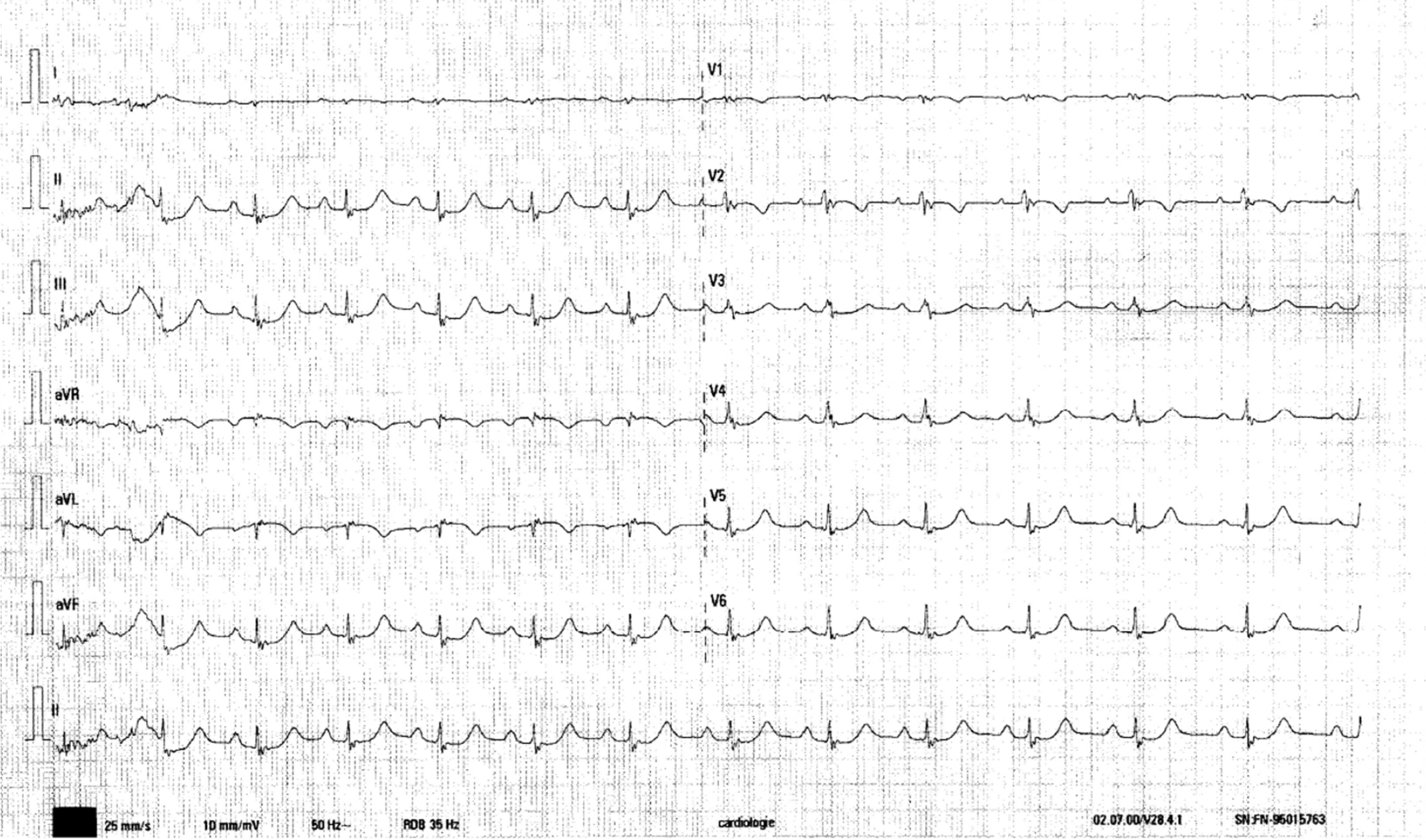
Figure 1
Twelve lead surface ECG showing sinus rhythm with low QRS voltage and QRS fragmentation, inverted T waves in V1–V2, aVL. Low-amplitude post-QRS deflections were observed in the precordial leads V4–V6, suggesting right ventricle involvement. ECG: Electrocardiogram.
Transthoracic echocardiography revealed a non-dilated left ventricle with localised, almost nodular region of increased thickness in the basal interventricular septum (11 mm) and preserved ejection fraction (approximately 55%), without regional wall motion abnormalities. Global longitudinal strain was mildly reduced (–17.9%), with more pronounced impairment in the basal septum and mid-anterolateral segments (Figure 2). Right ventricular size and global function were normal; however, a localised dyskinetic area was observed in the apical free wall.
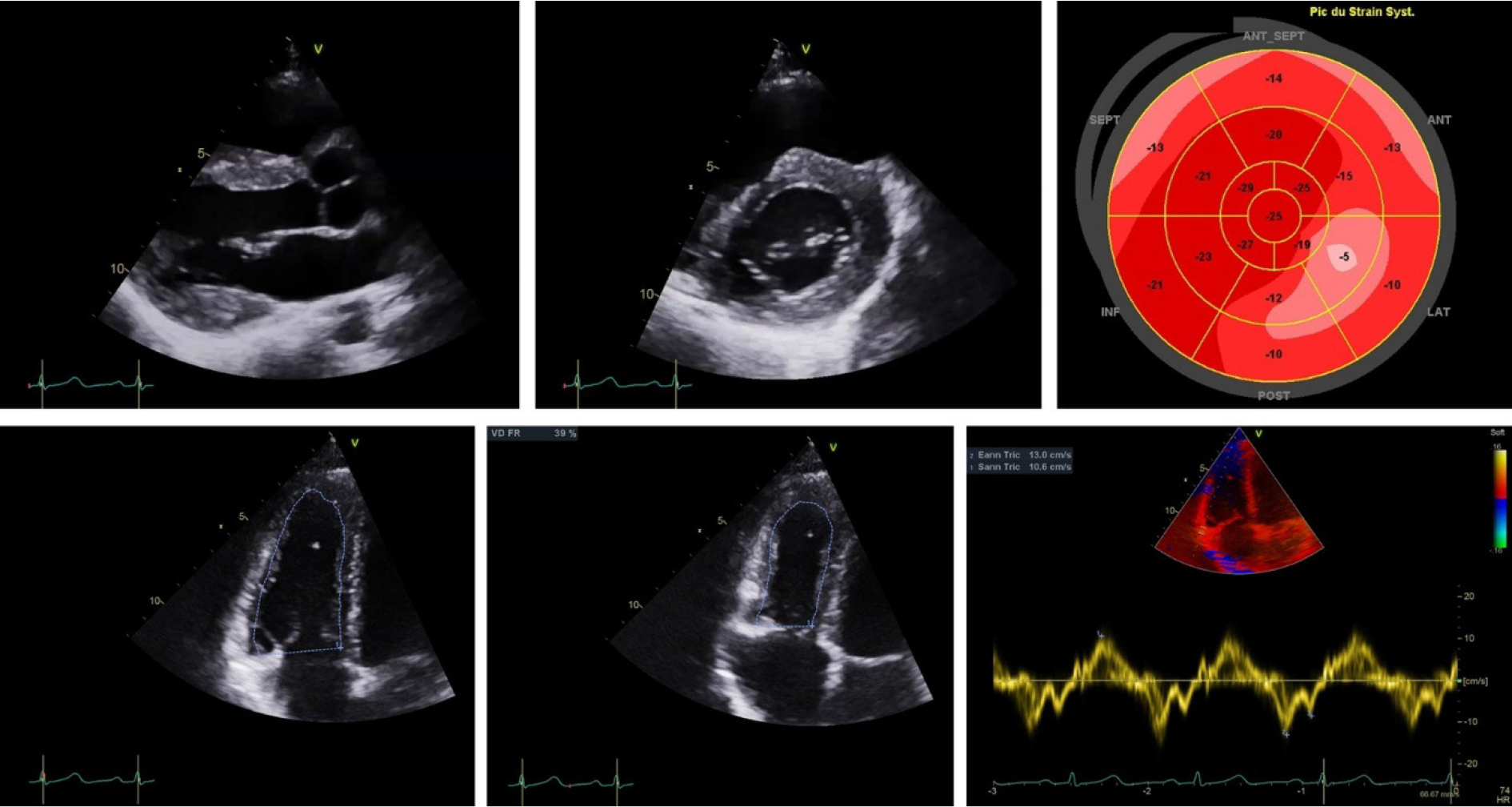
Figure 2
Transthoracic echocardiography: (a) Parasternal long axis view and (b) parasternal short axis view showing localised hypertrophy in the basal IVS segment. (c) Global longitudinal strain—showing mild longitudinal dysfunction (−17.9% with a patchy distribution of abnormal areas. (d) RV-centred apical four chambers view showing diastolic RV area and (e) systolic RV area, with an RV fractional area change of 39%. (f) Tissue Doppler tricuspid S wave velocity of 10.5 cm/s showing a normal right ventricle longitudinal function.
The combination of a nodular aspect of the interventricular septum, regional strain abnormalities, right ventricular mechanical abnormalities and ECG changes—particularly QRS fragmentation and T-wave inversion—raised suspicion for a cardiomyopathic process. The differential diagnosis included a genetic cardiomyopathy, particularly ACM with left ventricular involvement, as well as an inflammatory cardiomyopathy, such as cardiac sarcoidosis or myocarditis. The absence of a family history and the presence of borderline troponin elevation favoured an inflammatory aetiology.
3. Multimodality imaging and diagnosis
To further characterise myocardial tissue and clarify the differential diagnosis, hybrid 18F-fluorodeoxyglucose (18F-FDG) PET–CMR was performed (Figure 3).
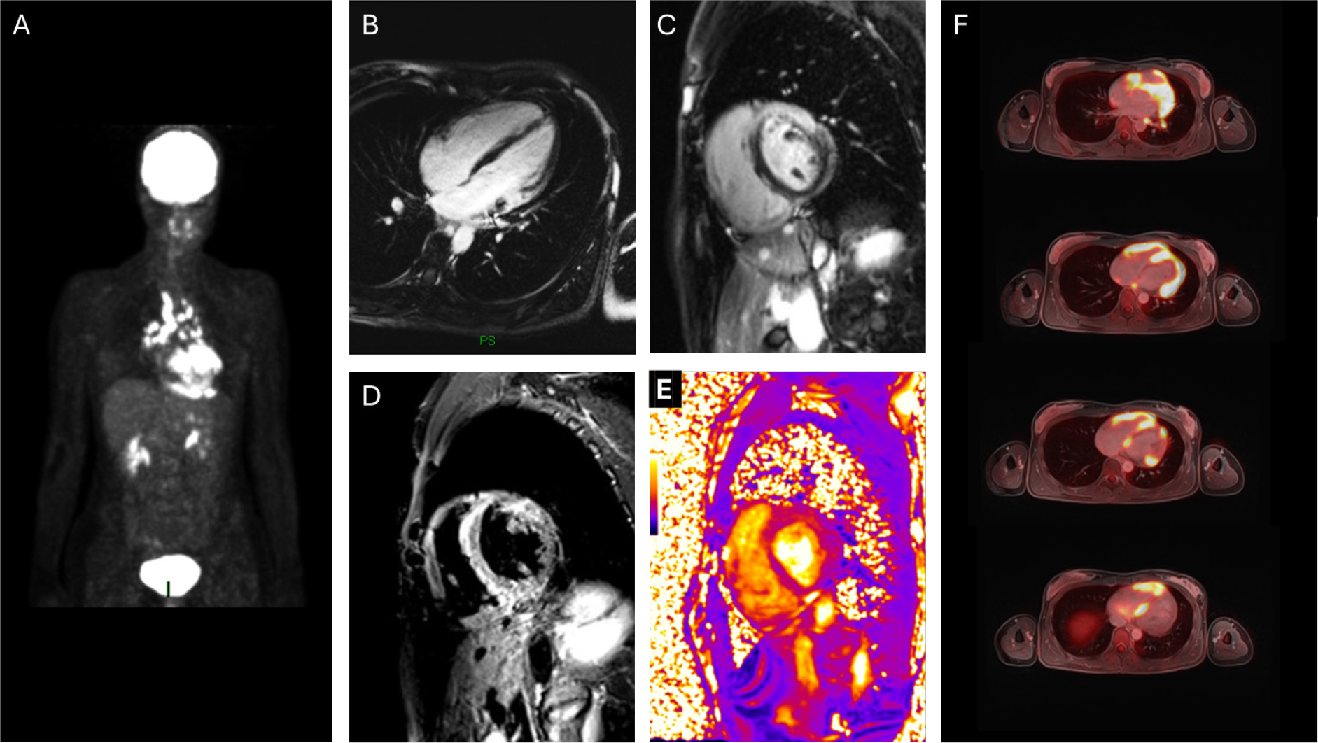
Figure 3
Hybrid 18F-FDG PET–CMR (3 T system) prior to initiation of immunosuppressive therapy. (a) Whole-body maximum intensity projection image demonstrating heterogeneous myocardial FDG uptake, without significant extracardiac hypermetabolic foci apart from small mediastinal lymph nodes. (b) Four-chamber view cine CMR showing non-dilated left ventricle with preserved systolic function (LVEF 53%) and no regional wall motion abnormalities. (c) and (d) LGE imaging showing heterogeneous mid-wall and subepicardial enhancement involving the basal and mid-anterior and anterolateral segments, with additional involvement of the basal inferolateral wall and focal enhancement of the right ventricular free wall. (e) Parametric mapping demonstrating increased T2 values (52–53 ms) in the basal inferolateral and anterior segments, consistent with active myocardial inflammation. (f) Fused axial FDG PET–CMR images demonstrating intense, heterogeneous FDG uptake in the inferolateral and anterior walls, basal interventricular septum and right ventricular free wall, co-localising with regions of LGE, confirming active inflammatory cardiomyopathy. 18F-FDG, l8F-fluorodeoxyglucose; LGE: Late gadolinium enhancement; PET–CMR: Positron emission tomography–cardiac magnetic resonance.
Prior to imaging, the patient followed a high-fat, low-carbohydrate diet for 24 h, followed by fasting, in order to suppress physiological myocardial FDG uptake.
Hybrid PET–CMR was performed on a 3 T Siemens Biograph mMR system. Blood glucose at the time of imaging was 4.9 mmol/L. A dose of 216 MBq of 18F-FDG was administered, and acquisition was performed after 60 min of rest.
Simultaneous PET–CMR acquisition included cine imaging, T1 and T2 parametric mapping, first-pass perfusion following administration of 0.2 mmol/kg gadoterate meglumine and late gadolinium enhancement (LGE) imaging using inversion recovery and PSIR sequences in standard cardiac planes. Whole-body imaging was also performed.
Cardiac magnetic resonance imaging demonstrated focal hypertrophic remodelling involving the basal inferolateral and anteromedial segments, associated with increased T2 values (52–53 ms), suggesting myocardial oedema and inflammation. Tissue characterisation revealed heterogeneous mid-wall and subepicardial LGE involving the anterior and anterolateral walls, predominantly in the basal and mid-segments, with additional involvement of the right ventricular free wall. Native T1 values were borderline elevated, with increased extracellular volume in the basal anterior segment.
FDG PET demonstrated intense radiotracer uptake throughout the myocardium, which appeared mildly heterogeneous, with focal areas of increased uptake in the antero-lateral wall, interventricular septum and in the basal portion of the right ventricle free wall. The concordance between metabolic activity and structural abnormalities strongly supported the presence of active inflammatory cardiomyopathy rather than a primary genetic disorder, with cardiac sarcoidosis considered to be the most likely diagnosis.
On whole-body imaging, intensely hypermetabolic mediastinal lymphadenopathy was also identified, with the findings consistent with sarcoidosis.
4. Histological confirmation
Given the absence of significant extracardiac hypermetabolic lesions suitable for biopsy, endomyocardial biopsy was performed targetting the right ventricle. Histopathological examination demonstrated epithelioid granulomas with multinucleated giant cells, confirming the diagnosis of cardiac sarcoidosis.
5. Treatment and follow-up
Immunosuppressive therapy was initiated with high-dose corticosteroids combined with methotrexate, along with beta-blocker therapy. Despite preserved left ventricular ejection fraction and absence of sustained ventricular arrhythmias on ambulatory monitoring, a wearable cardioverter-defibrillator was prescribed due to the presence of extensive myocardial involvement and active inflammation.
At 3-month follow-up, FDG PET showed a marked reduction of myocardial metabolic activity, indicating therapeutic response (Figure 4). Cardiac magnetic resonance performed at 6 months demonstrated persistence of LGE in previously involved regions without progression and normalisation of T2 values, consistent with resolution of active inflammation but residual myocardial fibrosis.
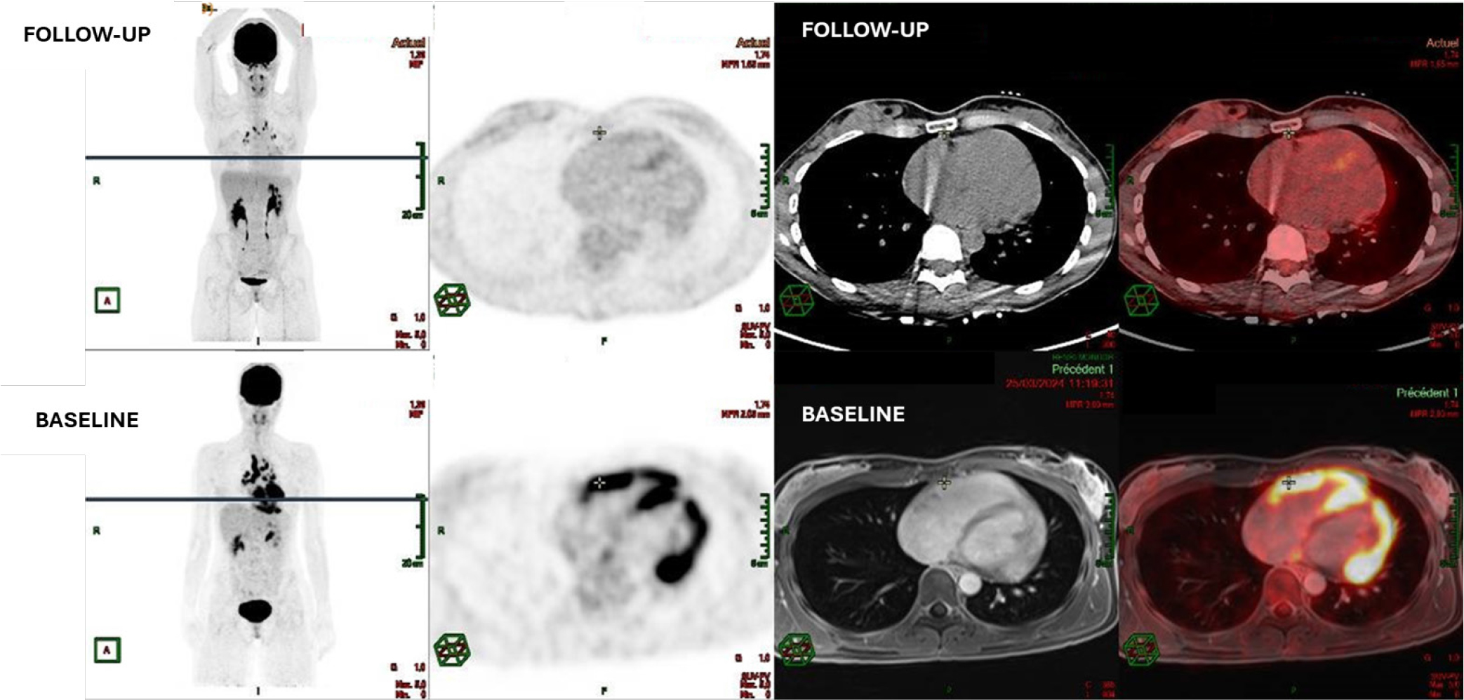
Figure 4
Follow-up 18F-FDG PET/CT compared with baseline hybrid PET–CMR in biopsy-proven cardiac sarcoidosis. Upper panels: Follow-up FDG PET/CT performed after initiation of immunosuppressive therapy demonstrating marked reduction of previously observed heterogeneous myocardial FDG uptake, with near-complete resolution of focal hypermetabolic activity. Lower panels: Baseline hybrid FDG PET–CMR prior to treatment showing intense, heterogeneous myocardial FDG uptake involving the anterior, inferolateral, basal interventricular septal and right ventricular free wall segments. Structural abnormalities with LGE identified on baseline CMR persisted despite metabolic remission, consistent with residual myocardial fibrosis. 18F-FDG, 18F-fluorodeoxyglucose; LGE: Late gadolinium enhancement; PET–CMR: Positron emission tomography–cardiac magnetic resonance.
Implantable cardioverter-defibrillator (ICD) therapy was discussed in accordance with the current guideline recommendations, representing a Class IIa indication. A shared decision-making process was undertaken, incorporating the patient’s clinical profile, arrhythmic risk markers and personal preferences.
In the absence of documented sustained ventricular arrhythmias, the patient declined ICD implantation at the time of diagnosis. Given the presence of active myocardial inflammation, extensive scar burden and syncope suggestive of a potential arrhythmic origin, a wearable cardioverter-defibrillator was strongly recommended as a bridging strategy. This was maintained throughout the 6-month follow-up period, with ongoing reassessment of ICD candidacy.
Discussion
Cardiac sarcoidosis represents a diagnostic challenge due to its heterogeneous clinical presentation and its ability to mimic other cardiomyopathies, particularly HCM and ACM. In young patients presenting with exertional syncope, echo and ECG abnormalities, genetic cardiomyopathies are frequently considered first. However, inflammatory cardiomyopathies, including cardiac sarcoidosis, should remain part of the differential diagnosis, particularly in the absence of family history or when imaging findings are atypical for primary genetic disease.
In the present case, several features initially suggested ACM, including right ventricular mechanical abnormalities and ECG findings, including QRS fragmentation and inverted T waves. According to the 2023 ESC Guidelines for the management of cardiomyopathies, cardiac magnetic resonance imaging represents a key diagnostic tool in differentiating genetic from inflammatory myocardial disease due to its ability to characterise myocardial tissue and detect fibrosis and oedema patterns inconsistent with ischaemic injury or physiological remodelling [1].
The addition of metabolic imaging with 18-FDG PET further increased diagnostic confidence allowing for identification of active myocardial inflammation and has demonstrated high sensitivity for cardiac sarcoidosis, particularly when combined with CMR. Hybrid PET–CMR provides complementary structural and metabolic information, improving diagnostic accuracy and facilitating identification of optimal biopsy targets [2,3]. In our patient, the complete co-localisation between FDG uptake and LGE strongly supported an inflammatory substrate rather than a primary cardiomyopathy, which was subsequently confirmed by endomyocardial biopsy.
Another important aspect illustrated by this case is the dissociation between inflammatory activity and myocardial fibrosis during follow-up. Immunosuppressive therapy resulted in marked reduction of metabolic activity on PET imaging, indicating suppression of inflammation. However, LGE persisted on follow-up CMR, reflecting residual myocardial fibrosis. The presence and extent of LGE have been consistently associated with increased risk of ventricular arrhythmias and adverse outcomes in cardiac sarcoidosis, independent of left ventricular ejection fraction [4].
Risk stratification for sudden cardiac death in cardiac sarcoidosis remains challenging, particularly in patients with preserved ventricular function. Current ESC guidelines recommend taking into consideration ICD implantation (Class IIa) in patients with cardiac sarcoidosis and significant myocardial fibrosis, even in the presence of preserved LVEF [5]. In our patient, the presence of exertional syncope, extensive LGE and active myocardial inflammation supported consideration of ICD implantation. A shared decision-making process was undertaken, integrating guideline recommendations, individual risk markers, and patient preferences, leading to the initial use of a wearable cardioverter-defibrillator with planned reassessment for ICD implantation. The persistence of LGE despite resolution of inflammatory activity represents an important prognostic marker, supporting continued surveillance and an individualised, patient-centred approach to device therapy.
Recent studies have also demonstrated that patients with both LGE on CMR and focal FDG uptake on PET (MR-positive/PET-positive pattern) have the highest risk of major adverse cardiac events, including ventricular arrhythmias and need for ICD implantation [3,6]. This emphasises the value of multimodality imaging not only for diagnosis but also for risk stratification and therapeutic guidance.
Overall, this case highlights the importance of considering cardiac sarcoidosis in the differential diagnosis of suspected genetic cardiomyopathy, particularly when imaging findings are atypical or when inflammatory features are present. It also underscores that resolution of inflammatory activity does not equate to normalisation of arrhythmic risk, which remains driven by residual myocardial fibrosis.
Conclusion
This case illustrates the diagnostic complexity of cardiac sarcoidosis in a young athlete presenting with exertional syncope and features suggestive of genetic cardiomyopathy.
Risk stratification for sudden cardiac death in cardiac sarcoidosis remains challenging. Beyond left ventricular ejection fraction, the presence and extent of LGE represent a major prognostic marker.
Our findings are consistent with previous reports highlighting the protean presentation of cardiac sarcoidosis, which may mimic primary cardiomyopathies or isolated arrhythmic disorders, often leading to delayed diagnosis. Multimodality imaging, particularly the combination of CMR and FDG PET, plays a pivotal role in identifying both myocardial fibrosis and active inflammation. Importantly, arrhythmic risk remains significant even in patients with preserved left ventricular function, supporting the need for careful risk stratification and consideration of device therapy despite apparent clinical stability.
Acknowledgment
None.
Notes
[1] Conflicts of interest Conflicts of Interest
The authors have each completed the International Committee of Medical Journal Editors Form for uniform Disclosure of Potential Conflicts of Interest. No authors have any potential conflict of interest to disclose.
[2] Contributed by Author Contributions
All authors have read and agreed to the published version of the manuscript
[3] Ethics compliance
We confirm that the article was conducted in compliance with ethical guidelines.
[4] Informed Consent Statement
Written informed consent has been obtained from the patients to publish this paper.