
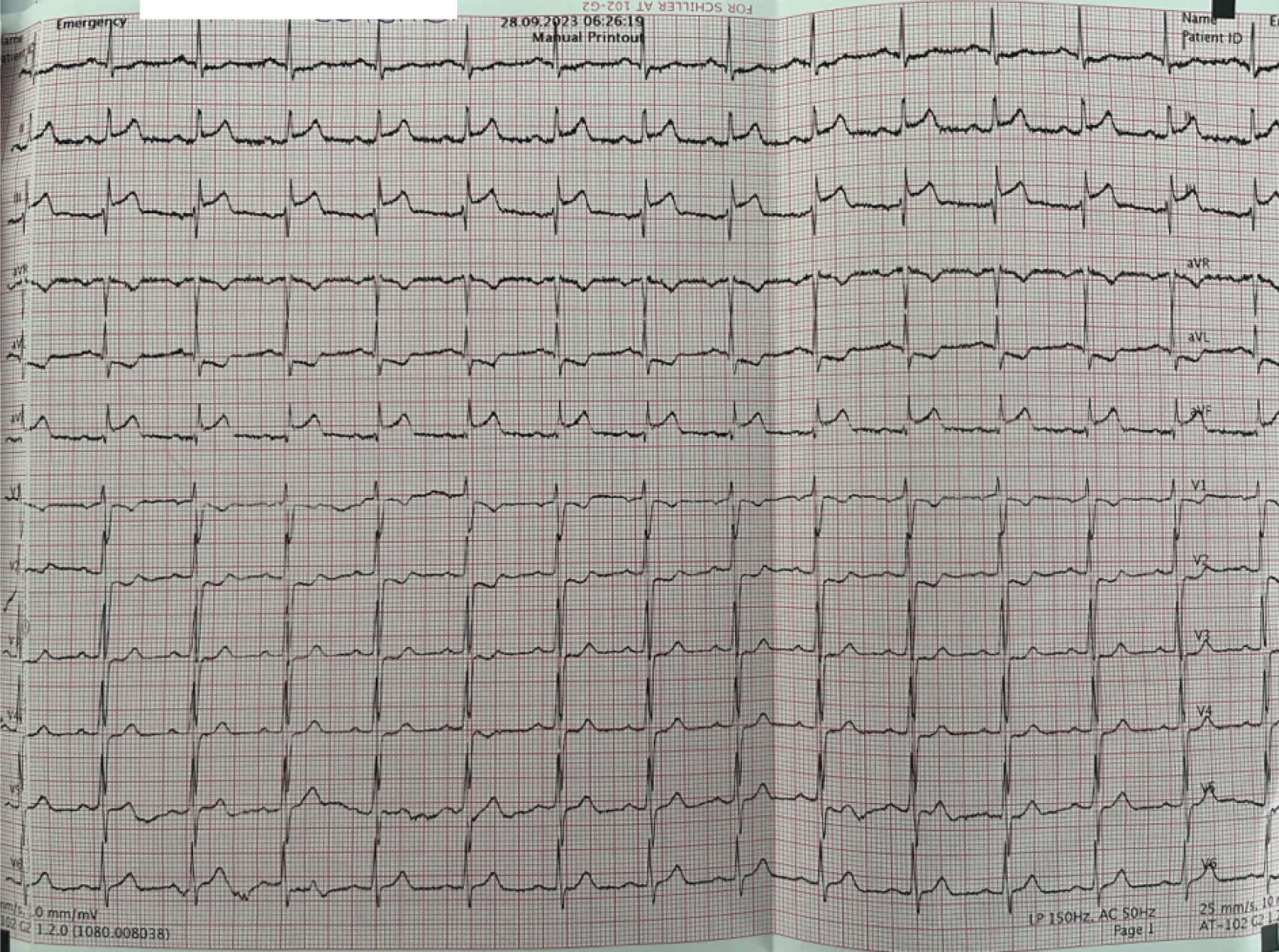
Figure 1
Presenting electrocardiogram: ST elevation in D2,D3, aVF and ST depression in V1-V2, D1,aVL.
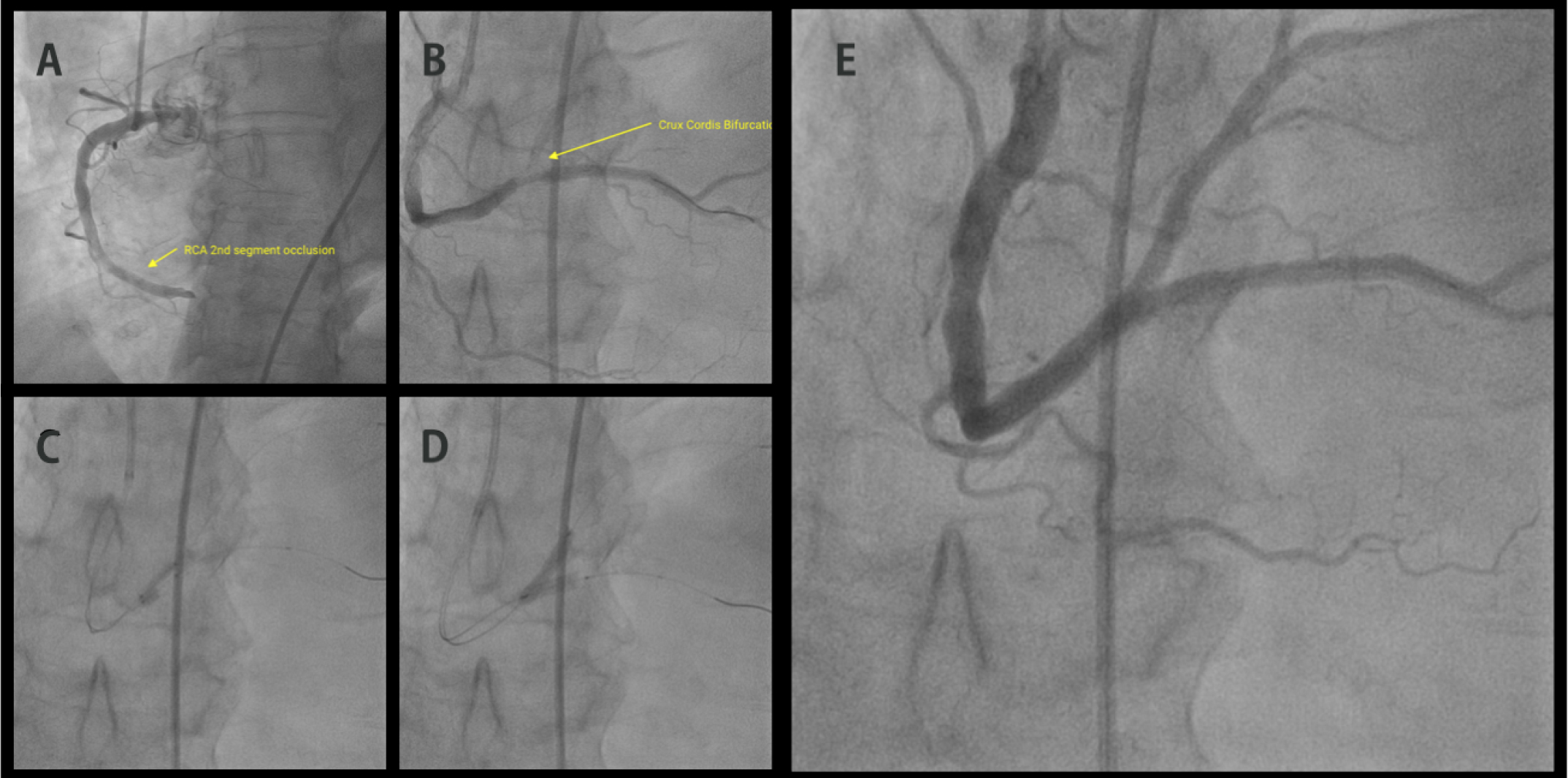
Figure 2
A. Second segment of the right coronary artery occluded by thrombus. B. Bifurcation lesion of the crux cordis after workhorse guide wire crossing. C. Simultaneous kissing stents of the retroposterior branch and posterior descending artery. D. Balloon inflation of the 2 stents. E. Final result showing good peripheral perfusion..
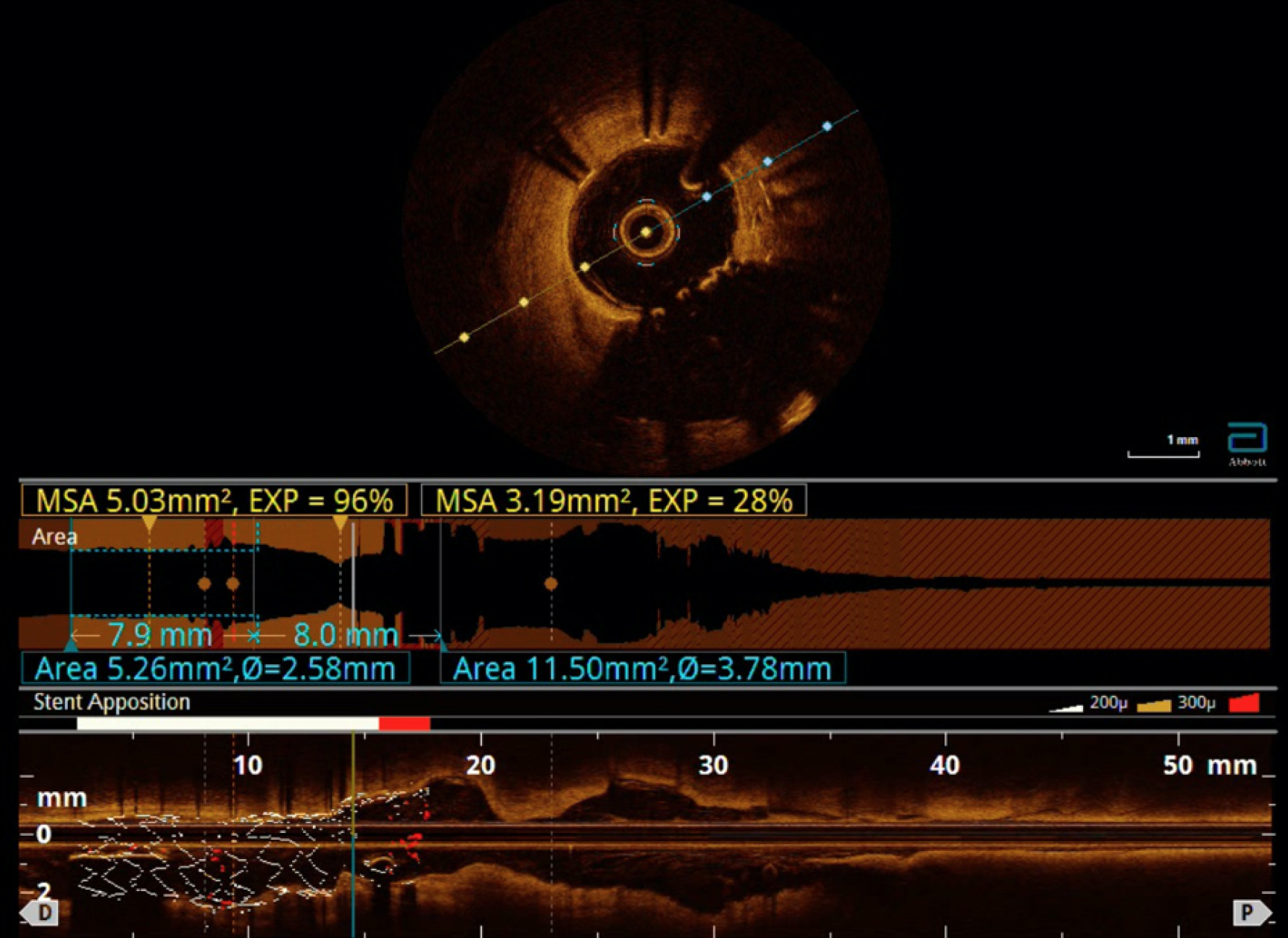
Figure 3
Optical coherence tomography (OCT) demonstrated functional neo carina with no malapposition and successful endothelialization of two stents.