
Table 1
Patient characteristics
| Characteristics | All sample (n=80) |
|---|---|
| Sex | M – 59 (73.8 %) F – 21 (26.3 %) |
| Environment | R – 44 (55.0 %) U – 36 (45.0 %) |
| Smoker | YES – 39 (48.8 %) NO – 41 (51.2 %) |
| Alcohol consumption | YES – 18 (22.5%) NO – 62 (77.5%) |
| Diabetes | YES – 25 (31.3 %) NO – 55 (68.8 %) |
| CKD | YES – 18 (22.5 %) NO – 62 (77.5 %) |
| Obesity | YES – 22 (27.5 %) NO – 58 (72.5 %) |
| NYHA class | I – 6 (7.5 %) II – 37 (46.3 %) III – 34 (42.5 %) IV – 3 (3.8 %) |
Table 2
Summary of investigated parameter values
| Minimum | Maximum | Mean | Standard deviation | |
|---|---|---|---|---|
| LVEDD (mm) | 40 | 91 | 57.804 | 9.14 |
| LVEF (%) | 10 | 55 | 32 | 8.31 |
| LVEDV (ml) | 97 | 586 | 224.01 | 88.1 |
| LVESV (ml) | 55 | 513 | 157.1 | 76.8 |
| E/A | 0.15 | 3.27 | 1.32 | 0.75 |
| E/E’ | 3.5 | 28 | 12.87 | 5.83 |
| E/E’ lateral | 1.5 | 26 | 10.93 | 5.34 |
| E/E’ septal | 5.07 | 34.3 | 15.01 | 7.48 |
| S’ lateral (cm/s) | 0.03 | 0.13 | 0.06 | 0.02 |
| S’ septal (cm/s) | 0.03 | 0.11 | 0.06 | 0.02 |
| MV Dec T (ms) | 64 | 685 | 197.92 | 88.16 |
| LAVI (mL/m2) | 18.52 | 72.08 | 31.63 | 5.83 |
| ePASP | 9 | 93 | 32.91 | 16.69 |
| Cardiac output (L) | 1.72 | 7.77 | 4.76 | 1.44 |
| Aortic Vmax (m/sec) | 0.8 | 4.9 | 1.8 | 1 |
| Mean AVG (mmHg) | 2.58 | 95 | 17.71 | 1 |
| SDNN (ms) | 32 | 165 | 81.48 | 29.87 |
| SDANN (ms) | 24 | 140 | 68.87 | 27.15 |
| SDNN Index (ms) | 17 | 102 | 40.59 | 18.23 |
| RMSSD (ms) | 7 | 93 | 30.45 | 18.48 |
| PNN50 (%) | 0 | 43 | 7.58 | 10.17 |
| Triangular Index (ms) | 5.5 | 54.2 | 19.8 | 8.7 |
| VLF (Hz) | 126.8 | 10314.3 | 1665.67 | 1835.99 |
| LF (Hz) | 23.8 | 1775.9 | 338.88 | 380.22 |
| HF (Hz) | 4.5 | 2202.4 | 195 | 275.96 |
| Deceleration capacity(ms) | -4.96 | 9.05 | 3,92 | 2.33 |
| Acceleration Capacity(ms) | -10.72 | -1.23 | -4.9 | 2.20 |
1 LVEDD – left ventricular end-diastolic diameter, LVEF – left ventricular ejection fraction, LVEDV – left ventricular end-diastolic voume, LVESV – left ventricular end-systolic volume, E/A – peak velocity blood flow from left ventricular relaxation in early diastole/ peak velocity flow in late diastole caused by atrial contraction, E/e’ – left ventricular transmitral early diastolic filling velocity/left ventricular early diastolic myocardial velocity, E/E’ lateral – peak velocity blood flow from left ventricular relaxation in early diastole/ lateral left ventricular early diastolic myocardial velocity, E/E’ septal – left ventricular transmitral early diastolic filling velocity/septal wall of left ventricular early diastolic myocardial velocity, MV Dec T – mitral valve deceleration time, S’ lateral – systolic excursion velocity of lateral wall of the left ventricle, S’ septal – systolic excursion velocity of the septum of left ventricle, LAVI – left atrial volume index, MAPSE – mitral annular plane systolic excursion, SDNN – standard deviation of RR intervals for the entire duration, SDANN – standard deviation of the averages of NN intervals in each 5 min segment across the entire recording, SDNN index – mean of the 5 min normal-to-normal intervals throughout the complete recording, RMSSD – square root of the mean of the squares of the successive differences between adjacent NN intervals, PNN50 – the ratio of NN50 to the total count of NN intervals, hF – high frequency, lF – low frequency, vlF – very low frequency.
Table 3
The relationship between Echocardiographic Parameters and Holter ECG Parameters
| SDNN | SDANN | SDNN Index | RMSSD | PNN50 | Triangular Index | VLF | LF | HF | Deceleration capacity | Acceleration Capacity | ||
|---|---|---|---|---|---|---|---|---|---|---|---|---|
| LVEDD (mm) | Spearman’s rho | 0.096 | 0.064 | 0.105 | 0.215 | 0.201 | 0.003 | -0.01 | -0.01 | 0.221 | -0.087 | -0.124 |
| p-value | 0.399 | 0.574 | 0.355 | 0.056 | 0.074 | 0.978 | 0.956 | 0.942 | 0.049 | 0.445 | 0.274 | |
| LVEF (%) | Spearman’s rho | 0.075 | 0.030 | 0.029 | -0.294 | -0.305 | 0.195 | -0.01 | 0.039 | -0.22 | 0.201 | -0.142 |
| p-value | 0.507 | 0.793 | 0.802 | 0.008 | 0.006 | 0.083 | 0.967 | 0.731 | 0.054 | 0.075 | 0.210 | |
| LVEDV (ml) | Spearman’s rho | 0.080 | 0.053 | 0.115 | 0.153 | 0.169 | 0.003 | 0.134 | 0.009 | 0.083 | -0.089 | -0.016 |
| p-value | 0.479 | 0.638 | 0.311 | 0.175 | 0.134 | 0.977 | 0.237 | 0.935 | 0.467 | 0.433 | 0.886 | |
| LVESV (ml) | Spearman’s rho | 0.028 | 0.028 | 0.045 | 0.209 | 0.220 | -0.097 | 0.070 | -0.04 | 0.111 | -0.191 | 0.074 |
| p-value | 0.807 | 0.805 | 0.691 | 0.063 | 0.050 | 0.390 | 0.539 | 0.750 | 0.325 | 0.090 | 0.513 | |
| E/A | Spearman’s rho | -0.19 | -0.116 | -0.218 | 0.127 | 0.139 | -0.233 | -0.21 | -0.15 | 0.109 | -0.381 | 0.226 |
| p-value | 0.098 | 0.303 | 0.052 | 0.261 | 0.220 | 0.037 | 0.061 | 0.173 | 0.337 | < .001 | 0.044 | |
| E/E’ | Spearman’s rho | -0.11 | -0.051 | -0.145 | 0.177 | 0.225 | -0.241 | -0.13 | -0.18 | 0.146 | -0.239 | 0.208 |
| p-value | 0.319 | 0.653 | 0.199 | 0.116 | 0.045 | 0.031 | 0.239 | 0.121 | 0.196 | 0.033 | 0.064 | |
| E/E’ lateral | Spearman’s rho | -0.13 | -0.065 | -0.140 | 0.123 | 0.218 | -0.250 | -0.13 | -0.16 | 0.129 | -0.115 | 0.091 |
| p-value | 0.240 | 0.565 | 0.216 | 0.278 | 0.053 | 0.024 | 0.247 | 0.152 | 0.254 | 0.311 | 0.424 | |
| E/E’ septal | Spearman’s rho | -0.15 | -0.099 | -0.142 | 0.211 | 0.226 | -0.243 | -0.17 | -0.18 | 0.142 | -0.358 | 0.236 |
| p-value | 0.187 | 0.383 | 0.210 | 0.060 | 0.044 | 0.030 | 0.135 | 0.111 | 0.208 | 0.001 | 0.035 | |
| S’ lateral (cm/s) | Spearman’s rho | 0.122 | 0.074 | 0.092 | -0.206 | -0.224 | 0.282 | -0.01 | 0.048 | -0.20 | 0.163 | -0.150 |
| p-value | 0.281 | 0.513 | 0.418 | 0.066 | 0.046 | 0.011 | 0.959 | 0.674 | 0.078 | 0.149 | 0.183 | |
| S’ septal (cm/s) | Spearman’s rho | 0.116 | 0.107 | 0.102 | -0.263 | -0.258 | 0.263 | 0.064 | 0.096 | -0.22 | 0.311 | -0.285 |
| p-value | 0.306 | 0.347 | 0.366 | 0.018 | 0.021 | 0.019 | 0.571 | 0.399 | 0.051 | 0.005 | 0.010 | |
| MV Dec T (ms) | Spearman’s rho | 0.221 | 0.125 | 0.222 | -0.022 | -0.010 | 0.251 | 0.290 | 0.221 | 0.027 | 0.309 | -0.293 |
| p-value | 0.049 | 0.270 | 0.048 | 0.847 | 0.931 | 0.025 | 0.009 | 0.049 | 0.813 | 0.005 | 0.008 | |
| LAVI (mL/m2) | Spearman’s rho | 0.049 | 0.013 | 0.118 | 0.304 | 0.278 | 0.001 | 0.100 | 0.015 | 0.261 | -0.208 | -0.080 |
| p-value | 0.669 | 0.910 | 0.296 | 0.006 | 0.010 | 0.990 | 0.376 | 0.895 | 0.019 | 0.065 | 0.479 | |
| ePASP | Spearman’s rho | -0.01 | -0.039 | -0.045 | 0.292 | 0.278 | -0.101 | -0.17 | -0.07 | 0.254 | -0.316 | 0.064 |
| p-value | 0.975 | 0.732 | 0.694 | 0.009 | 0.013 | 0.373 | 0.138 | 0.512 | 0.023 | 0.004 | 0.570 | |
| Cardiac output (L) | Spearman’s rho | 0.030 | 0.107 | -0.027 | -0.237 | -0.233 | -0.032 | 0.084 | 0.053 | -0.28 | 0.003 | 0.066 |
| p-value | 0.795 | 0.346 | 0.814 | 0.035 | 0.038 | 0.779 | 0.460 | 0.646 | 0.014 | 0.981 | 0.564 | |
| Aortic Vmax (m/sec) | Spearman’s rho | 0.004 | -0.069 | 0.019 | 0.024 | -0.011 | 0.009 | 0.072 | 0.103 | 0.008 | 0.151 | -0.119 |
| p-value | 0.975 | 0.543 | 0.864 | 0.833 | 0.921 | 0.934 | 0.524 | 0.365 | 0.943 | 0.181 | 0.295 | |
| Mean AVG (mmHg) | Spearman’s rho | -0.05 | -0.063 | 0.003 | 0.012 | -0.005 | -0.050 | 0.075 | -0.12 | -0.03 | 0.099 | -0.073 |
| p-value | 0.638 | 0.577 | 0.976 | 0.918 | 0.963 | 0.658 | 0.506 | 0.273 | 0.823 | 0.384 | 0.520 |
1 LVEDD – left ventricular end-diastolic diameter, LVEF – left ventricular ejection fraction, LVEDV – left ventricular end-diastolic voume, LVESV – left ventricular end-systolic volume, E/A – peak velocity blood flow from left ventricular relaxation in early diastole/ peak velocity flow in late diastole caused by atrial contraction, E/e’ – left ventricular transmitral early diastolic filling velocity/left ventricular early diastolic myocardial velocity, E/E’ lateral – peak velocity blood flow from left ventricular relaxation in early diastole/ lateral left ventricular early diastolic myocardial velocity, E/E’ septal – left ventricular transmitral early diastolic filling velocity/septal wall of left ventricular early diastolic myocardial velocity, MV Dec T – mitral valve deceleration time, S’ lateral – systolic excursion velocity of lateral wall of the left ventricle, S’ septal – systolic excursion velocity of the septum of left ventricle, LAVI – left atrial volume index, MAPSE – mitral annular plane systolic excursion, SDNN – standard deviation of RR intervals for the entire duration, SDANN – standard deviation of the averages of NN intervals in each 5 min segment across the entire recording, SDNN index – mean of the 5 min normal-to-normal intervals throughout the complete recording, RMSSD – square root of the mean of the squares of the successive differences between adjacent NN intervals, PNN50 – the ratio of NN50 to the total count of NN intervals, hF – high frequency, lF – low frequency, vlF – very low frequency.
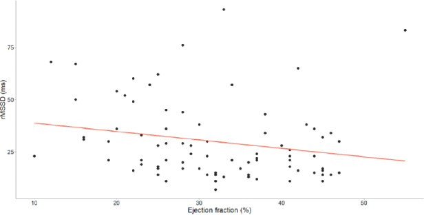
Figure 1
Correlation between left ventricular ejection fraction (%) and RMSSD (ms). RMSSD—square root of the mean of the squares of the successive differences between adjacent NN intervals.
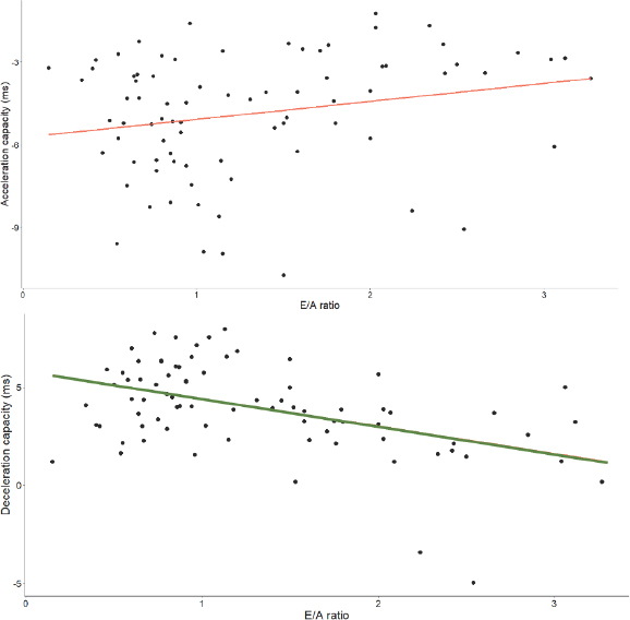
Figure 2
Association between the E/A ratio and both acceleration (ms) and deceleration capacity (ms).
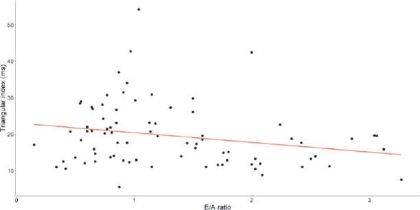
Figure 3
Correlation between the E/A ratio and the Triangular Index (ms).
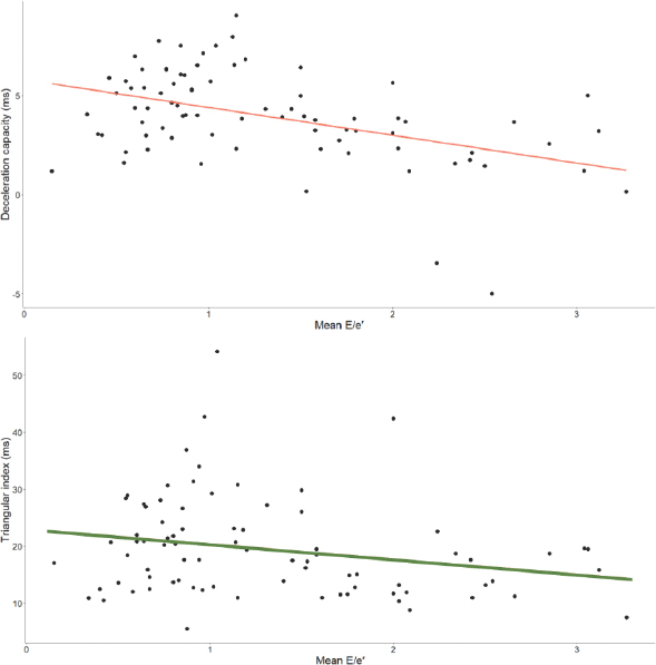
Figure 4
Correlation between the E/E’ ratio and both deceleration capacity (ms) and the Triangular Index (ms).
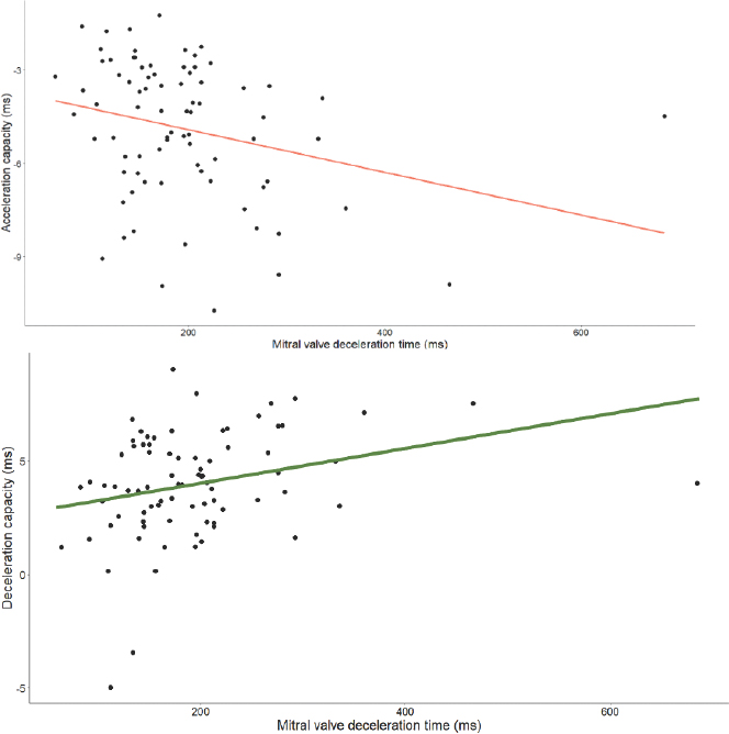
Figure 5
Association between mitral valve deceleration time (ms) and both acceleration (ms) and deceleration capacity (ms).
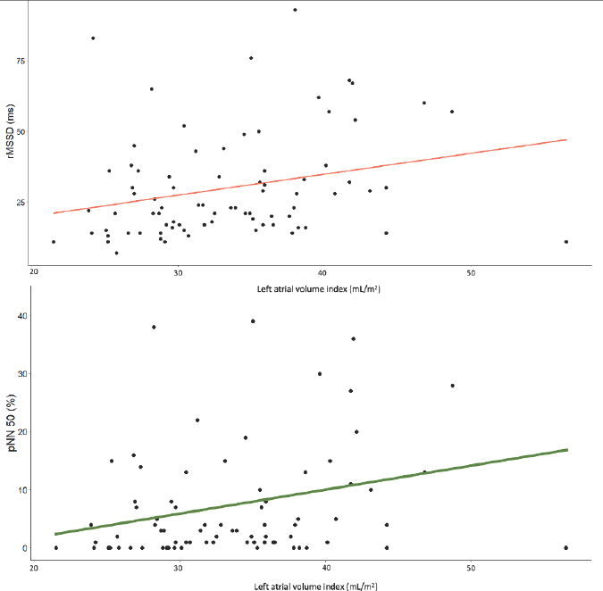
Figure 6
Association between the left atrial volume index (mL/m2) and RMSSD (ms) as well as PNN50 (%). RMSSD—square root of the mean of the squares of the successive differences between adjacent NN intervals, PNN50—ratio of NN50 to the total count of NN intervals.