Figure 1
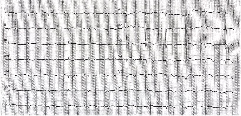
Figure 2
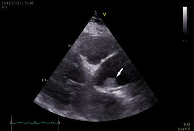
Figure 3
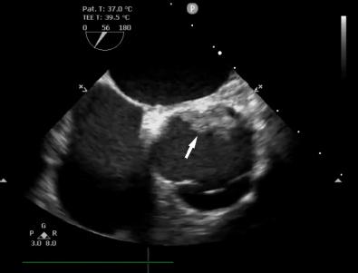
Figure 4A
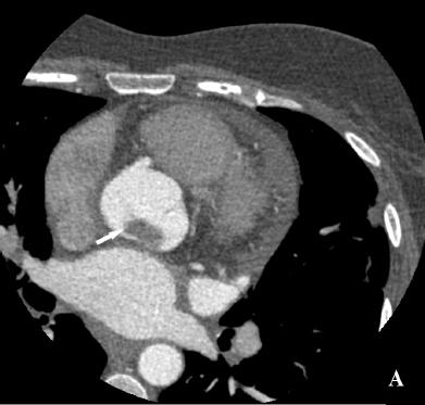
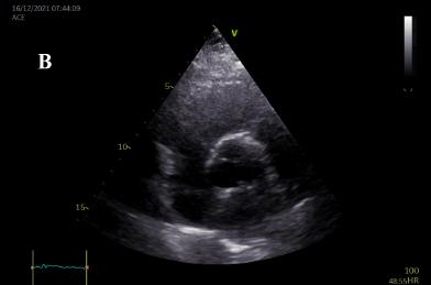
Figure 4B
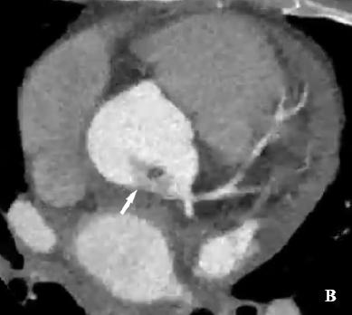
Figure 5
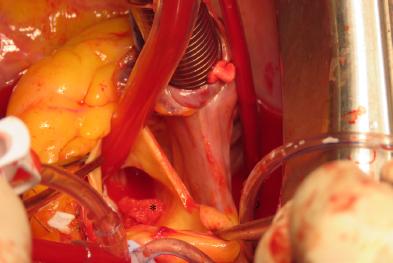
Figure 6
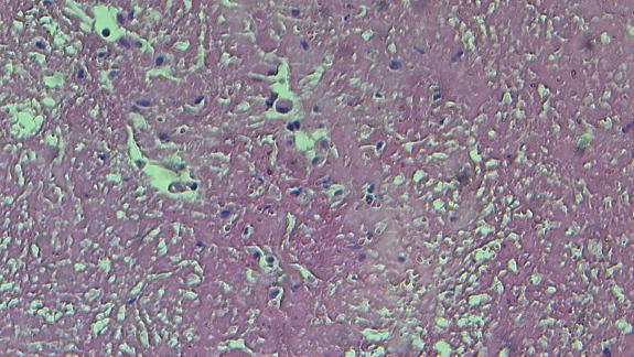
Figure 7A
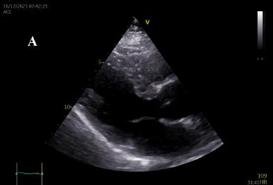
Figure 7B

© 2023 Mihai Teodor Bica, Claudia Irina Nitu, Andrei Iosifescu, Carmen Cristiana Beladan, Bogdan A. Popescu, published by Romanian Society of Cardiology
This work is licensed under the Creative Commons Attribution 4.0 License.