
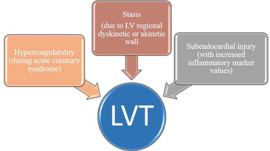
Figure 1
Virchow's triad adapted for left ventricular thrombosis (LVT) (modified after Delewi, et al.5).
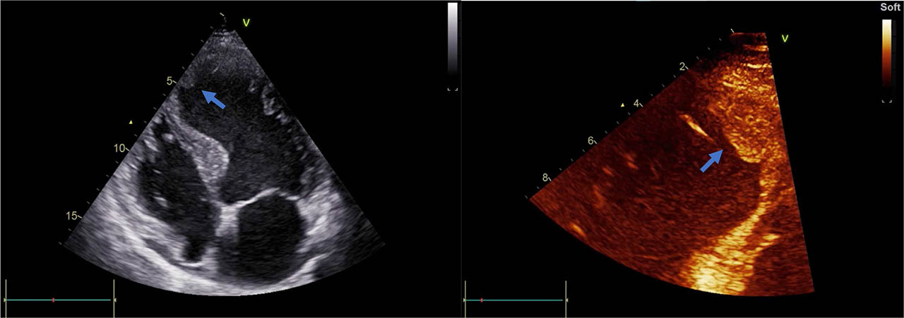
Figure 2
Transthoracic echocardiography of the left ventricle. Thrombus (arrows) attached to the apical septal wall in a patient with ischemic cardiomyopathy.
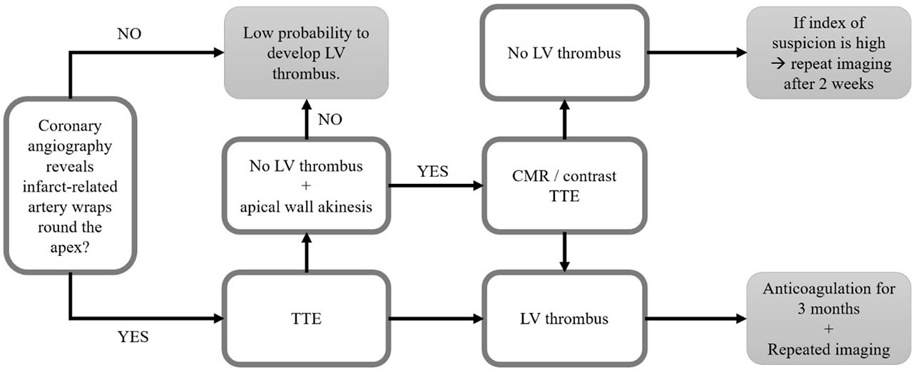
Figure 3
Algorithm for the diagnosis of LVT in reperfused STEMI patients. Abbreviations: LV – left ventricle, TTE – transthoracic echocardiography, CMR – cardiac magnetic resonance imaging (adapted after Bulluck, et al.12)
Table 1
Guidelines for management of left ventricular thrombosis
| LVT | Recommendation | |
|---|---|---|
| STEMI Guidelines | ||
| ACC/AHA-201313 | ||
| Patient with STEMI and | DAPT+VKA with INR target 2–2.5 | |
| ||
| ESC 2012 | ||
| LVT | VKA for a minimum of 3 months | |
| ESC 201715 | ||
| LVT | Anticoagulation should be administered for up to 6 months guided by repeated imaging | |
| Stroke Guidelines | ||
| AHA/ASA 201416 | ||
Patient with ischemic stroke or TIA:
|
| |
| AHA/ASA 202117 | ||
| Patient with stroke or TIA: | ||
|
| |
| CHEST guidelines | ||
| ACCP 201218 | ||
| Patient with anterior MI + LVT/high risk for LVT | ||
|
| Then discontinue warfarin and continue DAPT for up to 12 months as per ACS recommendation |
| Patient with LV systolic dysfunction + LVT without CAD | VKA for 3 months with INR target 2–3 | |
[i] Abbreviations: LVT, left ventricular thrombosis; STEMI-ST, elevation acute myocardial infarction; ACC/AHA, American College of Cardiology/American Heart Association; ESC, European Society of Cardiology; DAPT, dual antiplatelet therapy; VKA, vitamin K antagonist; INR, international normalized ratio; AHA/ASA, American Heart Association/American Stroke Association; TIA, transient ischemic attack; LMWH, low-molecular-weight heparin; MI, myocardial infarction; DOAC, direct oral anticoagulant; PCI, percutaneous coronary intervention; BMS, bare metal stent; DES, drug-eluting stent; ACS, acute coronary syndrome; CAD, coronary artery disease; LV, left ventricle.