
Introduction
Acute limb ischaemia (ALI) is defined as a sudden decrease in limb perfusion that threatens tissue viability and usually presents within 14 days of symptom onset, although contemporary series sometimes include subacute thrombotic occlusions when they remain technically accessible.1–5 ALI is clinically heterogeneous: the patient with a fresh cardiac embolus to the common femoral bifurcation differs fundamentally from the patient with thrombosis on severe chronic peripheral arterial disease, thrombosed bypass graft, occluded stent, or popliteal artery aneurysm.
Despite advances in revascularization, ALI remains associated with high morbidity, amputation risk, and early mortality.2,4–8 Historical randomized trials established surgery and catheter-directed thrombolysis (CDT) as competing revascularization strategies in selected patients, but both approaches have limitations: surgery may be physiologically demanding and anatomically extensive, whereas CDT is slower, bleeding-prone, and resource-intensive.9–12
Mechanical thrombectomy (MT) has therefore gained traction as a lysis-sparing or lysis-reducing endovascular option that can restore flow quickly and permit immediate angioplasty or stenting of the culprit lesion. Yet comparative data remain mostly retrospective, device-specific, and vulnerable to seegy whose value depends on anatomy, limb-threat severity, bleeding risk, and the feasibility of definitive treatment in the same session.
This interventional radiology-oriented review focuses on aspects of ALI that strongly influence procedural success but are often diluted in devicecentered discussions: bedside assessment, surgical indications, thrombolysis strategy, popliteal artery aneurysm-related ALI, management of calcified or residual culprit lesions after thrombectomy, postrevascularization antithrombotic treatment, and recognition of distal perfusion failure after technilection bias, and a recent systematic review concluded that superiority of percutaneous thrombectomy over CDT has not been proven.1 MT is therefore best viewed as one element within a broader ALI stratcally successful recanalization. Its central perspective is that MT should be interpreted within the full treatment spectrum rather than in isolation, and that successful ALI treatment requires attention to the lesion behind the thrombus as well as to the quality of distal reperfusion. Figure 1 and Supplementary Figure S1 are included as descriptive companion schemata to support reassessment, planning, and reporting rather than to function as treatment algorithms.
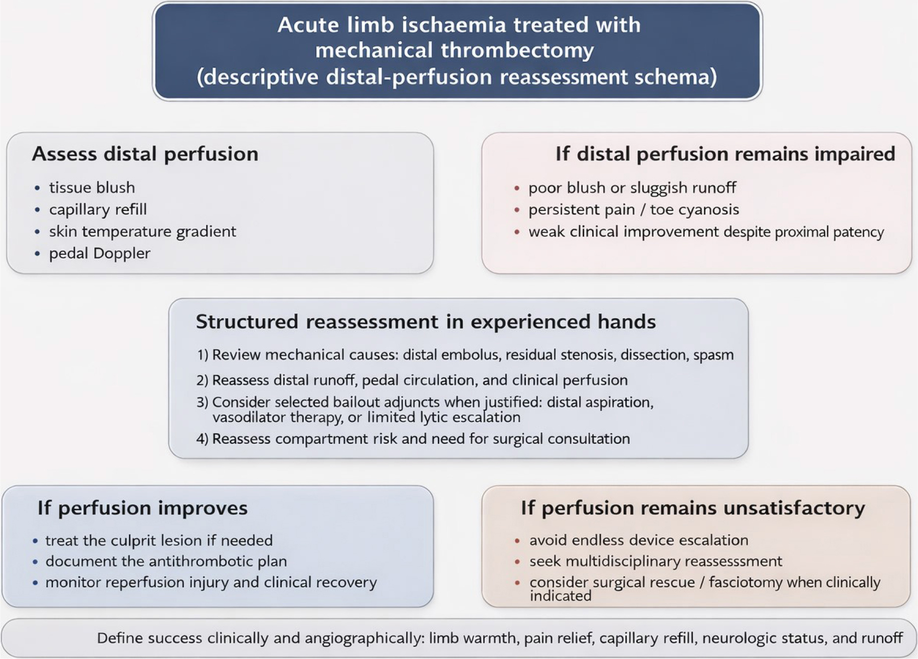
FIGURE 1.
Descriptive reassessment schema for distal perfusion after mechanical thrombectomy in acute limb ischaemia.
After restoration of macro-flow, distal perfusion should be reassessed clinically and angiographically. When distal perfusion remains impaired despite proximal patency, structured reassessment should focus first on treatable mechanical causes, including distal embolus, residual stenosis, dissection, or vasospasm, followed by review of runoff, pedal circulation, and clinical perfusion. Selected bailout adjuncts such as distal aspiration, vasodilator therapy, or limited thrombolytic escalation may be considered in experienced hands when clinically justified. The figure is intended as a descriptive companion schema to support reassessment and reporting, not as a validated treatment algorithm or standard-of-care pathway.
Search strategy and scope of review
To inform this narrative review, we performed a targeted search of PubMed/MEDLINE and Scopus for English-language articles published or available online from database inception to 1 March 2026, using combinations of terms such as “acute limb ischaemia”, “mechanical thrombectomy”, “catheter-directed thrombolysis”, “aspiration”, “rotational thrombectomy”, “compartment syndrome”, and “distal perfusion”. Priority was given to contemporary clinical practice guidelines, systematic reviews, randomized trials, major device registries, and representative contemporary series directly addressing ALI. Foundational historical trials were retained where necessary to frame the evolution of ALI management, while device-specific case series and small reports were included selectively when they provided clinically relevant context for rescue, distal, or off-label techniques that remain sparsely studied in peripheral practice. The structure of the review was informed by contemporary vascular reporting standards and quality-improvement frameworks, especially guideline-based staging, procedural reporting, and outcome definitions.2
Grey literature, conference abstracts, and non-English publications were not systematically included, and the potential for publication and language bias should therefore be acknowledged. Evidence was interpreted hierarchically, with guideline-level, randomized, and systematic-review evidence distinguished from registry data, case series, expert interpretation, and analogy-based extrapolation from coronary or stroke literature. Formal studyquality scoring was not performed because the aim was a structured narrative synthesis rather than a PRISMA-style systematic review.
Clinical framework: definition, staging, and pathophysiology
Definition and staging
ALI should first be framed clinically. Rutherford staging (Table 2) remains the most practical bedside classification because it integrates sensory loss, motor deficit, and arterial/venous Doppler findings, and therefore directly informs urgency and salvageability.2–3,13 The initial assessment should document pain, pallor, temperature gradient, capillary refill, neurologic deficit, duration of symptoms, previous peripheral interventions or bypasses, anticoagulation status, atrial fibrillation, and the likelihood of embolic versus in-situ thrombosis. A threatened limb with rest pain, sensory deficit, or evolving motor deficit should not be delayed by unnecessary imaging.11–13
Mechanisms and ‘the lesion behind the thrombus’
In practice, the visible thrombus is often only part of the problem. Native-vessel thrombosis commonly overlies severe atherosclerotic stenosis, plaque rupture, stent restenosis, or bypass graft failure, while embolic ALI may arise from atrial fibrillation, mural thrombus, or aneurysmal disease.2,4–5,14 Popliteal artery aneurysm deserves specific mention because it is a classic cause of tibial embolization and poor runoff. In this setting, computed tomography angiography (CTA) or duplex ultrasound should assess aneurysm morphology, runoff, and distal target vessels. The practical sequence is usually rapid revascularization to improve runoff and limb viability, followed by definitive aneurysm exclusion or repair once outflow has been restored and anatomy clarified; purely definitive aneurysm repair without addressing the distal embolic burden risks persistent ischaemia.5,14
Reperfusion injury and compartment syndrome
Ischaemia duration and tissue-level injury determine the risk of reperfusion syndrome, acidosis, myoglobin release, acute kidney injury, and compartment syndrome.2,5,15–17 Severe pain out of proportion, tense muscle compartments, pain with passive stretch, worsening neurologic deficit, or rising creatine kinase after revascularization should prompt immediate reassessment. Compartment syndrome is not a secondary detail in ALI management: delayed fasciotomy is associated with worse limb outcomes, and any endovascular-first program must incorporate a low threshold for urgent vascular surgical review and decompression when clinically indicated.15–17
Therapeutic landscape in acute lower limb ischaemia (ALI): surgery, catheter-directed thrombolysis (CDT), and mechanical thrombectomy
Open surgery
Open thrombectomy/embolectomy and bypass remain central components of ALI management rather than historical comparators only.2,9–12,18–19 Open surgery is particularly relevant for common femoral bifurcation emboli, embolic occlusions requiring immediate flow restoration, thrombosed bypass grafts not suitable for endovascular salvage, complex multilevel occlusion in unstable anatomy, and situations in which there is associated aneurysm, infected material, or irreversible ischaemia requiring amputation planning. Surgery also remains important as a rescue option when endovascular crossing fails or when compartment syndrome and tissue loss necessitate combined operative management.2,9–12,18–19
Catheter-directed thrombolysis (CDT)
CDT remains a valid and often highly effective therapy, especially in thrombosed native vessels, bypass grafts, or stents when the limb is viable or marginally threatened and there is time for infusion.2,9–12,20–21 Its advantages are controlled thrombus dissolution, improved visualization of the underlying lesion, and the ability to treat distal runoff that may be difficult to access mechanically. Its disadvantages are equally important: prolonged treatment time, need for monitored care, bleeding risk, repeated imaging, and limited suitability in patients with recent surgery, trauma, stroke, uncontrolled hypertension, or other major contraindications. In contemporary practice, CDT should not be portrayed as an outdated comparator; rather, it remains a key component of the treatment spectrum, either as stand-alone therapy or as an adjunct after MT.2,9–12,20–21
Mechanical thrombectomy (MT)
MT aims to reduce thrombus burden quickly and, in selected patients, shorten time-to-flow and total lytic dose. Potential advantages include single-session treatment, reduced intensive care unit (ICU) utilization, and immediate correction of the culprit lesion by angioplasty or stenting. Potential disadvantages include distal embolization, hemolysis, vessel injury, and incomplete extraction in organized thrombus. MT is best understood as an important tool within ALI management rather than as a universally superior strategy.2,9–12,18–21
Comparative effectiveness
At the comparative-effectiveness level, the evidence remains unsettled. The landmark Rochester, STILE, and TOPAS trials were foundational for the surgery-versus-thrombolysis debate, but they were conducted in an earlier era of vascular imaging, endovascular devices, and peri-procedural care, and they enrolled clinically mixed populations that included embolic occlusions, thrombosed native arteries, bypass graft occlusions, and, in some cohorts, subacute rather than strictly hyperacute presentations.9–12 Their contemporary value is therefore conceptual rather than prescriptive: they frame trade-offs between speed of reperfusion, bleeding risk, durability, and the consequences of treatment delay, but they do not directly determine device-first practice with current aspiration, rotational, hydrodynamic, or pharmacomechanical platforms. Observational studies and device registries report high technical success for modern MT systems, but case mix, Rutherford class, thrombus chronicity, adjunctive therapy, and thresholds for conversion to CDT or surgery vary substantially. The 2026 systematic review and meta-analysis by Acosta et al. screened 9,801 records, reviewed 153 full-text articles, and identified only seven comparative observational studies; the available data allowed only limited pooled analysis and a single meta-regression of three observational studies.1 Any apparent advantage of thrombectomy-first strategies over CDT therefore remains unproven, with very low certainty of evidence, substantial risk of bias, and limited generalizability across devices and clinical patterns.
Mechanical thrombectomy in acute lower limb ischaemia (ALI): device classes and technical principles
Mechanical thrombectomy devices for ALI can be grouped pragmatically by mechanism and workflow (Table 1). This classification is useful for interventional radiology (IR) practice, but available evidence should not be mistaken for proof that any single platform is superior across all anatomic patterns or clinical scenarios. In practice, device choice should be driven by vessel caliber, thrombus burden, distal runoff, suspected clot composition, and the likelihood that definitive adjunctive treatment will be required immediately after thrombus removal.22,23 In broad terms, aspiration is often most attractive when fresh embolic material, distal reach, or catheter trackability is the dominant issue; rotational thrombectomy is often strongest in larger femoropopliteal mixed thrombotic burdens or occluded stents and grafts where rapid debulking is needed; hydrodynamic and pharmacomechanical approaches may be considered when bulky thrombus requires aggressive debulking and the operator accepts haemolysis- or lytic-related trade-offs; and CDT remains preferable when thrombus extends beyond safe mechanical reach or when staged clarification of the underlying lesion is advantageous.20–28
aspiration thrombectomy (manual or pump-assisted),
rotational thrombectomy (mechanical fragmentation with aspiration),
hydrodynamic systems (high-velocity saline jets creating localized suction), and
rheolytic or pharmacomechanical systems (jetbased thrombectomy, sometimes combined with lytic delivery).
TABLE 1.
Pragmatic interventional radiology taxonomy of mechanical thrombectomy approaches in acute lower limb ischaemia (ALI)
| Approach class | Typical use | Main advantages/limitations | Practical notes |
|---|---|---|---|
| Aspiration | Fresh embolic or thrombotic occlusions; distal aspiration-first work+- | Advantages: simple, scalable, distal reach, lysis-sparing. Limitations: organized clot, embolization, clogging. | Adjuncts: angioplasty/stent, micro-dose intra-arterial lytic, vasodilators. Typically ≥2.0 mm distally; ≥3.0 mm for large-bore systems. Low–moderate aspirate, often 50–300 mL but higher if prolonged. |
| Rotational | Large femoropopliteal mixed thrombus needing rapid debulking | Advantages: rapid debulking, handles mixed thrombus. Limitations: vessel trauma, embolization, device cost. | Adjuncts: angioplasty/stent, distal aspiration, selective protection. Typically ≥3.0 mm; not suited for small below-the-knee arteries. Usually <100–200 mL blood loss/aspirate. |
| Hydrodynamic | High-burden thrombus when lysis-sparing debulking is desired | Advantages: effective in high thrombus burden, trackable. Limitations: hemolysis, learning curve, embolization. | Adjuncts: angioplasty/stent, limited catheterdirected thrombolysis rescue. Typically ≥3.0 mm (device-dependent). Low–moderate aspirate; haemolysis risk may exceed visible blood loss. |
| Rheolytic/pharmacomechanical | Thrombectomy with local lytic facilitation when a combined debulking strategy is desired | Advantages: debulking plus lytic delivery, versatile. Limitations: hemolysis, bradycardia, bleeding if lytic is used. | Adjuncts: heparin optimisation, limited catheter-directed thrombolysis, intensive care unit protocol if lytic is used. Approximately ≥2.5–3.0 mm (catheter-dependent). Monitor for haemolysis/bradycardia. |
| Rescue (off-label) | Below-the-knee/pedal rescue for distal emboli when dedicated devices are unsuitable | Advantages: can engage distal emboli with neuro-like control. Limitations: sparse evidence, vessel injury risk. | Adjuncts: distal aspiration, vasodilators, bailout surgery. Approximately ≥2.0 mm with appropriate sizing. Minimal device-related blood loss; bleeding risk mainly reflects adjunctive therapy. |
Aspiration thrombectomy
Aspiration thrombectomy uses large-lumen catheters connected to manual aspiration or pump systems to remove thrombus en bloc or in fragments. It is versatile, intuitive, and particularly attractive in fresh embolic occlusions and mixed thrombus burdens because it can be adapted from iliac-femoral to distal runoff vessels. Prospective data from the STRIDE study support feasibility and good shortterm outcomes, but aspiration is not complication-free: distal embolization, blood loss, incomplete extraction of organized thrombus, and difficulty maintaining catheter-vessel apposition in tortuous or small-caliber segments remain important limitations.22,23
Rotational thrombectomy
Rotational systems such as Rotarex combine fragmentation with aspiration and can achieve rapid debulking of thrombotic material, often allowing immediate angioplasty or stenting of the culprit lesion.24–26 They may be particularly useful in long thrombotic segments or occluded stents and grafts, but they can also generate distal embolic debris and should be used cautiously in small distal vessels or when runoff is already critically limited. Device profile, vessel diameter, and lesion chronicity matter substantially.24–26
Hydrodynamic and rheolytic thrombectomy
Hydrodynamic and rheolytic systems provide another lysis-sparing option, especially in bulky thrombus.27,28 Their appeal lies in rapid lumen gain, yet they bring device-specific risks including hemolysis, bradyarrhythmia, renal stress, and distal embolization. Pharmacomechanical systems can also blur the conceptual boundary between MT and thrombolysis because some workflows incorporate adjunctive lytic delivery. When discussing these devices, it is more precise to describe them as part of a continuum of clot-removal strategies rather than categorically ‘lysis-free’ solutions.27,28
Off-label stent-retrievers for below-the-knee (BTK) rescue
IR practice frequently encounters distal thrombus propagation into tibial or pedal arteries, where dedicated peripheral thrombectomy devices may be constrained by size and trackability. Small reports describe off-label stent-retriever use for selected below-the-knee embolic occlusions, but the evidence remains limited to case reports and very small series.29–31 Such approaches should be reserved for individualized bailout situations in experienced hands rather than viewed as routine peripheral MT strategies.
The “single-session paradigm”
One practical attraction of MT is the possibility of a single-session strategy: thrombus removal, treatment of the culprit stenosis, and optimization of inflow and runoff during the same procedure. This can be highly effective when the limb is threatened, anatomy is technically accessible, the culprit lesion can be definitively treated without major delay, and there is no competing reason to favour prolonged thrombolysis or immediate open surgery. By contrast, staged therapy is often preferable when distal runoff cannot be safely restored mechanically, when thrombus extends diffusely into small-calibre runoff vessels, when bleeding risk still permits CDT, when substantial residual thrombus would make immediate stenting unreliable, or when thrombus dissolution is needed before durable treatment of the underlying lesion. The single-session paradigm should therefore be framed as a situational advantage, not as a default goal.
Technical pitfalls and tips
Several technical principles are worth emphasizing. The initial device should match vessel calibre, thrombus burden, distal runoff, and suspected clot composition rather than operator habit alone. When aspiration is chosen for a fresh total occlusion, bringing the aspiration catheter to the thrombus face and maintaining aspiration during careful crossing may reduce uncontrolled distal embolization compared with free wiring beyond the clot, although this remains an operator-level technical principle rather than a formally validated ALI standard. Repeated blind fragmentation or maceration without aspiration capture can likewise worsen distal embolization, particularly when runoff is already precarious. In small runoff vessels, vigorous continuous suction may create a practical “vacuum-collapse” effect in which the compliant vessel wall is drawn against the catheter tip, reducing effective aspiration and increasing the risk of intimal trauma or apparent loss of distal flow. In such circumstances, pulsed aspiration, smaller-profile systems, and deliberate repositioning are often more sensible than maximal suction. Completion angiography should extend beyond the treated segment to distal runoff and pedal circulation, because opening the conduit artery may overestimate the true reperfusion result.22–28
Calcified culprit lesions require special consideration after thrombus clearance. Standard balloon angioplasty may be sufficient in some cases, but heavy eccentric or concentric calcium can lead to underexpansion, recoil, and early re-occlusion. In selected anatomies, plaque modification strategies such as specialty balloons or intravascular lithotripsy may be reasonable adjuncts, although ALI-specific evidence is sparse and treatment must be individualized.14,20,21
Likewise, the definitive treatment of the culprit lesion after thrombus removal may include plain balloon angioplasty, bare-metal stenting, covered stenting, or, in selected femoropopliteal lesions, drug-coated technology. These decisions are driven primarily by the underlying peripheral arterial disease (PAD) anatomy and durability goals rather than by ALI alone.14,20,21 The key point is that thrombectomy should be viewed as part of revascularization strategy, not as the endpoint.
Finally, access and closure strategy should be planned early. Ultrasound-guided puncture, minimizing sheath escalation where possible, and a realistic haemostasis plan reduce access-site complications in patients who frequently require intensive anticoagulation and antiplatelet therapy.20,21
Practical interventional radiology (IR) workflow: from triage to post-procedure care
Triage and imaging
Initial assessment should establish (i) Rutherford category, (ii) embolic versus thrombotic mechanism, (iii) duration and severity of symptoms, (iv) contraindications to lysis and anticoagulation, and (v) the probability of compartment syndrome or irreversible tissue injury (Table 2).2–5,23 Bedside Doppler, neurologic examination, serum lactate, potassium, creatine kinase, haemoglobin, and creatinine are useful because they inform both urgency and procedural risk. CTA is the dominant crosssectional modality because it defines inflow, outflow, occlusion length, aneurysm, bypass anatomy, and calcified culprit lesions; however, in a clearly threatened limb it should accelerate strategy, not delay reperfusion.2–5,23
TABLE 2.
Rutherford clinical categories of acute limb ischaemia (simplified)
| Category | Clinical findings | Doppler signals | Treatment urgency (typical) |
|---|---|---|---|
| I (Viable) | No sensory loss; no motor deficit | Arterial audible; venous audible | Urgent but not emergent; imaging + plan |
| IIa (Marginally threatened) | Minimal sensory loss; no motor deficit | Arterial often inaudible; venous audible | Urgent revascularization |
| IIb (Immediately threatened) | More than toes sensory loss; mild–moderate motor deficit | Arterial inaudible; venous audible | Emergent revascularization |
| III (Irreversible) | Profound anesthesia; paralysis/rigor | Arterial and venous inaudible | Primary amputation/palliation |
Access strategy and anticoagulation
Access choice should prioritize speed, support, and bailout options: contralateral crossover for iliac and common femoral inflow disease, ipsilateral antegrade access for more distal segments, or upper-extremity access only when anatomically advantageous. Systemic heparinization is generally recommended unless contraindicated, with activated clotting time (ACT)-based reassessment in longer procedures.2,23
Crossing the lesion and first-pass strategy
Crossing strategy is a major determinant of distal embolization. A soft, torqueable wire and support catheter should be used to traverse thrombotic occlusions with minimal forward force and with constant awareness of subintimal passage. The first-pass strategy should reflect the balance between speed and distal protection: aspiration is often attractive for fresh embolic or mixed thrombus, whereas longer organized thrombotic occlusions may require a different platform or staged therapy. After lumen gain, the operator must actively look for the lesion behind the thrombus and determine whether adjunctive angioplasty, stenting, or surgery is required.2,20–23
Adjunctive thrombolysis: lysis-sparing vs. lysis-free
Adjunctive thrombolysis should not be reduced to a simple ‘MT versus lysis’ dichotomy. In real-world ALI practice, thrombolysis may still be used as primary CDT, as a short adjunct after incomplete MT, or as limited intra-arterial bolus therapy for residual distal thrombus or sluggish runoff. The clinically relevant distinction is therefore often not lysis-free versus lysis-based treatment, but whether lytic therapy is prolonged and primary, brief and adjunctive, or highly selective for a residual distal problem. Emerging reports also describe ultrasound-guided direct thrombolytic injection as a highly selective strategy for acute and early-subacute arterial occlusions, emphasizing that targeted local lytic use may occasionally complement rather than compete with catheter-based revascularization.38 Some limbs therefore favour an MT-first, lysis-reducing strategy, whereas others still favour CDT as the primary endovascular option.
Post-revascularization antithrombotic management
Antithrombotic management after revascularization deserves more emphasis than it often receives in device-oriented discussions because it influences re-occlusion, embolic recurrence, and limb-related outcomes. Embolic ALI related to atrial fibrillation or another cardiac source typically requires systemic anticoagulation once bleeding risk permits, whereas native-vessel thrombosis on atherosclerotic disease usually requires antiplatelet therapy and broader secondary prevention. Stented segments may additionally require temporary dual antiplatelet therapy according to lesion location, stent type, and bleeding risk. For the interventional radiologist, the practical point is that the procedure does not end with thrombus extraction: the post-procedural plan should explicitly document the antithrombotic strategy, source investigation when relevant, lipid-lowering treatment, smoking cessation counselling, and early surveillance imaging. It should also make clear whether anticoagulation is already therapeutic, being bridged, or awaiting embolic work-up, whether antiplatelet treatment is intended to be single or dual, and which service will own follow-up of the medical plan.2,14,20–21
Post-procedure care
After revascularization, surveillance should focus on recurrent thrombosis from residual lesion, distal embolization, acute kidney injury, reperfusion syndrome, and compartment syndrome. Clinical reassessment remains essential: a warm foot, improving pain, improving capillary refill, and recovering sensorimotor findings matter more than an attractive angiogram alone. Any deterioration after the procedure should trigger repeat imaging and urgent multidisciplinary review.2–5
Evidence and outcomes: what we can (and cannot) conclude
Current evidence supports MT as a useful endovascular option, but not as a proven replacement for CDT or surgery. Comparative studies remain small, heterogeneous, and heavily influenced by case mix, Rutherford class, thrombus mechanism, and device-specific selection. The strongest recent synthesis remains the 2026 systematic review and meta-analysis by Acosta et al., which found no proven superiority of pharmacomechanical, rotational, or aspiration-first strategies over CDT after adjustment for available confounders and emphasized the very low certainty of evidence.1 In practice, outcomes still appear to depend more on correct patient selection, rapid restoration of meaningful perfusion, and complete treatment of the underlying lesion than on thrombectomy technology alone.
Complications: recognition, classification, and mitigation
Pathophysiology: mechanical vs. functional obstruction
Complications after MT are not limited to perforation or access-site bleeding. From a pathophysiologic standpoint, it is useful to distinguish a predominantly mechanical component - distal embolic obstruction and microembolization of thrombus or atheromatous debris - from a predominantly functional component - vasospasm, endothelial swelling, reperfusion oedema, capillary sludging, and other forms of microvascular dysfunction. In practice these mechanisms often coexist, so a limb may have an apparently patent conduit artery yet persistently poor tissue perfusion.32–40
Redefining ‘procedural success’ as a perfusion endpoint
For that reason, procedural success in ALI should be judged clinically as well as angiographically. Completion angiography, runoff, and pedal Doppler are necessary, but so are warmth, capillary refill, pain relief, neurologic stabilization, and awareness of compartment pressure. These bedside findings remain the most practical immediate surrogates of nutritive perfusion in routine ALI practice and help prevent an attractive angiogram from being mistaken for a definitively salvaged limb.2,20,21
Considerations for reassessment and reporting when distal perfusion remains impaired
When distal perfusion remains unsatisfactory after apparently successful MT, the key issue is not to define a new syndrome prematurely, but to recognize that angiographic endpoints may overestimate true tissue reperfusion. Poor blush, sluggish runoff, persistent distal pain, toe cyanosis, or recurrent small filling defects despite restoration of proximal patency should prompt closer clinical and angiographic reassessment.32–37,39–40
Initial reassessment should focus on treatable mechanical causes such as untreated distal embolus, residual stenosis, dissection, or catheter-induced spasm, followed by selective review of distal runoff and, when clinically justified, limited catheter-based adjuncts such as distal aspiration, vasodilators, or brief thrombolytic escalation.20,21,32–37,41
Any vasodilator or thrombolytic use in this setting is supported mainly by broader endovascular experience and analogy to coronary or stroke literature rather than by established ALI trials. Small intra-arterial doses of nitroglycerin or verapamil may be considered when spasm appears contributory, while selective low-dose alteplase boluses or a short infusion may occasionally be used for residual distal thrombus when bleeding risk is acceptable.32–38,41 These manoeuvres are best understood as individualized bailout measures in experienced hands, not evidence-based protocols.
If perfusion remains unsatisfactory despite endovascular correction, the next step is not endless device escalation but multidisciplinary reassessment, including the possibility of surgical thrombectomy, distal exploration, bypass, or fasciotomy depending on anatomy and clinical findings.2,5,15–17
Pharmacologic adjuncts as descriptive rescue measures for reporting and future study
Any pharmacologic rescue framework for impaired distal perfusion should therefore be regarded as descriptive and investigational only. Catheter-directed vasodilator therapy and selective micro-dose thrombolysis may occasionally be used in experienced hands, but the present literature is insufficient to support protocolized recommendations in ALI.32–38,41,42 These options are included mainly to encourage consistent reporting of indication, agent, dose, route, response, and complications in future studies.
Peripheral ‘no-reflow’ after thrombectomy: a reporting concept for future study
Impaired distal perfusion after recanalization may be viewed as a peripheral ALI analogue of the no-reflow phenomenon that is well recognized in coronary intervention and other vascular beds.32–37 The analogy is useful because it reminds the operator that conduit patency is not the same as nutritive tissue perfusion. However, the peripheral construct remains less well defined and has not been systematically studied in ALI; at present it is best regarded as a descriptive construct for reassessment, reporting, and future study.
Its clinical relevance is straightforward: some limbs remain cold, painful, or poorly perfused despite technically successful thrombectomy because distal embolization, oedema, spasm, microvascular dysfunction, or compartment pressure continues to limit tissue flow.32–37,43–44 Recognizing this possibility may prevent premature procedural closure and encourage closer clinical reassessment.
For documentation and future studies, suspicion of distal perfusion failure after macro-recanalization could be recorded when there is discordance between proximal angiographic success and distal tissue response, such as poor blush, sluggish runoff, persistent toe cyanosis, poor capillary refill, or ongoing ischaemic pain without another clear explanation. This is proposed as a pragmatic reporting aid and research construct, not as a validated diagnostic category or treatment algorithm.
Figure 1 and Table 3 should therefore be read as descriptive companion schemata intended to support reassessment, documentation, and prospective study rather than as treatment algorithms or standards of care.
TABLE 3.
Descriptive examples of adjuncts that may be reported in selected bailout cases when distal perfusion remains impaired after thrombectomy
| Agent/class | Rationale | Typical route | Key risks | Reporting note |
|---|---|---|---|---|
| Nitroglycerin | Vasodilation; spasm relief | Intra-arterial via catheter (small bolus) | Hypotension | May be reported when focal spasm is suspected; reassess clinically and angiographically. |
| Verapamil | Calcium-channel vasodilator | Intra-arterial bolus via catheter | Hypotension/bradycardia | Primarily extrapolated from coronary practice; ALI-specific evidence remains limited. |
| Heparin optimization | Reduce ongoing thrombus propagation | Intravenous bolus; activated clotting time-guided | Bleeding | Supportive antithrombotic optimization rather than a no-reflowspecific therapy. |
| Alteplase (microdose) | Resolve distal microthrombus/sludge | Intra-arterial bolus 2 mg every 5-10 min (max 10 mg) | Bleeding | May be described in selected cases with suspected residual distal thrombus; distinct from infusion CDT; not protocolized. |
| How to report bailout adjunct use | Encourage consistent documentation of indication, agent, dose, route, response, and complications | Via working/thrombectomy catheter when used | Bleeding, hypotension, bradycardia; avoid in unstable patients or when surgical rescue is more appropriate | Illustrative descriptive reporting construct only; not a recommended sequence. |
Future directions for interventional radiology research in acute lower limb ischaemia (ALI)
CTA thrombus mapping as a planning and reporting concept
CTA can also serve as a planning tool before intervention. Thrombus length, vessel diameter, runoff quality, aneurysm, heavy calcification, and abrupt embolic cutoff versus diffuse in-situ disease can help frame whether aspiration, rotational thrombectomy, CDT, surgery, or a hybrid approach is most likely to succeed quickly and safely.2,4,5,14 The point of CTA “thrombus mapping” is therefore not to impose a density-based device rule, but to make pre-procedural discussion more explicit and reproducible by bringing anatomy, runoff, clot appearance, access feasibility, and the likelihood of staged versus single-session therapy into the same planning frame.
A useful pre-procedural IR report might comment on inflow/access feasibility, occlusion segment and length, suspected embolic versus thrombotic pattern, runoff vessel patency, aneurysm, bypass or stent anatomy, and lesion calcification.42–43 Reporting thrombus density or related imaging descriptors may be explored in research settings, but their procedural utility in ALI is not yet established and should not override stronger anatomic and clinical determinants.44–45 Supplementary Figure S1 should therefore be read as a conceptual planning and reporting schema, not as a validated device-selection rule.
Standardized technical reporting
Standardized reporting is urgently needed if ALI studies are to become comparable. Beyond technical success, reports should capture Rutherford class, symptom duration, embolic versus thrombotic mechanism, device type, number of passes, adjunctive thrombolysis, percutaneous transluminal angioplasty (PTA)/stenting, runoff change, distal embolization, acute kidney injury, compartment syndrome/fasciotomy, 30-day major amputation, 30-day mortality, and reintervention.1,38–39
A pragmatic registry dataset for interventional radiology (IR)-led studies
Prospective registries are probably the most realistic short-term path toward better evidence. Table 4 illustrates a compact dataset that can be incorporated into routine IR documentation while still capturing clinically meaningful endpoints, mechanism-specific failure modes, adjunctive treatments, and complications.38,39
TABLE 4.
Suggested minimum dataset for prospective ALI thrombectomy registries
| Domain | Variables (minimum set) | Why it matters | Capture point |
|---|---|---|---|
| Baseline | Age, sex, atrial fibrillation, coronary artery disease, chronic kidney disease, diabetes, smoking; antithrombotics | Risk stratification, bleeding risk, etiology | Pre-procedure |
| Ischemia | Symptom onset (hours), Rutherford category, baseline Doppler, lactate/creatine kinase | Ischemia time drives limb loss/reperfusion injury | Pre-procedure |
| Imaging | CTA/MRA segment map; calcification; runoff score (if used) | Device selection; predicts below-the-knee work | Pre-procedure |
| Procedure | Access; device class/size; passes; adjunctive angioplasty/stent; lytic (bolus/infusion, dose); vasodilators; distal embolization/below-the-knee rescue | Reproducibility; identifies failure modes | Intraprocedure |
| Outcomes | Technical success; time-to-flow; intensive care unit need; bleeding; complications class; 30-day limb salvage; 30-day mortality; reinterventions; 6-12-month major amputation and patency/target lesion reintervention | Clinically meaningful endpoints | Post-procedure & follow-up |
Trial design opportunities
High-yield research questions include MT-first versus CDT-first strategies in Rutherford IIa and IIb ALI, aspiration versus rotational first-line treatment in long thrombotic segments, the role of adjunctive thrombolysis after incomplete MT, and better definition of clinically relevant distal perfusion failure after technically successful revascularization.1,38,39
Equally important is greater integration of endovascular and surgical thinking. Future trials and registries should not treat surgery as a historical control, but as an active comparator and rescue option within contemporary multidisciplinary limb salvage pathways.2,9–12,18–21
Limitations of the review
This article is a targeted interventional radiology-oriented narrative synthesis rather than a formal systematic review. It integrates guideline-based management, major trials, contemporary device literature, and the recent systematic review by Acosta et al., but remains susceptible to selection bias and uneven evidence quality across device classes. The sections on distal perfusion reassessment, peripheral no-reflow, CTA-based planning, and rescue adjuncts are intended primarily as descriptive reporting considerations and directions for future study, not as validated clinical algorithms.
Conclusions
Mechanical thrombectomy is an important component of modern ALI management, but current evidence does not justify portraying it as categorically superior to CDT or surgery.1,2,9–12 For interventional radiology practice, its value depends on correct staging, appropriate positioning within the broader treatment spectrum, treatment of the culprit lesion behind the thrombus, individualized post-revascularization antithrombotic planning, and recognition that limb salvage ultimately depends on restored tissue perfusion rather than conduit patency alone. Distal perfusion reassessment after thrombectomy and CTA-based pre-procedural mapping are best understood at present as structured descriptive frameworks that may support a more microcirculation-aware and reportable approach to ALI without being mistaken for validated algorithms.