FIGURE 1.
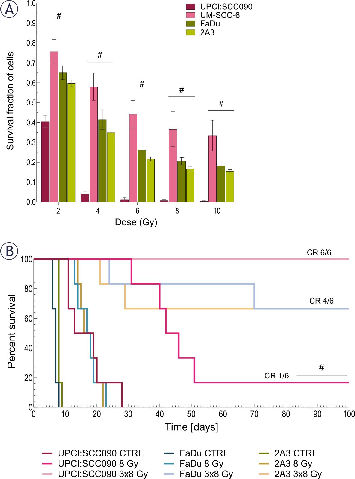
FIGURE 2.
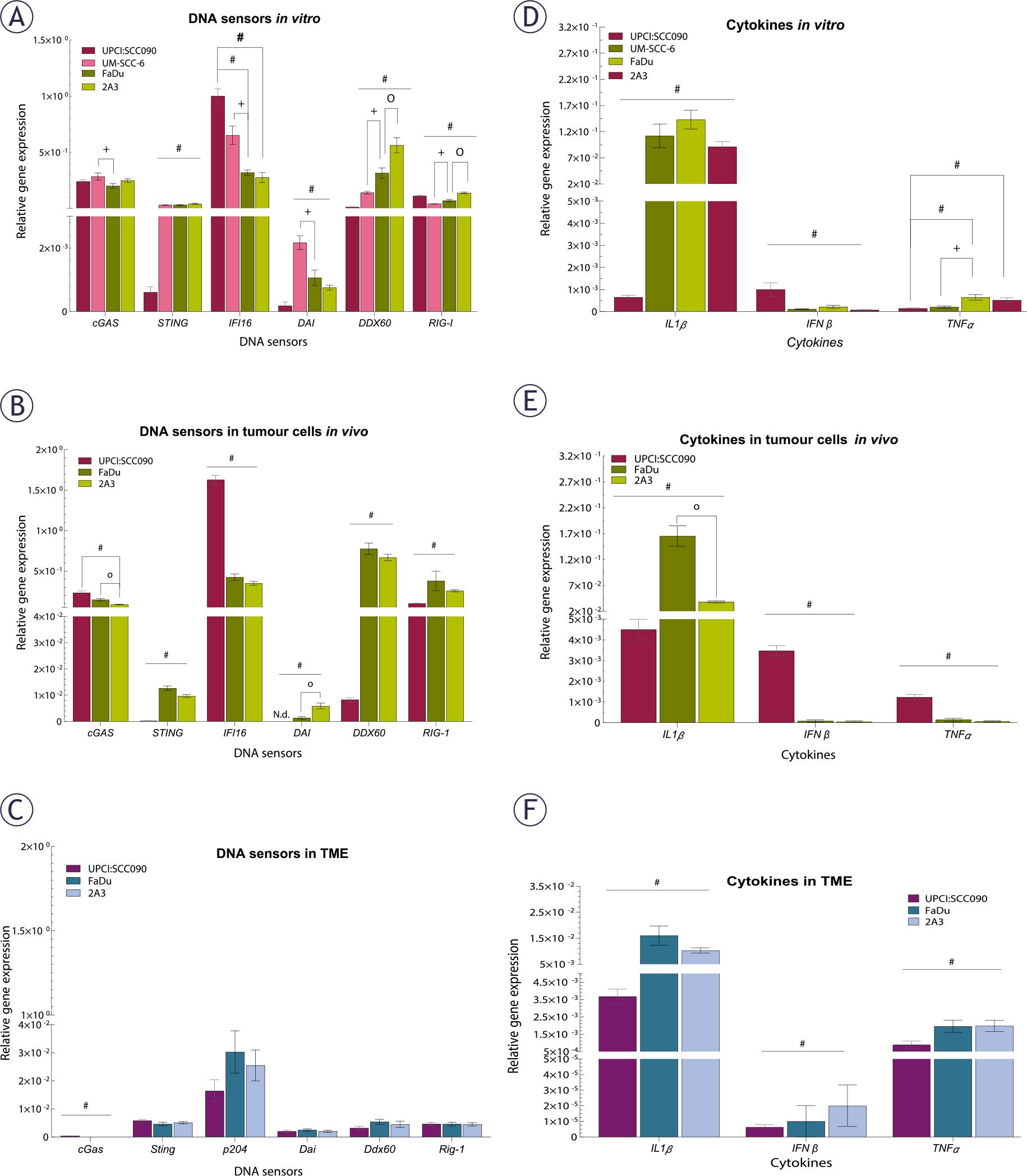
FIGURE 3.
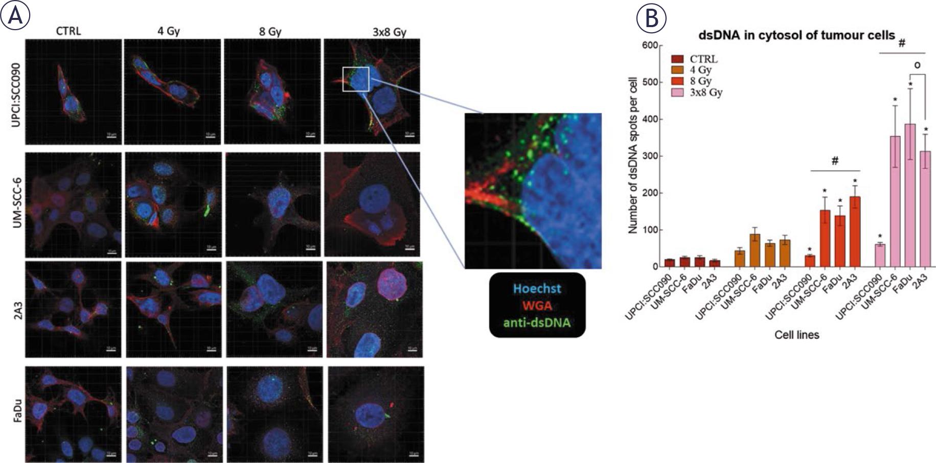
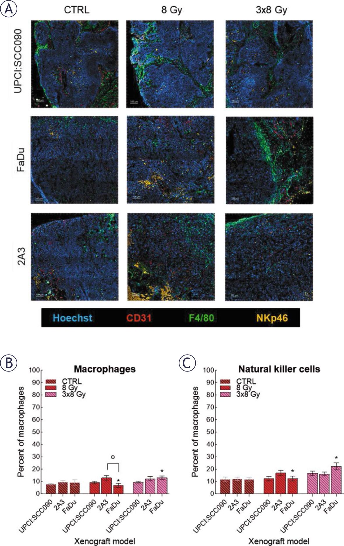
FIGURE 4.
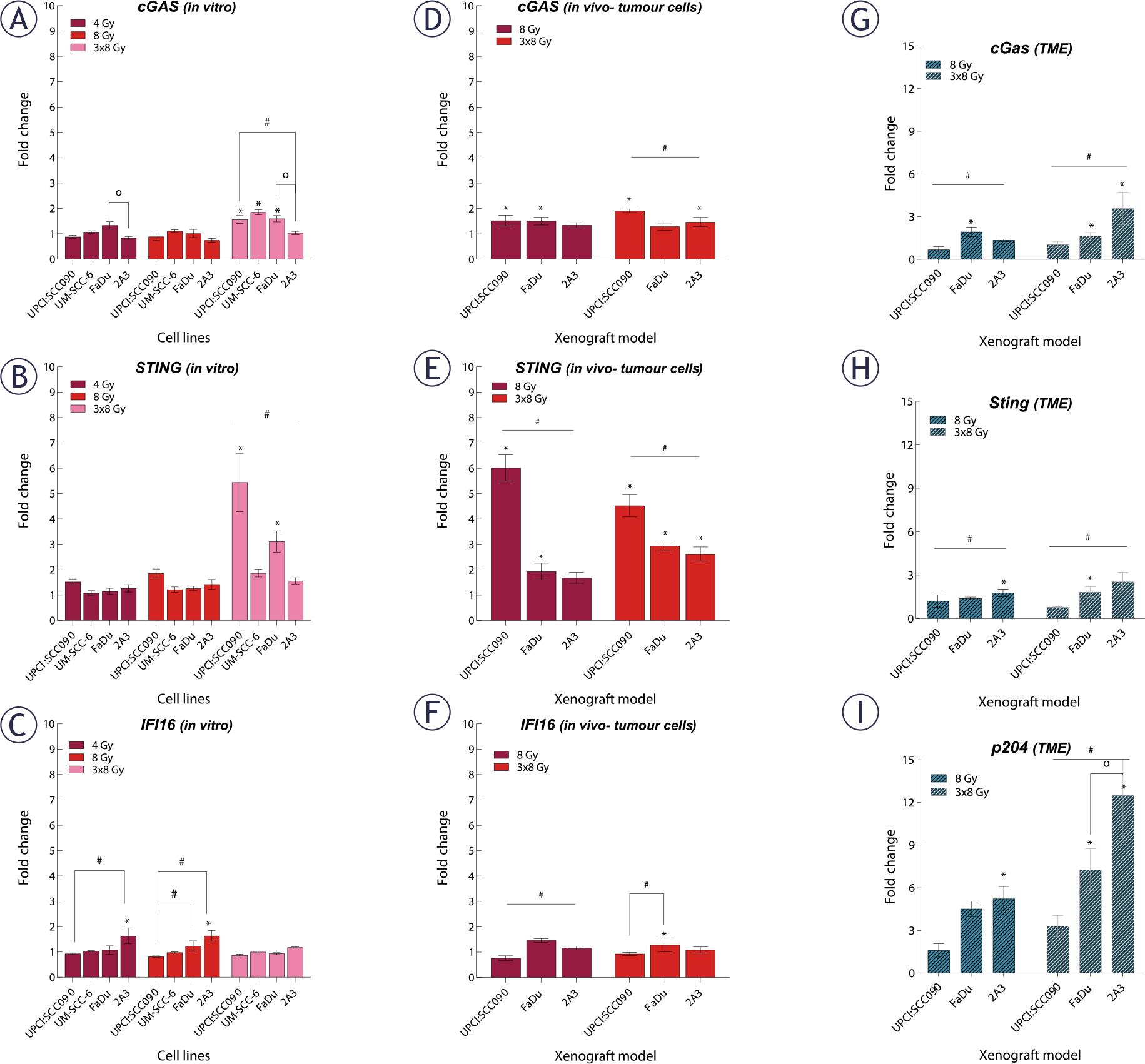
FIGURE 5.
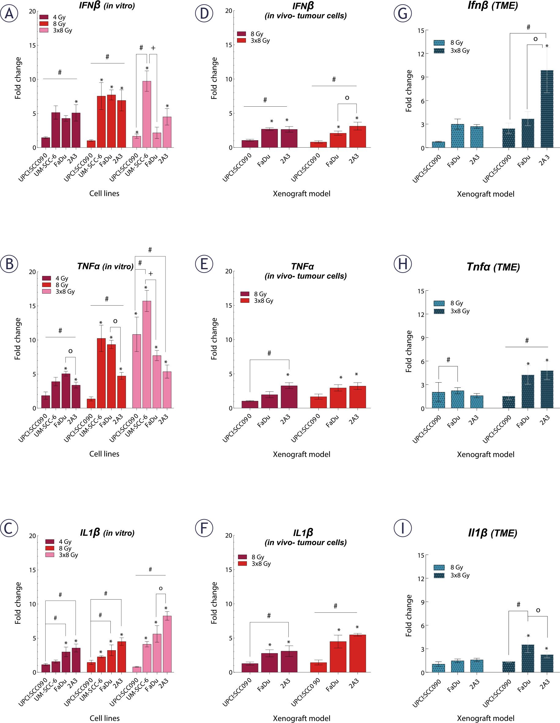
FIGURE 6.

© 2025 Kristina Levpuscek, Tanja Jesenko, Tilen Komel, Simona Kranjc Brezar, Gregor Sersa, Maja Cemazar, Primoz Strojan, published by Association of Radiology and Oncology
This work is licensed under the Creative Commons Attribution 4.0 License.