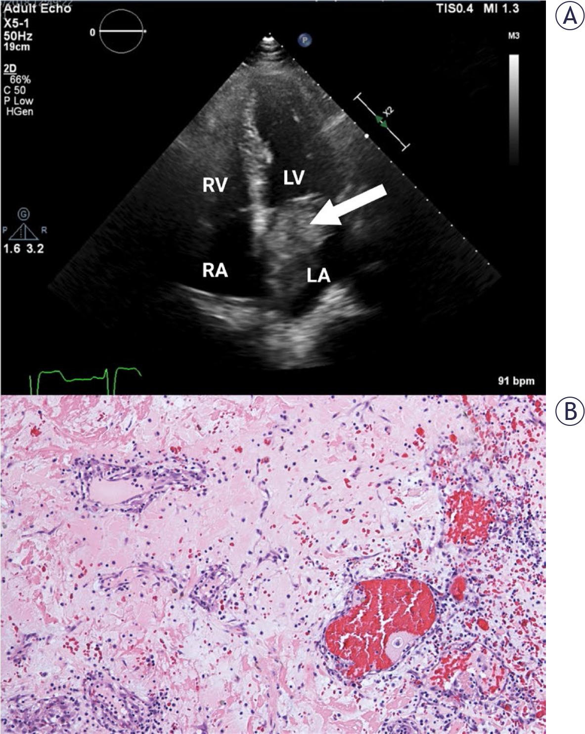
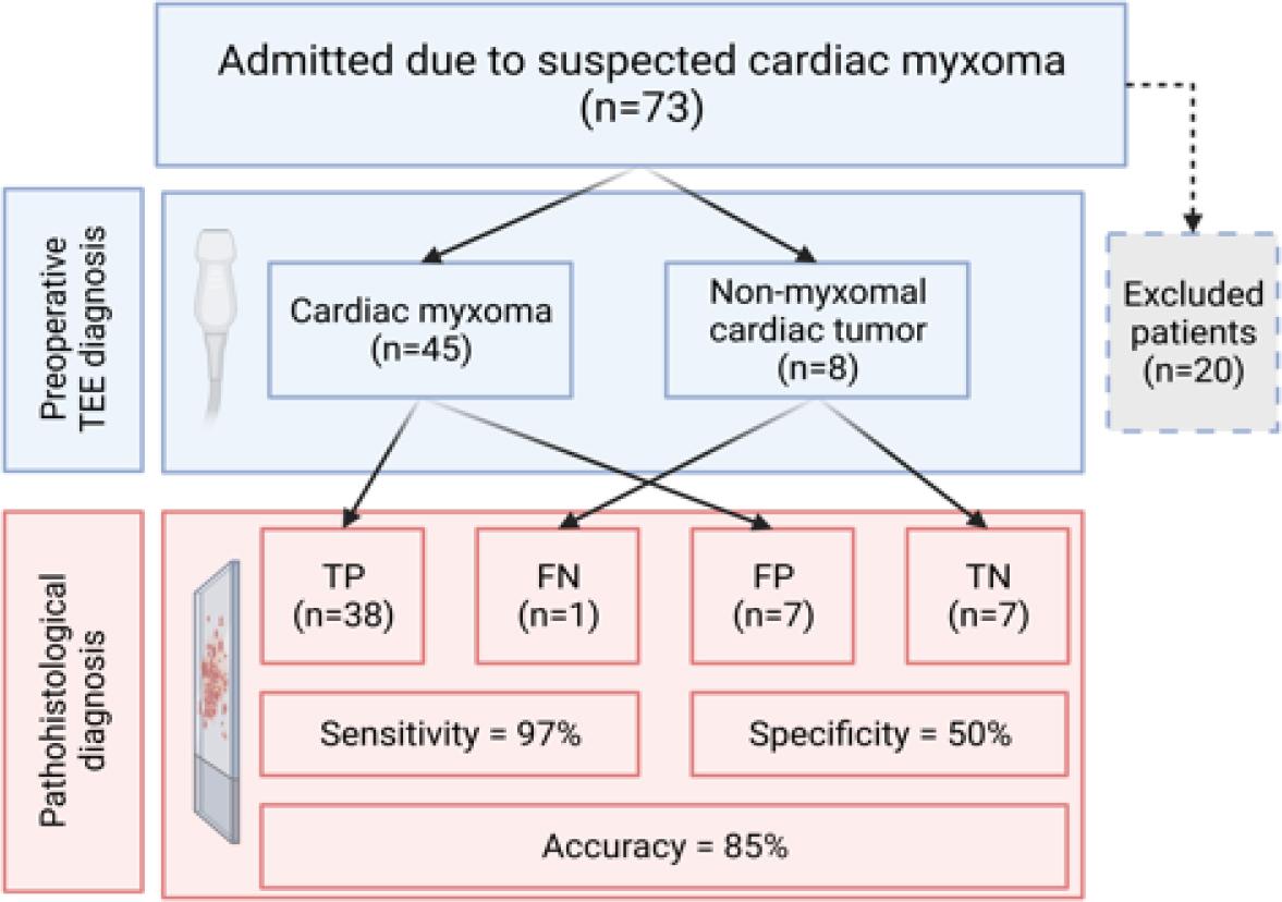
Have a personal or library account? Click to login
Accuracy of transthoracic echocardiography in diagnosis of cardiac myxoma: single center experience Cover

Accuracy of transthoracic echocardiography in diagnosis of cardiac myxoma: single center experience

Open Access
|Jan 2025

Figures & Tables

FIGURE 1.

(A) Transthoracic echocardiography, apical 4-chamber view. Cardiac mass in left atrium is attached to the interatrial septum in the region of the fossa ovalis (arrow). Histopathological characterization confirmed cardiac myxoma. (B) Abundant myxoid stroma with clusters of myxoma cells forming cords and ring structures (HE 100×).
LA = left atrium; LV = left ventricle; RA = right atrium; RV = right ventricle
(A) Transthoracic echocardiography, apical 4-chamber view. Cardiac mass in left atrium is attached to the interatrial septum in the region of the fossa ovalis (arrow). Histopathological characterization confirmed cardiac myxoma. (B) Abundant myxoid stroma with clusters of myxoma cells forming cords and ring structures (HE 100×). LA = left atrium; LV = left ventricle; RA = right atrium; RV = right ventricle

FIGURE 2.

Flow diagram showing the diagnostic accuracy of preoperative TTE in patients with suspected CM. The predictive value and accuracy of preoperative TTE was calculated using the results of pathohistological examination as the gold
FN = false negative; FP = false positive; TP = true positive; TN = true negative; TTE = transthoracic echocardiography
Flow diagram showing the diagnostic accuracy of preoperative TTE in patients with suspected CM. The predictive value and accuracy of preoperative TTE was calculated using the results of pathohistological examination as the gold FN = false negative; FP = false positive; TP = true positive; TN = true negative; TTE = transthoracic echocardiography

Comparison of demographic and echocardiographic characteristics between pathohistologically confirmed cardiac myxoma (CM) and non-myxoma (NM) groups

CharacteristicCM (n= 39)NM (n = 14)p value
Age (years)63.1 ± 13.666.6 ± 15.10.434
Sex (female)25 (64)10 (71)0.620
Location
    Typical28 (72)0P < 0.001
    Atypical11 (28)14 (100)P < 0.001
Size (mm)37.9 ± 18.324.3 ± 13.20.017

Preoperative echocardiographic characteristics of the cardiac myxoma (CM) and non-myxoma (NM) group

Echocardiographic characteristicsCM (n=45)NM (n =8)
Mean size (mm)35.3 ± 18.6 (range: 10–81)30.3 ± 16.9 (range: 8–50)
Location
  Left atrium36 (80)2 (25)
    Fossa ovalis27 (75)
    Mitral valve4 (11)1 (50)
    Posterior interatrial septum3 (8)
    Free left atrial wall1 (3)1 (50)
    Left atrial appendage1 (3)
  Right atrium8 (18)4 (50)
  Left ventricle1 (2)1 (12)
  Aortic valve 1 (12)
Mobility
  Mobile32 (71)7 (88)
  Non-mobile5 (11)
  No data available8 (18)1 (12)
Surface
  Smooth or lobulated28 (62)1 (12)
  Villous8 (18)3 (38)
  No data available9 (20)4 (50)
Obstruction (present)11 (24)2 (25)
  Mitral valve10 (21)1 (50)
  Tricuspid valve1 (9)1 (50)
DOI: https://doi.org/10.2478/raon-2025-0007 | Journal eISSN: 1581-3207 | Journal ISSN: 1318-2099
Language: English
Page range: 63 - 68
Submitted on: Feb 10, 2024
|
Accepted on: Dec 8, 2024
|
Published on: Jan 22, 2025
In partnership with: Paradigm Publishing Services
Publication frequency: 4 issues per year

© 2025 Polona Kacar, Nejc Pavsic, Mojca Bervar, Zvezdana Dolenc Strazar, Katja Prokselj, published by Association of Radiology and Oncology
This work is licensed under the Creative Commons Attribution 4.0 License.