
Figure 1.
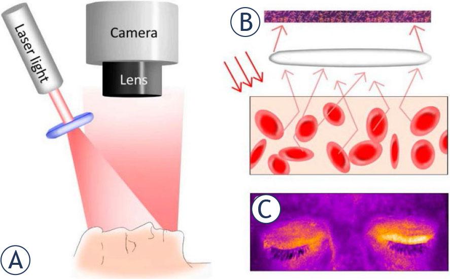
Figure 2.
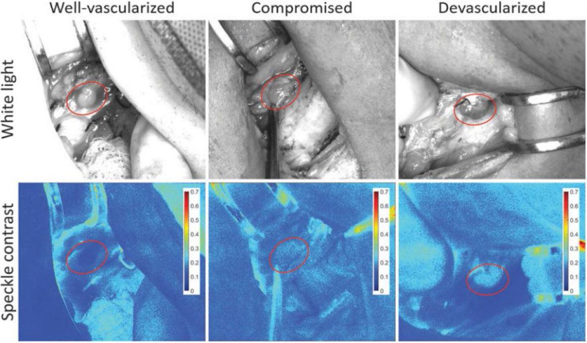
Figure 3.
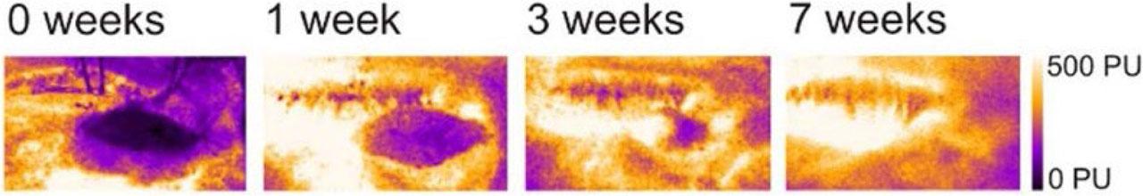
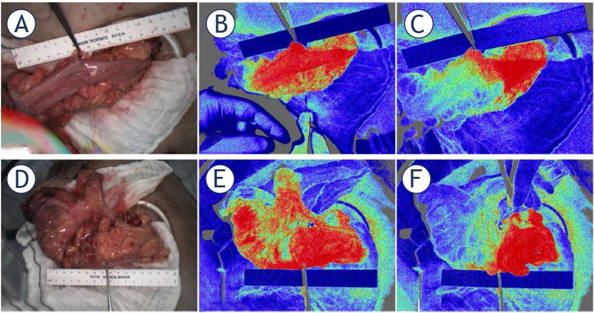
Figure 4.
Included articles reporting the use of laser speckle contrast imaging (LSCI) to quantify perfusion in clinical applications in oncology
| Reference | Year of publication | Number of patients | Oncologic setting |
|---|---|---|---|
| Brain | |||
| Parthasarathy et al.21 | 2010 | 3 | Tumor resection |
| Richards et al.22 | 2014 | 10 | Tumor resection |
| Richards et al.27 | 2017 | 8 | Tumor resection |
| Klijn et al.25 | 2013 | 8 | Tumor resection |
| Ideguchi et al.28 | 2017 | 12 | Tumor resection |
| Breasts | |||
| Tesselaar et al.29 | 2017 | 15 | Adjuvant radiotherapy for stage I-II breast cancer |
| Zötterman et al.30 | 2020 | 23 | Deep inferior epigastric artery perforator (DIEP) flap surgery |
| Endocrine glands | |||
| de Paula et al.31 | 2021 | 42 | Non-functioning adrenal incidentaloma |
| Mannoh et al.32 | 2017 | 28 | Thyroidectomy/parathyroidectomy |
| Mannoh et al.33 | 2021 | 72 | Thyroidectomy |
| Mannoh et al.34 | 2023 | 21 | Thyroidectomy/parathyroidectomy |
| Skin | |||
| Tchvialeva et al.35 | 2012 | 214 lesions | Malignant melanoma, squamous cell carcinoma, basal cell carcinoma, melanocytic nevus, seborrheic keratosis |
| Reyal et al.36 | 2012 | 12 | Basal cell carcinoma |
| Zhang et al.37 | 2019 | 12 (total 143) | Facial nerve palsy due to nerve tumor (also including other etiology) |
| Zieger et al.38 | 2021 | 9 | Basal cell carcinoma |
| Tenland et al.39 | 2019 | 13 | Oculoplastic reconstructive surgery (tarsoconjunctival flaps) |
| Berggren et al.40 | 2019 | 9 | Oculoplastic reconstructive surgery (tarsoconjunctival flaps) |
| Tenland et al.41 | 2021 | 12 | Oculoplastic reconstructive surgery after squamous cell carcinoma, basal cell carcinoma, and intradermal nevus |
| Berggren et al.42 | 2021 | 7 | Oculoplastic reconstructive surgery after squamous cell carcinoma and basal cell carcinoma |
| Berggren et al.43 | 2021 | 7 | Oculoplastic reconstructive surgery after squamous cell carcinoma and basal cell carcinoma |
| Berggren et al.44 | 2021 | 1 | Oculoplastic reconstructive surgery |
| Berggren et al.45 | 2022 | 7 | Oculoplastic reconstructive surgery after squamous cell carcinoma and basal cell carcinoma |
| Stridh et al.46 | 2024 | 1 | Cutaneous angio-sarcoma |
| Gastrointestinal tract (open surgical setting) | |||
| Eriksson et al.47 | 2014 | 10 | Liver resection |
| Milstein et al.48 | 2016 | 11 | Esophagectomy |
| Ambrus et al.49 | 2017 | 45 | Esophagectomy |
| Ambrus et al.50 | 2017 | 25 | Ivor-Lewis esophagectomy |
| Di Maria et al.51 | 2017 | 2 | Colorectal resection |
| Jansen et al.52 | 2018 | 26 | Esophagectomy |
| Kojima et al.53 | 2019 | 8 | Colorectal resection |
| Kaneko et al.54 | 2020 | 36 | Colorectal resection (34 due to colorectal carcinoma) |
| Gastrointestinal tract (laparoscopic/thoracoscopic setting) | |||
| Heeman et al.55 | 2019 | 10 | Colorectal resection |
| Kojima et al.56 | 2020 | 27 | Colorectal resection |
| Slooter et al.57 | 2020 | 24 | Esophagectomy |
| Heeman et al.58 | 2023 | 67 | Hemicolectomy and sigmoid resection |
| Nwaiwu et al.59 | 2023 | 40 | Colectomy, also non-oncological interventions (Roux-en-Y gastric bypass and sleeve gastrectomy) |