
Figure 1
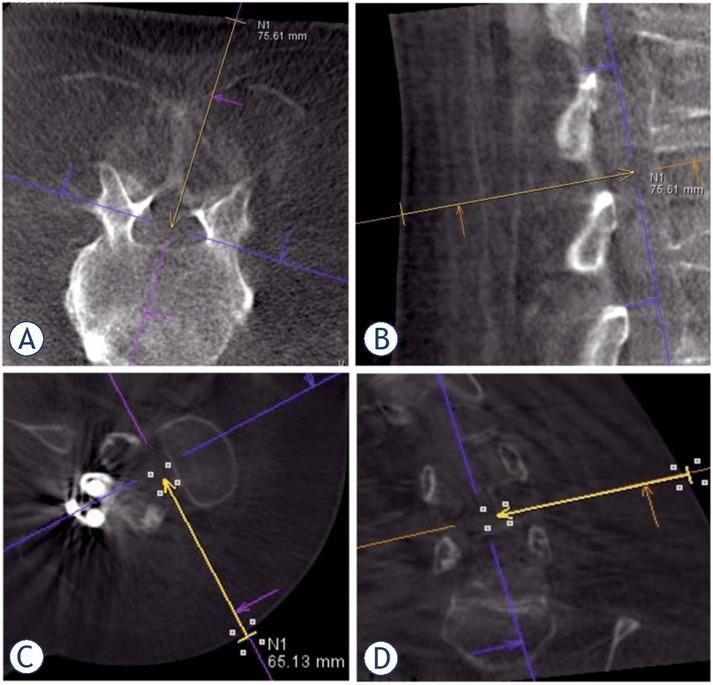
Figure 2
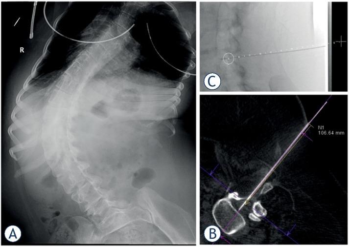
Figure 3
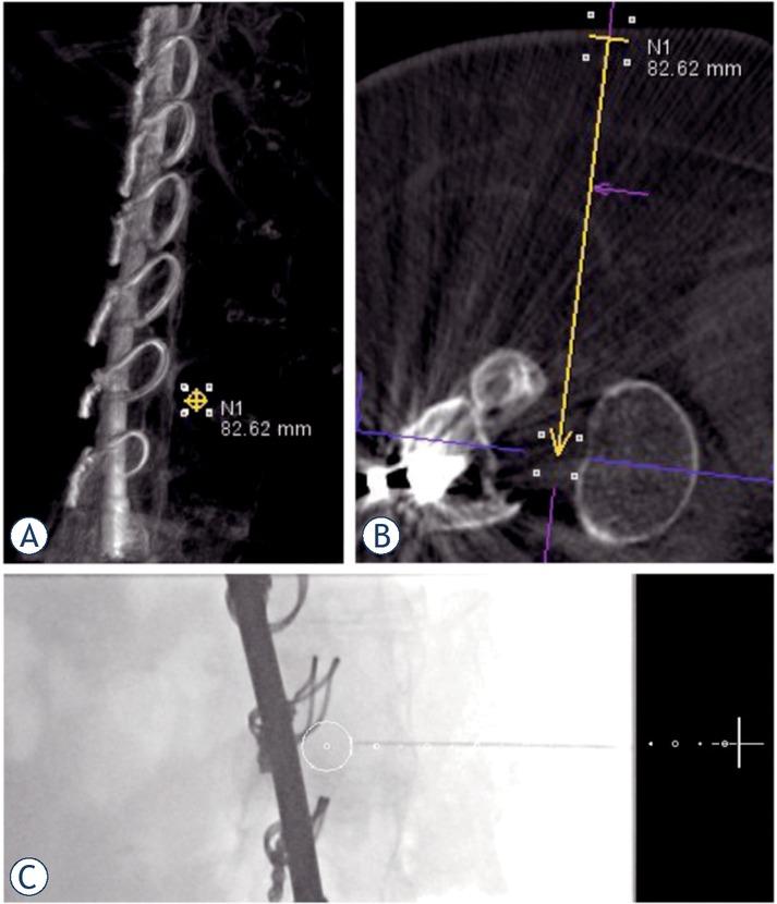
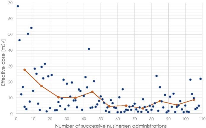
Figure 4
Adverse events for cone-beam CT (CBCT)-guided intrathecal nusinersen delivery patients and classical lumbar puncture patients
| CBCT- guided (n = 108) | Conventional lumbar (n = 112) | P-value | |
|---|---|---|---|
| Headache occurence (%) | 18 (17) | 42 (37) | |
| Headaches (range) VAS, median | 2 (0–10) | 4.5 (0–10) | 0.12 |
| Headaches median (range) duration day, | 0.05 (0–5) | 2 (0–6) | 0.05 |
| Low back pain occurrence (%) | 11 (10) | 40 (36) | |
| Low (range) back pain VAS, median | 0 (0–2) | 2.75 (6) | < 0.01 |
| Low median back (range) pain duration day, | 0 (0–4) | 2.45 (0–14) | < 0.01 |
Patient characteristics for cone-beam CT (CBCT)-guided intrathecal nusinersen delivery patients and classical lumbar puncture patients
| CBCT-guided | Classical lumbar | P-value | |
|---|---|---|---|
| Male sex (%) | 10 (50) | 12 (67) | |
| Age median at first (range) administration, year | 33.5 (20–62) | 44.5 (19–69) | 0.62 |
| BMI, median (range) kg/m2 | 23.4 (14.2–41.8) | 24.7 (14.3–33.6) | 0.08 |
| SMA type 2 (%) | 13 (65) | 0 | |
| SMA type 3 (%) | 7 (35) | 15 (83) | |
| SMA type 4 (%) | 0 | 3 (17) | |
| Posterior fusion instrumentation scoliosis (%) due to | 10 (50) | 0 | |
| Severe scoliosis (%) | 17 (85) | N/A |
Procedure summary
| Interlaminar | Transforaminal | Total | |
|---|---|---|---|
| L1-L2 (%) | 0 (0) | 4 (4) | 4 (4) |
| L2-L3 (%) | 7 (6) | 48 (44) | 55 (50) |
| L3-L4 (%) | 10 (9) | 35 (32) | 45 (41) |
| L4-L5 (%) | 2 (2) | 3 (3) | 4 (5) |
| Number of procedures (%) | 19 (18) | 89 (82) | 108 (100) |
| Duration mean ± SD per min procedure, | 63 ± 21 | 60 ± 26 | 62 ± 25 |