
Table 1
Patient characteristics and postoperative complications
| Age at diagnosis | Localisation | Size [cm] | Histology | Grading | Days to resection | Resection status | Postoperative complication | Follow up |
|---|---|---|---|---|---|---|---|---|
| 85 | forearm | 7.5 | NOS | 2 | 45 | 1 | hematoma | alive, NED |
| 91 | lower leg | 5.4 | NOS | no surgery | lost to follow up | |||
| 82 | thigh | 7.0 | myxofibrosarcoma | 2–3 | 25 | 0 | alive, NED | |
| 84 | forearm | 6.0 | epitheliod myxofibrosarcoma | 3 | 18 | 0 | alive, NED | |
| 91 | thigh | 5.5 | NOS | 3 | 15 | 2 | local and distant recurrence | |
| 79 | thoracic wall | 7.7 | liposarcoma | 2 | 29 | 0 | alive, NED | |
| 80 | gluteus | 10.0 | NOS | 3 | 30 | 0 | alive, NED | |
| 84 | thigh | 3.7 | leiomyosarcoma | 3 | 34 | 0 | alive, NED | |
| 83 | thigh | 10.0 | liposarcoma | 3 | 21 | 1 | wound healing complication | local and distant recurrence |
| 80 | thigh | 8.0 | NOS | 3 | 31 | 0 | wound healing complication, seroma | alive, distant recurrence lower leg, curative treatment |
| 90 | thigh | 8.5 | leiomyosarcoma | 2 | 29 | 0 | wound healing complication | alive, NED |
| 85 | axilla | 9.2 | liposarcoma | 2 | 31 | 1 | alive, NED | |
| 82 | thigh | 17.0 | liposarcoma | 2 | 23 | 0 | alive, NED | |
| 87 | thoracic wall | 5.0 | NOS | 3 | 20 | 0 | alive, NED | |
| 82 | thoracic wall | 9.0 | NOS | 3 | 23 | 0 | wound healing complication | alive, NED |
| 91 | upper arm | 5.2 | NOS | 3 | 31 | 0 | recurrence distant | |
| 81 | thigh | 8.3 | myxoid fibrosarcoma | 3 | 31 | 0 | alive, NED | |
| 81 | upper arm | 8.3 | NOS | 2 | 32 | 0 | alive, NED |
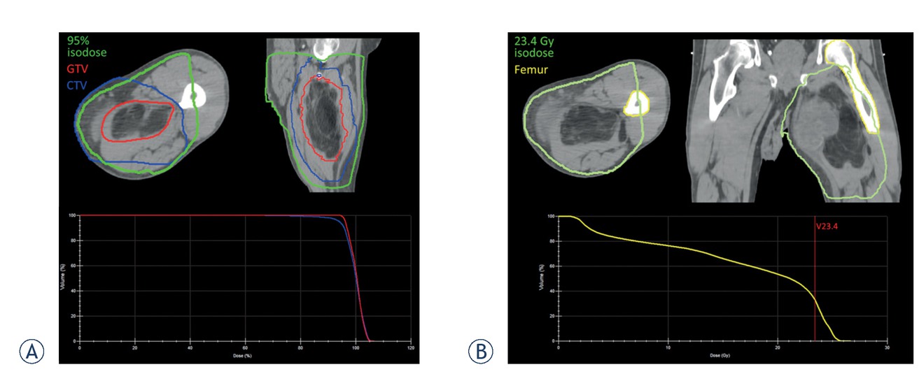
Figure 1
Example of a radiation plan for a thigh sarcoma. The 3D conventional radiotherapy plan shows a good dose coverage for the target volumes (even for this case of the largest tumour in our series with 17 cm) (A). The dose constraints for bones concerning pathologic fractures described below were kept (B).
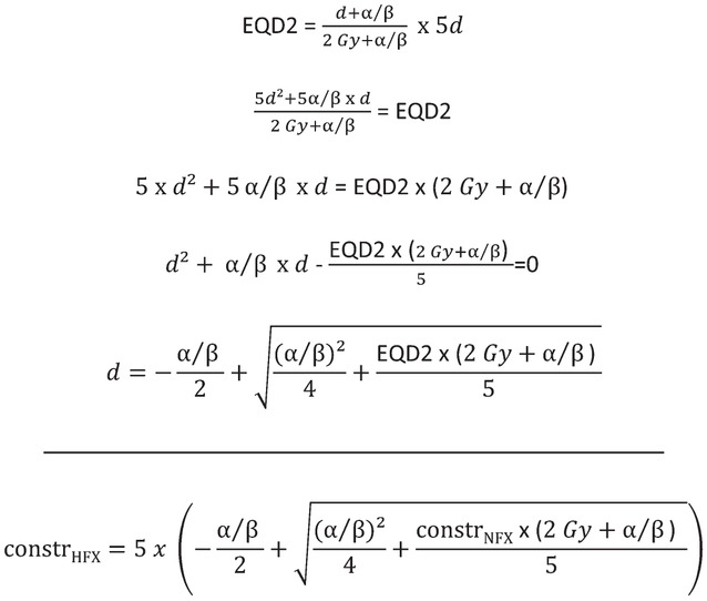
Figure 2
Starting from the equation for equivalent dose 2 Gy (EQD2) for a hypofractionated radiation regimen in 5 fractions, the quadratic equation for the single dose in five fractions corresponding to the known EQD2 was derived. Solving the quadratic equation leads to the dose per fraction for five fractions corresponding to the given EQD2 (assuming a known α/β value for tumour control or side effects in OARs, respectively). Dose per fraction for the constraints for normofractionated radiotherapy was fixed to 2 Gy, although dose per fraction varies with the number of fractions for the same total dose (e.g. 40 Gy circumferential dose would refer to a dose per fraction of 1.6 Gy for 25 fractions in preoperative radiotherapy or 1.2 Gy for postoperative radiotherapy in 33 fractions). As dose constraints for normofractionated radiotherapy normally are not corrected for number of fractions in clinical plan evaluation, they were not corrected to EQD2 for the transfer to the hypofractionated regimen. constrHFX = constraint for hypofractioanted radiotherapy. constrNFX = constraint for normofractioanted radiotherapy.
Table 2
Dose constraints
| Bone | α/β = 1.8 Gy | α/β = 2.8 Gy | ||
|---|---|---|---|---|
| V40 < 64% | V23.4 < 64% | V24.8 < 64% | [18] | |
| Dmean < 37 Gy | Dmean < 22.4 | Dmean < 23.6 Gy | [18] | |
| D2 < 59 Gy | D2 < 29.3 Gy | D2 < 31.3 Gy | [18] | |
| Circumferential < 50 Gy | Circumferential < 26.4 Gy | Circumferential < 28.3 Gy | Institutional standard | |
| Soft tissue | α/β = 2.0 Gy | |||
| Circumferential < 40 Gy | Circumferential < 23.7 Gy | Institutional standard |
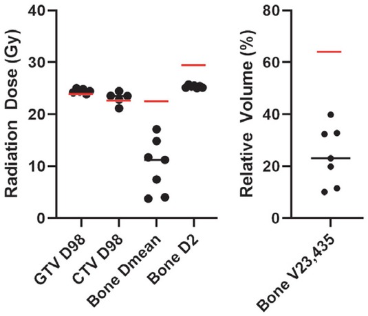
Figure 3
Radiation planning parameters were evaluated for seven of nine patients with sarcomas of the lower extremity. For bone constraints concerning pathologic fracture the whole femur or the whole tibia were contoured for thigh and calf sarcomas, respectively. Black bars indicate the median values, red bars indicate the assumed constraints as described in Tbl 2 and in the main text (for bone with α/β = 1.8 Gy, worst case scenario). Gross tumour volume (GTV) coverage was reached in all cases. D98 for clinical target volume (CTV) fell short in one patient with a large calf sarcoma with a CTV reaching the skin in large areas. Re-calculated dose constraints for pathologic fracture were not reached in any case.