
TABLE 1
Demographic data of the patients and healthy controls enrolled in the study
| Number | Mean | |
|---|---|---|
| (n) | (SD) | |
| Number | 40 | |
| Crohn’s disease | 16 | |
| Female/male | 8/8 | |
| Age (years) | 38.6 | |
| (13.7) | ||
| Disease duration (years) Age at diagnosis (years) | 7.1 (6.5) 31.4 | |
| (14.5) | ||
| Smokers | 5 | |
| Ex-smokers | 3 | |
| Nonsmokers | 8 | |
| Location of Crohn’s disease | 16 | |
| I. L1 (ileal) | ||
| II. L2 (colonic) | ||
| III. L3 (ileocolonic) | ||
| IV. L4 (isolated upper disease) | ||
| Concomitant medications | 13 | |
| I. Aminosalicylates | ||
| II. Corticosteroid | ||
| III. Azathioprine | ||
| IV. Azathioprine + corticosteroids | ||
| V. None | ||
| Ulcerative colitis | 14 | |
| Female/male | 8/6 | |
| Age (years) | 39.2 (12) | |
| Disease duration (years) | 5.6 (2.8) | |
| Age at diagnosis (years) | 33.5 | |
| (11.7) | ||
| Smokers | 1 | |
| Ex-smokers | 3 | |
| Nonsmokers | 10 | |
| Extent of ulcerative colitis | 14 | |
| I. E1 (proctitis) | ||
| II. E2 (left sided) | ||
| II. E3 (extensive) | ||
| Concomitant medications | 14 | |
| I. Aminosalicylates | ||
| II. Corticosteroid | ||
| III. Azathioprine | ||
| IV. Azathioprine + corticosteroids | ||
| V. None | ||
| Healthy controls | 10 | |
| Female | 6 | |
| Male | 4 | |
| 58.3 | ||
| Age (years) | (9.4) |
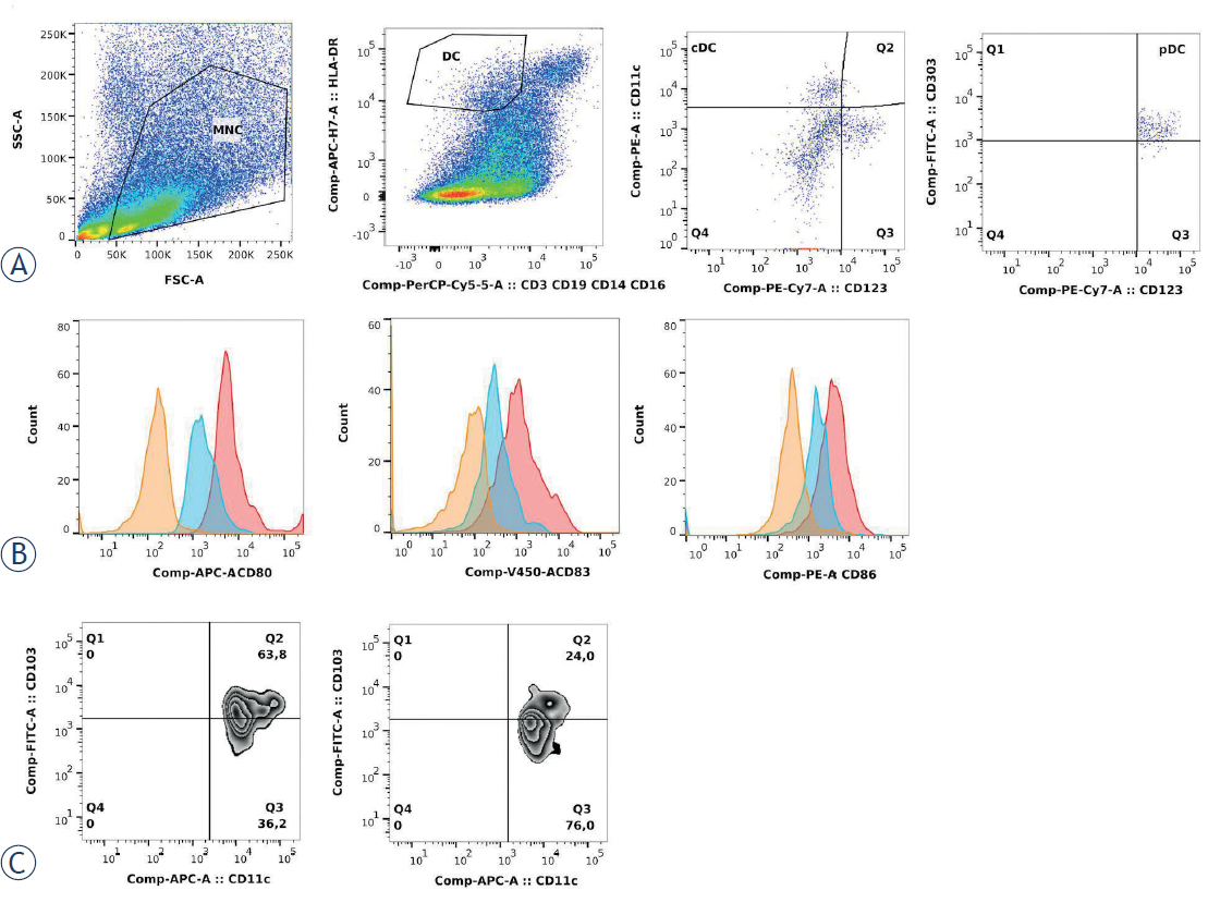
FIGURE 1
Dendritic cell (DC) phenotypes: DCs were isolated from the lamina propria by enzymatic degradation and mechanical dissociation. Data were analysed using FlowJo software. (A) Mononuclear cells were identified within the total cell population (based on the forward/side scatter properties). Total DCs were identified among the mononuclear cells as HLA-DR+ and lineage negative (CD14, CD16, CD3, CD19) cells. The DCs were further divided into two major subpopulations, defined as conventional DCs (lineage negative, HLA-DR+/CD11chi/CD123-CD303-) and plasmacytoid DCs (lineage negative, HLA-DR+/CD11c-/CD123+/CD303+). (B) Conventional DCs were further investigated for CD80, CD83, and CD86 expression. DCs isolated from the inflamed mucosa show enhanced expression of CD80, CD86 and CD83 (red histogram) compared to DCs isolated from the uninflamed mucosa of healthy donors (blue histogram) and unstained cells (yellow histogram). (C) Examples of conventional DCs expressing CD103 from the uninflamed mucosa (left) and inflamed mucosa (right).
TABLE 2
Phenotypes of the measured dendritic cells (DC)
| Abbreviation | Phenotype | |
|---|---|---|
| Plasmacytoid DC | pDC | HLA-DR+, CD123+, CD303+, CD11c-, CD3-, CD14-, CD16-, CD19- |
| Conventional DC | cDC | HLA-DR+, CD11c+, CD123-, CD303-, CD3-, CD14-, CD16-, CD19- |
| Mature conventional DC | CD86+ DC | CD80+, CD86+, CD83+, HLA-DR+, CD11c+, CD123-, CD303-, CD3-, CD14-, CD16-, CD19- |
| Activated mature conventional DC | HLA-DR DC | HLA-DRhi, CD83+, CD80+, CD86+, CD11c+, CD123-, CD303-, CD3-, CD14-, CD16-, CD19- |
| Intestinal CD103+ DCs important in maintaining intestinal immune homeostasis | CD103+ DC | CD103+, HLA-DR+, CD11c+, CD123-, CD303-, CD3-, CD14-, CD16-, CD19- |
TABLE 3
Clinical, biochemical and endoscopic data of the patients before and after treatment with TNFa inhibitors
| Before treatment | After treatment | |
|---|---|---|
| (week 0) | (week 12) | |
| ULCERATIVE COLITIS (n = 14) | ||
| SCCAI | 8.4 (0.9) | 3.1 (0.5) |
| CRP (mg/l) | 8.8 (2.9) | 7.8 (2.4) |
| Fecal calprotectin (mg/kg) | 351 (41.7) | 254 (58.2) |
| Endoscopic Mayo Score | 2.4 (0.1) | 1.5 (0.3) |
| CROHN’S DISEASE (n = 16) | ||
| HBSI | 9.5 (1.3) | 3.3 (0.8) |
| CRP (mg/l) | 17.5 (3.1) | 10.5 (3.6) |
| Fecal calprotectin (mg/kg) | 341 (40) | 209 (48) |
| SES-CD | 12.9 (1.1) | 5.5 (1.8) |
[i] The data are presented as the mean and standard error of the mean. CRP = C-reactive protein; HBSI = Harvey Bradshaw severity index; SCCAI = simple clinical colitis activity index; SES-CD = simple endoscopic score for Crohn disease
TABLE 4
Subpopulations of dendritic cells (DC) in the inflamed inflammatory bowel disease (IBD) mucosa and the mucosa of healthy controls (HC) before the treatment with TNFa inhibitors
| IBD-inflamed | HC | p value | |
|---|---|---|---|
| pDC (%) | 1.7 (0.3) | 0.3 (0.1) | 0.005 |
| cDC (%) | 7.8 (0.9) | 0.5 (0.1) | < 0.001 |
| CD86 (MFI) | 1248 (231) | 430 (43) | 0.04 |
| HLA-DR (MFI) | 10918 (767) | 11808 (711) | 0.52 |
| CD103+ (%) | 47.1 (3.1) | 66.3 (3.3) | 0.002 |
| CD103- (%) | 52.8 (3.2) | 32.1 (4.1) | 0.001 |
[i] Proportion and median fluorescence intensity (MFI) of plasmacytoid DCs (pDCs) and conventional DCs (cDCs) in the inflamed IBD mucosa (n = 30) and the mucosa of healthy controls (HC) (n = 10) before the treatment with TNFα inhibitors. The results are presented as the mean with the standard error of the mean (SEM). To compare means, we used an unpaired Student’s t-test.
TABLE 5
Subpopulations of dendritic cells (DCs) in the uninflamed and inflamed inflammatory bowel disease (IBD) mucosa before the treatment with TNFa inhibitors
| IBD-uninflamed | IBD-inflamed | p value | |
|---|---|---|---|
| pDC (%) | 1.7 (0.2) | 1.7 (0.3) | 0.9 |
| cDC (%) | 4.5 (0.6) | 7.8 (0.9) | 0.003 |
| CD86 (MFI) | 1483 (212) | 1248 (231) | 0.53 |
| HLA-DR (MFI) | 11965 (767) | 10918 (767) | 0.33 |
| CD103+ (%) | 57.3 (3.5) | 47.1 (3.1) | 0.03 |
| CD103- (%) | 42.3 (3.4) | 52.8 (3.2) | 0.02 |
[i] Proportion and median fluorescence intensity (MFI) of plasmacytoid DCs (pDCs) and conventional DCs (cDCs) in the inflamed and uninflamed IBD mucosa (n = 30) before the treatment with TNFα inhibitors. The results are presented as the mean with the standard error of the mean (SEM).
TABLE 6
Subpopulations of dendritic cells (DCs) in inflamed inflammatory bowel disease (IBD) mucosa before and after the treatment with TNFa inhibitors
| IBD-before | IBD-after | p value | |
|---|---|---|---|
| pDC (%) | 1.7 (0.3) | 1.3 (0.2) | 0.32 |
| cDC (%) | 7.8 (0.9) | 4.5 (0.9) | 0.01 |
| CD86 (MFI) | 1248 (231) | 826 (219) | 0.29 |
| HLA-DR (MFI) | 10918 (767) | 12141 (616) | 0.09 |
| CD103+ (%) | 47.1 (3.1) | 48.1 (4.5) | 0.86 |
| CD103- (%) | 52.8 (3.2) | 50.7 (4.8) | 0.75 |
[i] Proportion and median fluorescence intensity (MFI) of plasmacytoid DCs (pDCs) and conventional DCs (cDCs) in the inflamed IBD mucosa (n = 29) before and after the treatment with TNFα inhibitors. The results are presented as the mean with the standard error of the mean (SEM). To compare means, we used a paired Student’s t-test.
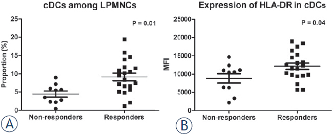
FIGURE 2
Predictors of the response to treatment. Inflammatory bowel disease patients who responded to TNFa inhibitors had a significantly higher proportion of (A) conventional dendritic cells (cDCs) with (B) higher expression of HLA-DR in the inflamed mucosa before treatment compared to nonresponders. cDC values are given as the percentage of cDCs among the lamina propria mononuclear cells and the expression of HLA-DR is shown as the mean fluorescence intensity (MFI) among cDCs. Horizontal lines indicate mean levels.
TABLE 7
Subpopulations of dendritic cells (DCs) in the inflamed inflammatory bowel disease (IBD) mucosa in responders and nonresponders before the treatment with TNFa inhibitors
| Responders | Nonresponders | p value | |
|---|---|---|---|
| IBD | |||
| pDC (%) | 1.9 (0.4) | 1.3 (0.4) | 0.3 |
| cDC (%) | 9.2 (1.0) | 4.4 (0.8) | 0.01 |
| CD86 (MFI) | 1436 (332) | 785 (177) | 0.2 |
| HLA-DR (MFI) | 12152 (868) | 8838 (1286) | 0.04 |
| CD103+ (%) | 51.0 (3.7) | 42.5 (5.5) | 0.2 |
| CD103- (%) | 48.8 (3.8) | 57.5 (5.5) | 0.2 |
TABLE 8
Subpopulations of dendritic cells (DCs) in the inflamed ulcerative colitis and Crohn’s disease mucosa in responders and nonresponders before the treatment with TNFa inhibitors
| Responders | Nonresponders | p value | |
|---|---|---|---|
| Ulcerative colitis | |||
| pDC (%) | 2.5 (0.6) | 1.7 (0.6) | 0.9 |
| cDC (%) | 10.1 (1.5) | 4.7 (1.2) | 0.04 |
| CD86 (MFI) | 1595 (523) | 552 (40) | 0.2 |
| HLA-DR (MFI) | 12182 (3613) | 6693 (3614) | 0.01 |
| CD103+ (%) | 61.4 (7.0) | 34.8 (6.5) | 0.02 |
| CD103- (%) | 38.4 (7.1) | 65.2 (2.7) | 0.02 |
| Crohn’s disease | |||
| pDC (%) | 1.6 (0.4) | 0.9 (0.3) | 0.2 |
| cDC (%) | 9.7 (1.6) | 4.2 (1.0) | 0.04 |
| CD86 (MFI) | 1452 (434) | 1019 (335) | 0.5 |
| HLA-DR (MFI) | 13152 (1319) | 10983 (1368) | 0.3 |
| CD103+ (%) | 50.8 (4.9) | 50.2 (9.9) | 0.9 |
| CD103- (%) | 49.2 (4.5) | 49.8 (9.9) | 0.9 |
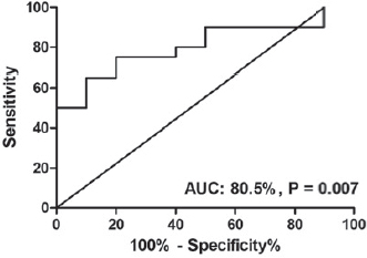
FIGURE 3
ROC curve analysis identified the proportion of conventional dendritic cells as a predictor of the response to TNFa inhibitors. Patients with a proportion of conventional dendritic cells above 7% responded to treatment.
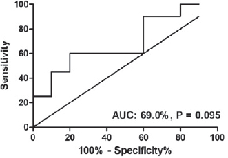
FIGURE 4
ROC curve analysis of conventional dendritic cell HLA-DR expression in the colonic mucosa before treatment for the prediction of the response to the induction treatment with TNF-α inhibitors. All patients with an HLA-DR MFI above 12450 responded to the treatment.