
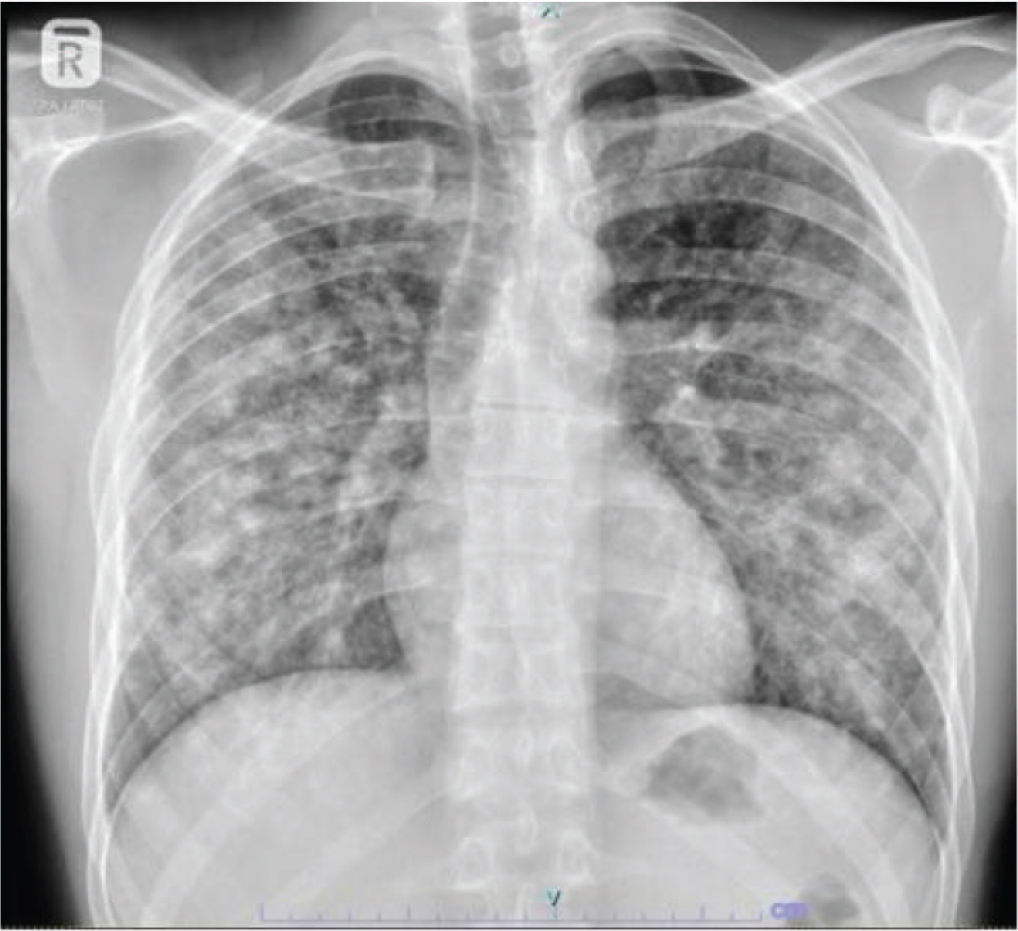
Figure 1.
Chest X-ray showing non-homogeneous air-space opacities.
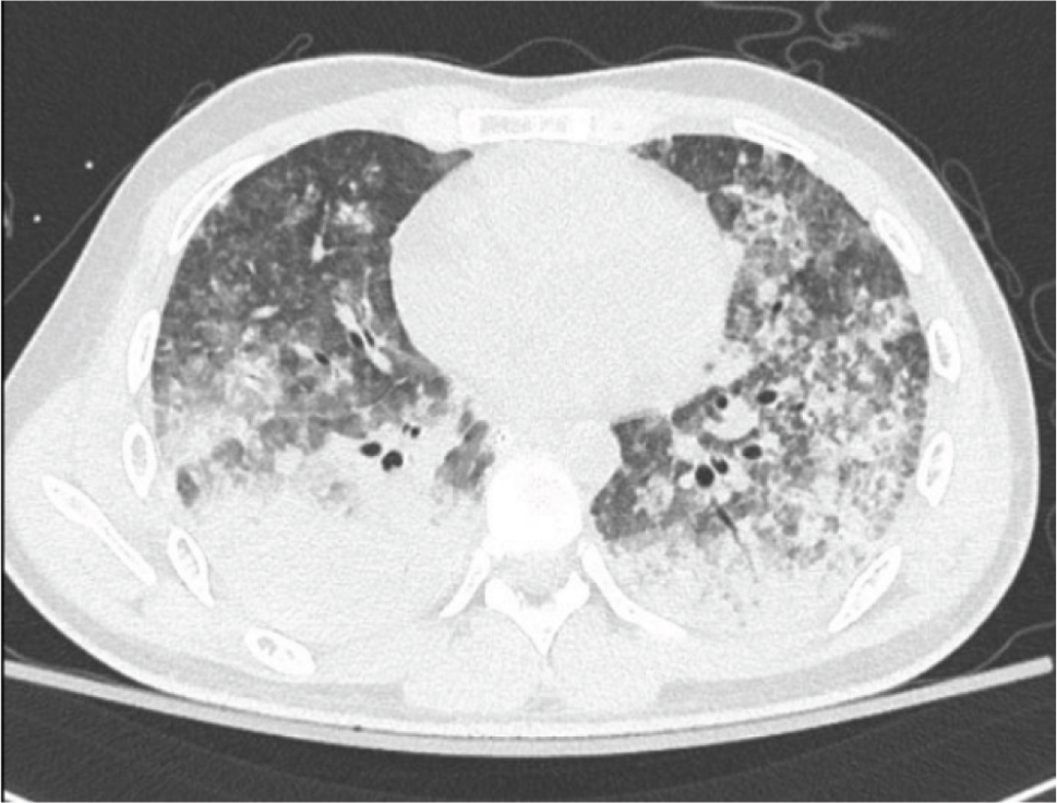
Figure 2.
Extensive consolidation, ground-glass densities and centrilobular nodules in both lungs.
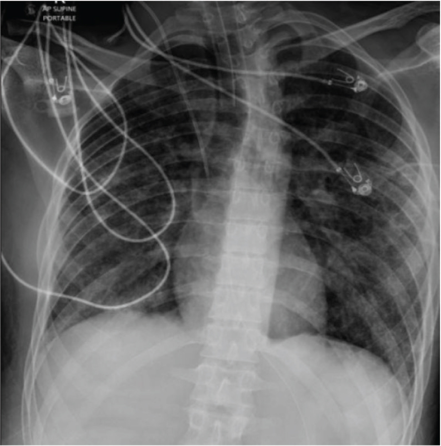
Figure 3.
Chest X-ray shows near clearance of bilateral non-homogeneous opacities in both lung fields.
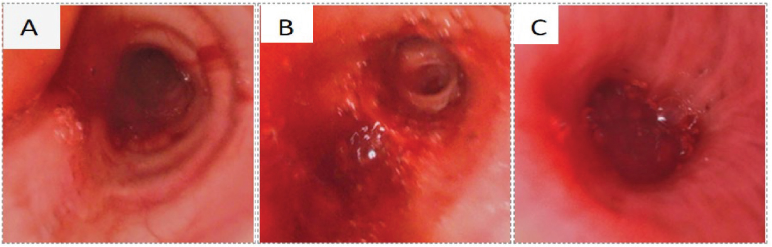
Figure 4.
Bronchoscopy picture showing fresh blood in the (A) trachea, (B) left lower lobe and (C) right lower lobe.
Table 1.
Investigation data.
| Parameters | Results |
|---|---|
| Haemoglobin | 12.8 g/dL |
| Total leucocyte count | 21.7 × 10⁹/L |
| Neutrophils | 91% |
| Platelet count | 260 × 109/L |
| CRP | 312 mg/L |
| Procalcitonin | 47 ng/mL |
| Serum sodium | 136 mEq/L |
| Serum potassium | 3.7 mEq/L |
| Renal profile | Normal |
| Liver function test | Normal |
| Blood culture | Negative |
| Tracheal aspirate culture | Negative |
| Urine culture | Negative |
| Legionella antigen | Negative |
| Viral panel | Negative |
| Leptospira IgM/PCR | Negative |
| TB PCR | Negative |
| QuantiFERON-TB Gold | Negative |
| c-ANCA | Negative |
| p-ANCA | Negative |
| HIV | Negative |
| Hepatitis markers | Negative |
| BAL appearance | Bloody |
| BAL TB PCR | Negative |
| BAL bacterial and fungal culture | Negative |
| BAL cytology | Plenty of RBC and few alveolar macrophages |
| Anti-GBM antibody | 1.5 IU/mL |
[i] BAL, bronchoalveolar lavage. c-ANCA, cytoplasmic antineutrophil cytoplasmic antibody; CRP, C-reactive protein; HIV, human immunodeficiency virus; IgM/PCR, immunoglobulin M/polymerase chain reaction; p-ANCA, perinuclear antineutrophil cytoplasmic antibody; RBC, red blood cells; TB PCR, tuberculosis polymerase chain reaction.
Table 2.
Clinical timeline of presentation, management and outcome.
| Time point | Clinical events and management |
|---|---|
| Approximately 1 week before admission | Dry cough, sore throat, malaise; increased vaping to approximately 10 sessions/day. |
| Hospital day 0 | High-grade fever and bilateral pulmonary opacities; antibiotics and supportive care started. |
| Within 3 hr | Severe hypoxaemia, supraventricular tachycardia, shock, intubation; fresh blood noted in endotracheal tube. |
| Early ICU course | Antibiotics, vasopressors, hydrocortisone for presumed septic shock; haemoglobin fell to 5.3 g/dL; six units of packed red cells transfused. |
| Hospital days 1–10 | Extensive negative microbiological/autoimmune evaluation; gradual clinical and radiological improvement; first extubation after 10 days. |
| Shortly after extubation | Recurrent fever, blood clots in secretions, tachypnoea, inability to clear secretions; re-intubation. |
| After re-intubation | Bronchoscopy showed blood in bilateral distal airways; bloody BAL obtained, but sequential aliquots and hemosiderin quantification were unavailable. |
| Three days later | Improvement with combined supportive care, antibiotics, transfusion, airway clearance and corticosteroids; successful extubation. |
| Fourth week of hospital stay | Clinically stable; cessation advised. Discharged from hospital |
| 1 month follow up | A video consultation was performed, and the patient remained asymptomatic and reported complete cessation of vaping and cigarette smoking. No recurrent respiratory symptoms were reported. Follow-up HRCT and pulmonary function testing could not be performed |