
Introduction
The use of electronic cigarettes (e-cigarettes) and vaping products has risen in recent years, particularly among adolescents and young adults (1). E-cigarette or vaping product use–associated lung injury (EVALI) is a serious condition characterised by acute respiratory symptoms and lung injury following vaping or e-cigarette use. Although initially marketed as safer alternatives to traditional cigarettes, these products are now associated with significant health risks, especially pulmonary complications, with EVALI being among the most severe. Diffuse alveolar haemorrhage (DAH) is a rare but potentially life-threatening syndrome presenting with dyspnoea, haemoptysis, anaemia and diffuse pulmonary infiltrates. DAH can result from various underlying conditions, including primary lung diseases, infections, cardiac or renal disorders and systemic autoimmune diseases (2). We report an uncommon presentation of suspected EVALI with clinical and radiological features suggestive of DAH. However, owing to the absence of sequential bronchoalveolar lavage (BAL) and the possibility of alternative explanations, neither DAH nor a causal relationship with vaping could be definitively established.
Case report
A 24-year-old man with no prior medical history presented to the emergency department with a 2-day history of high-grade fever, following nearly a week of dry cough, sore throat and malaise. He reported no known sick contacts but had recently travelled with his family to a coastal town. He had smoked approximately 20 cigarettes daily for 5 years and began regular vaping 4 months prior. During the week preceding symptom onset, he reported increasing the frequency of vaping to approximately 10–12 sessions per day, with an estimated 15–20 puffs during each session. The vaping device had been purchased from a local retail shop. According to the patient’s history, he used a commercially available disposable device (Kardinal Quik®; Kardinal Industries (M) Sdn. Bhd., Melaka, Malaysia; approximately 6000-puff capacity) in the lemon cola flavour. The product contained approximately 6 mL of e-liquid and was non-refillable. The manufacturer-listed ingredients included glycerol, propylene glycol, flavouring agents and nicotine. The available clinical record did not document the nicotine concentration or the exact timing of the final vaping exposure before symptom onset. Tetrahydrocannabinol (THC) or cannabinoid exposure was not specifically tested. However, based on the manufacturer’s product information for the vaping device used by the patient, THC or cannabinoids were not listed as ingredients. As the product was not independently analysed, exposure to unlisted constituents cannot be entirely excluded. In the emergency department, he was febrile at 102°F, with a heart rate of 117 beats per minute, blood pressure of 114/76 mmHg, respiratory rate of 20 breaths per minute and oxygen saturation of 96% on room air. Chest auscultation revealed bilateral coarse crepitations and scattered rhonchi. Chest radiography showed extensive bilateral reticulonodular and non-homogeneous air-space opacities, suggestive of multilobar pneumonia (Figure 1). He was admitted for intravenous antibiotics, nebulised bronchodilators and supportive care.
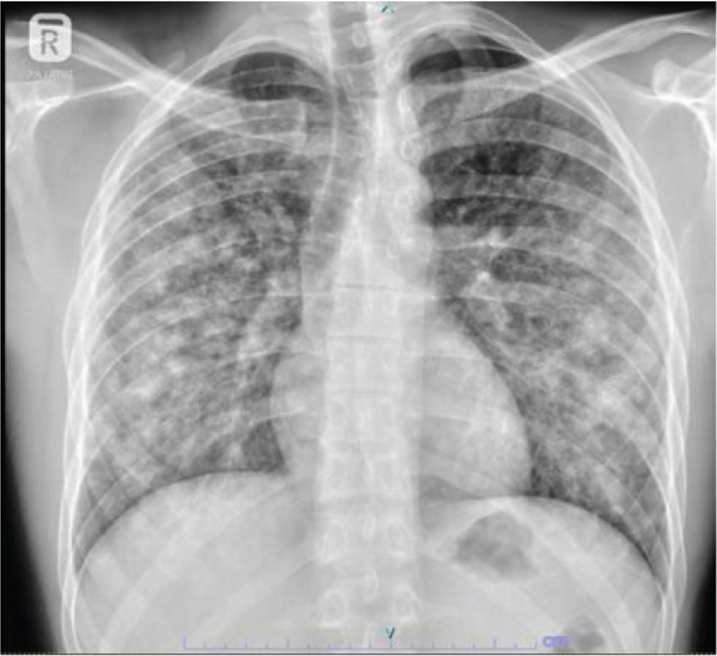
Figure 1.
Chest X-ray showing non-homogeneous air-space opacities.
Initial laboratory results showed haemoglobin of 12.8 g/dL, leukocytosis with a white blood cell count of 21.7 × 10⁹/L and neutrophil predominance, markedly elevated procalcitonin at 47 ng/mL, and C-reactive protein (CRP) of 312 mg/L. Serum electrolytes, renal and liver function tests were normal (Table 1). Within 3 hr of admission, his condition deteriorated rapidly. He developed worsening dyspnoea, a respiratory rate of 50 breaths per minute and acute desaturation to SpO2 74%. Electrocardiogram showed supraventricular tachycardia at 180 beats per minute, unresponsive to escalating intravenous adenosine. He was transferred to the intensive care unit (ICU) for cardioversion. He remained severely hypoxic, with respiratory rates up to 60 breaths per minute and persistent oxygen saturation around 80% despite high-flow oxygen via non-rebreather mask at 15 L/min. Due to impending respiratory failure, he was intubated. Fresh blood was observed in the endotracheal tube, raising concern for pulmonary haemorrhage. In the ICU, he required dual inotropic support for haemodynamic instability. Antibiotics were escalated to meropenem and ceftaroline while awaiting microbiological investigations, including leptospirosis and methicillin-resistant Staphylococcus aureus (MRSA) screening. The patient received intravenous hydrocortisone 100 mg every 12 hr for 3 days, followed by tapering to 50 mg every 12 hr for 5 days and subsequently 50 mg once daily for an additional 3 days. Corticosteroids were initially commenced for presumed septic shock and continued in view of the suspected diagnosis of EVALI. His haemoglobin dropped acutely to 5.3 g/dL, requiring transfusion of six units of packed red blood cells. High-resolution computed tomography (HRCT) of the chest revealed extensive consolidation, ground-glass densities and centrilobular nodules in both lungs (Figure 2). Computed tomography (CT) pulmonary angiography excluded pulmonary thromboembolism. A 2D echocardiogram showed normal ejection fraction and no pulmonary arterial hypertension. Further laboratory investigations (Table 1), including blood, tracheal aspirate and urine cultures, Legionella antigen, leptospirosis IgM/PCR, rapid viral panel, TB PCR, QuantiFERON-TB Gold, tropical fever panel, antinuclear antibody, antineutrophil cytoplasmic antibodies, HIV serology and hepatitis viral markers, were all negative. Over the next several days, his condition improved. Inflammatory markers declined, haemodynamics stabilized and he was weaned off inotropes. As gas exchange improved and chest radiographs showed progressive clearance of bilateral opacities (Figure 3), ventilator support was reduced, and he was extubated after 10 days. Shortly after extubation, he developed copious secretions with blood clots and persistent cough. He became restless, experienced intermittent shivering and had a temperature spike to 102.2°F. High-flow nasal cannula restored his oxygen saturation, but he remained tachypnoeic at 52 breaths per minute and appeared fatigued. Due to impending respiratory failure and inability to clear secretions, he was re-intubated. Bronchoscopy revealed fresh blood in the left main bronchus and segmental airways of the right and left lower lobes (Figure 4). Careful bronchoscopic examination demonstrated fresh blood extending into the segmental bronchi of both lower lobes without visible tracheal or upper airway mucosal injury, making procedure-related airway trauma a less likely explanation and supporting a distal rather than isolated upper airway source of bleeding. The blood was aspirated, and BAL from bilateral lower lobes appeared bloody and was sent for further analysis (Table 1). Sequential BAL aliquots, typically recommended to confirm DAH, were not feasible due to his clinical instability and hemosiderin-laden macrophages were not quantified. However, the presence of fresh blood during intubation, a bloody BAL return from the segmental airways of both lower lobes, an acute fall in haemoglobin, and rapidly evolving bilateral pulmonary opacities raised a strong clinical suspicion for DAH, which was considered a probable manifestation of suspected EVALI after exclusion of alternative causes. He continued chest physiotherapy, regular suctioning and supportive care. His condition improved, and he was weaned off the ventilator and extubated after 3 days. Repeat bronchoscopy showed normal airways without evidence of pulmonary haemorrhage. He recovered fully and was discharged in stable condition after a month-long hospital stay. At 1-month follow-up, conducted via video consultation because of the patient’s long travel distance from the treating centre, he remained asymptomatic and reported complete cessation of both vaping and cigarette smoking. There was no recurrence of respiratory symptoms. Follow-up HRCT and pulmonary function testing could not be performed because an inperson assessment was not feasible. The clinical timeline of the patient’s presentation, management and outcome is summarised in Table 2.
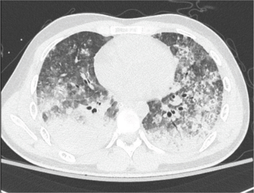
Figure 2.
Extensive consolidation, ground-glass densities and centrilobular nodules in both lungs.
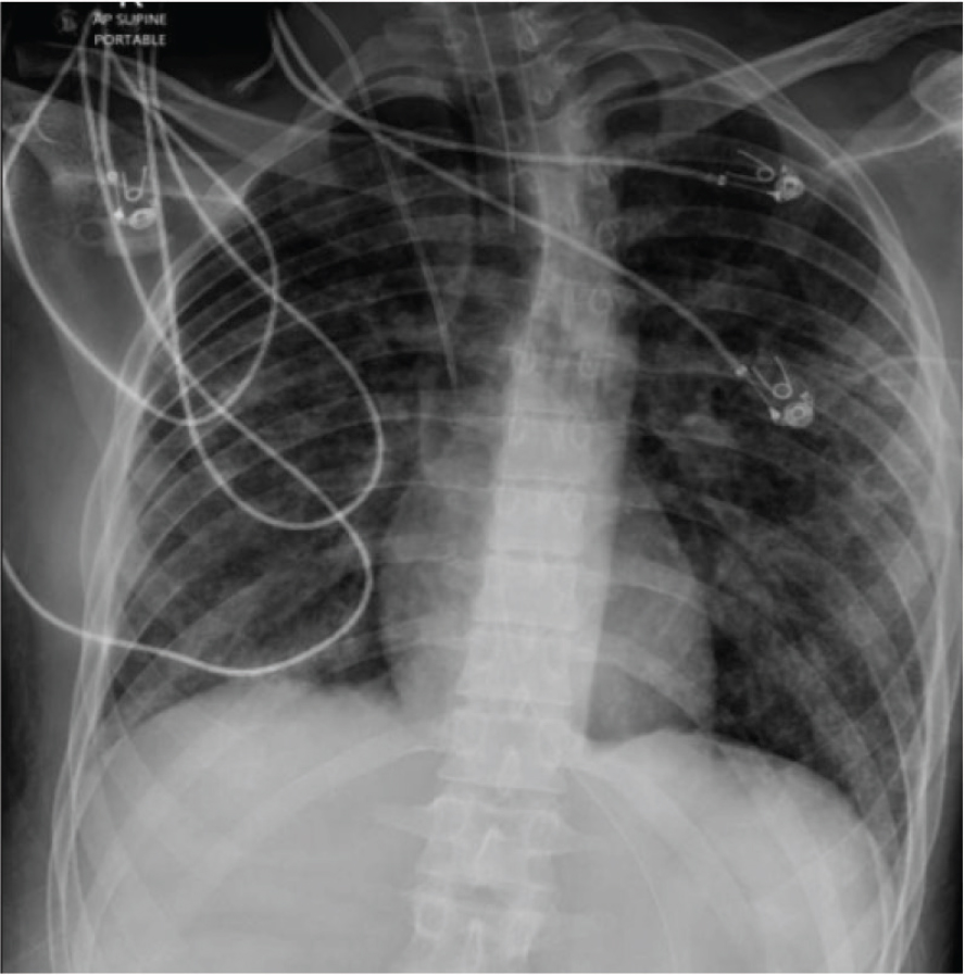
Figure 3.
Chest X-ray shows near clearance of bilateral non-homogeneous opacities in both lung fields.
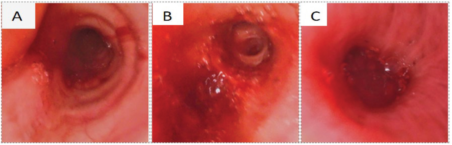
Figure 4.
Bronchoscopy picture showing fresh blood in the (A) trachea, (B) left lower lobe and (C) right lower lobe.
Table 1.
Investigation data.
| Parameters | Results |
|---|---|
| Haemoglobin | 12.8 g/dL |
| Total leucocyte count | 21.7 × 10⁹/L |
| Neutrophils | 91% |
| Platelet count | 260 × 109/L |
| CRP | 312 mg/L |
| Procalcitonin | 47 ng/mL |
| Serum sodium | 136 mEq/L |
| Serum potassium | 3.7 mEq/L |
| Renal profile | Normal |
| Liver function test | Normal |
| Blood culture | Negative |
| Tracheal aspirate culture | Negative |
| Urine culture | Negative |
| Legionella antigen | Negative |
| Viral panel | Negative |
| Leptospira IgM/PCR | Negative |
| TB PCR | Negative |
| QuantiFERON-TB Gold | Negative |
| c-ANCA | Negative |
| p-ANCA | Negative |
| HIV | Negative |
| Hepatitis markers | Negative |
| BAL appearance | Bloody |
| BAL TB PCR | Negative |
| BAL bacterial and fungal culture | Negative |
| BAL cytology | Plenty of RBC and few alveolar macrophages |
| Anti-GBM antibody | 1.5 IU/mL |
[i] BAL, bronchoalveolar lavage. c-ANCA, cytoplasmic antineutrophil cytoplasmic antibody; CRP, C-reactive protein; HIV, human immunodeficiency virus; IgM/PCR, immunoglobulin M/polymerase chain reaction; p-ANCA, perinuclear antineutrophil cytoplasmic antibody; RBC, red blood cells; TB PCR, tuberculosis polymerase chain reaction.
Table 2.
Clinical timeline of presentation, management and outcome.
| Time point | Clinical events and management |
|---|---|
| Approximately 1 week before admission | Dry cough, sore throat, malaise; increased vaping to approximately 10 sessions/day. |
| Hospital day 0 | High-grade fever and bilateral pulmonary opacities; antibiotics and supportive care started. |
| Within 3 hr | Severe hypoxaemia, supraventricular tachycardia, shock, intubation; fresh blood noted in endotracheal tube. |
| Early ICU course | Antibiotics, vasopressors, hydrocortisone for presumed septic shock; haemoglobin fell to 5.3 g/dL; six units of packed red cells transfused. |
| Hospital days 1–10 | Extensive negative microbiological/autoimmune evaluation; gradual clinical and radiological improvement; first extubation after 10 days. |
| Shortly after extubation | Recurrent fever, blood clots in secretions, tachypnoea, inability to clear secretions; re-intubation. |
| After re-intubation | Bronchoscopy showed blood in bilateral distal airways; bloody BAL obtained, but sequential aliquots and hemosiderin quantification were unavailable. |
| Three days later | Improvement with combined supportive care, antibiotics, transfusion, airway clearance and corticosteroids; successful extubation. |
| Fourth week of hospital stay | Clinically stable; cessation advised. Discharged from hospital |
| 1 month follow up | A video consultation was performed, and the patient remained asymptomatic and reported complete cessation of vaping and cigarette smoking. No recurrent respiratory symptoms were reported. Follow-up HRCT and pulmonary function testing could not be performed |
Discussion
Electronic cigarettes are battery-operated devices that deliver inhaled aerosols by heating a liquid mixture containing nicotine, chemicals, flavouring agents and other additives. When activated, the device heats the e-liquid, producing vapour for inhalation. EVALI has become a significant public health concern, especially among young adults. According to the Centers for Disease Control and Prevention (CDC), the 2019 outbreak resulted in 2807 hospitalised cases and deaths across the United States, including all 50 states, the District of Columbia and two U.S. territories, as of 18 February 2020 (3). Sixty-eight deaths were confirmed during this period (3, 4). Most cases occurred between mid-2019 and early 2020, after which incidence declined, likely due to increased regulatory scrutiny and identification of harmful substances in illicit vaping products (5, 6). Following this decline, the CDC discontinued routine national reporting of EVALI cases. Nonetheless, EVALI remains a relevant clinical concern due to ongoing vaping worldwide and the lack of long-term epidemiological data. E-cigarettes generate inhaled aerosol by heating a liquid mixture containing nicotine, flavouring agents, solvents and additives. This vapour exposes users to various chemicals, including potentially harmful substances such as diethylene glycol and formaldehyde (7). Inhalation of nicotine and these aerosols raises concerns about cardiovascular stress, airway irritation and reproductive risks, making these products unsuitable during pregnancy (8). Studies have shown that e-cigarette aerosols can irritate airways, trigger bronchospasm or worsen asthma symptoms (7). Although e-cigarettes are legally available to those over 18 in many countries, many devices and cartridges are obtained through informal or illicit markets. These products are often modified, exposing users to unpredictable and potentially toxic chemical mixtures (9). E-liquids may be substituted with formulations containing THC or other psychoactive agents, which were strongly linked to EVALI during the 2019 outbreak (5, 10). Despite the removal of vitamin E acetate and THC-containing cartridges from many commercial products after the outbreak, sporadic EVALI cases continue to be reported, suggesting that other constituents, contaminants, or device modifications may still contribute to lung injury (9, 11). The CDC defines EVALI by the following criteria: (1) use of e-cigarette or vaping products within 90 days of symptom onset, (2) pulmonary infiltrates on chest radiograph or CT, (3) no alternative plausible diagnosis and (4) no evidence of infection, or infection that does not fully explain the illness (3). The present case fulfilled the clinical CDC case definition for a probable case of EVALI, including recent vaping exposure, bilateral pulmonary infiltrates and extensive evaluation excluding an alternative diagnosis that more convincingly explained the presentation. Recent evidence suggests EVALI does not represent classical exogenous lipoid pneumonia. Histopathological analyses often show no features of lipid pneumonia, limiting the diagnostic value of lipid-laden macrophages in BAL. Instead, these findings support EVALI as a toxic inhalational injury causing airway-centered chemical pneumonitis, with lipid accumulation indicating exposure rather than disease (12). DAH represents a rare but serious manifestation of vaping-related lung injury. DAH is characterised by haemoptysis (though this may be absent), acute respiratory failure, anaemia and diffuse pulmonary opacities. The syndrome can arise from diverse underlying conditions, including primary pulmonary disease, infection, cardiac or renal disorders and systemic autoimmune diseases (2). The mechanisms underlying EVALI are multifactorial. Proposed pathways include direct toxic injury from volatile organic compounds and aldehydes, chemical pneumonitis from lipid-based additives, immune-mediated inflammation and surfactant dysfunction associated with vitamin E acetate found in BAL specimens during the 2019 outbreak (9, 10). These mechanisms can result in various injury patterns, including organising pneumonia, acute lung injury, and, rarely, DAH. In the present case, probable DAH was considered based on the combination of clinical, radiographic and bronchoscopic findings, despite the inability to obtain sequential BAL aliquots or quantify hemosiderin-laden macrophages. Thoracic CT showed areas of consolidation interspersed with areas of ground-glass attenuation and preserved normal areas. Fresh blood observed during intubation, a bloody BAL return from segmental airways of bilateral lower lobes, an acute drop in haemoglobin, and rapidly evolving bilateral infiltrates on imaging strongly suggested ongoing alveolar bleeding. Extensive microbiological and autoimmune investigations – including tests for leptospirosis, tropical infections, tuberculosis, c-ANCA, p-ANCA associated vasculitis and other systemic conditions – were negative. In critically ill patients, DAH is often diagnosed based on a combination of clinical features, imaging findings and bronchoscopic evidence when sequential BAL is not feasible due to respiratory instability (2). Although the precise mechanism underlying DAH in EVALI remains uncertain, several pathways have been proposed. Heating e-liquids containing propylene glycol, glycerol, nicotine, flavouring agents and other additives can generate reactive carbonyl compounds and oxidative stress, resulting in epithelial and endothelial injury, innate immune activation and disruption of the alveolar-capillary barrier. These processes may lead to diffuse alveolar damage or airway-centered chemical pneumonitis, creating conditions that could facilitate alveolar haemorrhage in susceptible individuals (13). In the present case, the temporal association with heavy vaping and the absence of a more convincing alternative diagnosis support suspected EVALI presenting with probable DAH, although a definitive causal relationship cannot be established. Notably, markedly elevated procalcitonin levels in our patient raised initial concern for severe bacterial infection; however, similar elevations have been reported in non-infectious inflammatory lung injury, reflecting systemic inflammatory response rather than true infection (14). Nevertheless, concomitant bacterial infection cannot be completely excluded in this patient, particularly because empirical broad-spectrum antibiotics were initiated before microbiological results became available. These limitations warrant cautious interpretation of the diagnosis. It underscores the need for a detailed exposure history, systematic exclusion of alternative diagnoses and cautious diagnostic language when standard confirmatory testing is incomplete.
The patient’s course highlights the potential severity of vaping-associated lung injury, including rapid respiratory decompensation, need for mechanical ventilation, haemodynamic instability and repeated intubation. Notably, the patient’s recovery occurred following comprehensive intensive care management, including mechanical ventilation, blood transfusion, empirical broad-spectrum antimicrobial therapy, corticosteroids initiated for presumed septic shock and supportive care, consistent with reported experiences in EVALI management (15).
Conclusion
This case contributes to the growing body of literature describing the varied pulmonary manifestations of EVALI. While most cases present with features of acute lung injury or organising pneumonia, this case highlights that DAH should be considered among the differential manifestations of suspected EVALI. However, because standard confirmatory investigations could not be completed, the diagnosis should be interpreted cautiously and alternative explanations remain possible.
Acknowledgements
None.
Notes
[3] Contributed by Authors’ Contribution
All authors have read and approved the submitted manuscript and have substantially contributed to its conception, data collection, analysis, interpretation, drafting, and revision.
[4] Conflicts of interest Declaration of conflict of interest
There is no conflict of interest.
[5] Ethics approval
Ethics Committee approval was not required.
[6] Informed Consent Statement
Informed consent was obtained from all subjects involved in the study.