
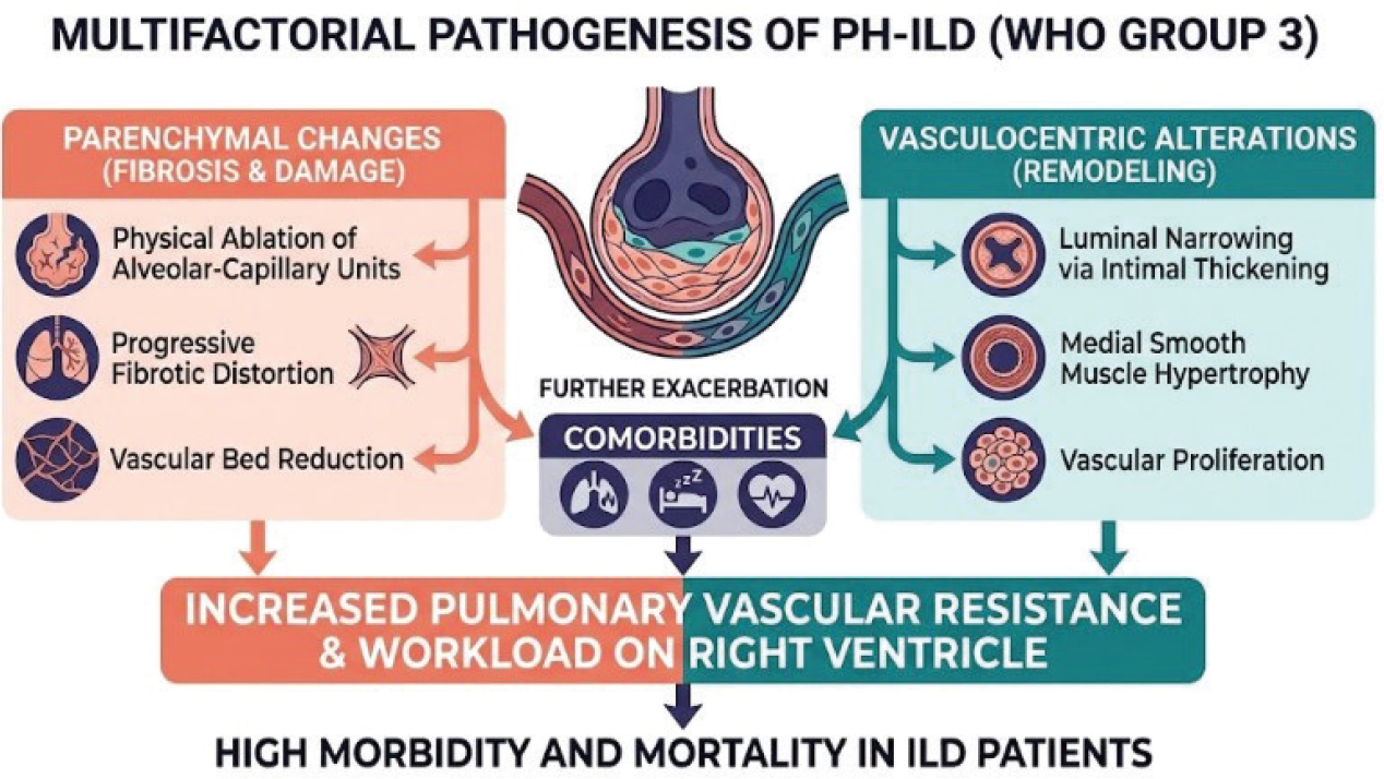
Figure 1.
Pathogenic factors associated with the development of PH in ILD [2]. Reproduced from Kattih et al. [2] under the terms of the Creative Commons Attribution (CC BY) License (https://creativecommons.org/licenses/by/4.0/). © 2025 by the authors. Licensee MDPI, Basel, Switzerland.
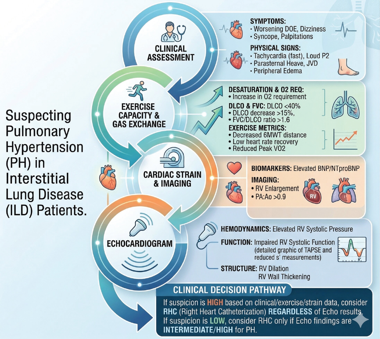
Figure 2.
Clinical, physiological, and imaging parameters indicative of pulmonary hypertension (PH) in individuals with interstitial lung disease (ILD). Patients at high risk are often identified by clinical manifestations such as worsening exertional dyspnea, syncope, and physical signs like an accentuated P2 heart sound or jugular venous congestion. From a physiological standpoint, a DLCO under 40%, an FVC/DLCO ratio surpassing 1.6, and diminished performance on the six-minute walk test (6MWT) are critical warning signs. These findings, when coupled with a CT-derived PA:A ratio over 0.9, elevated BNP or NT-proBNP levels, and echocardiographic evidence of right ventricular (RV) enlargement or low TAPSE, strongly indicate the necessity for gold-standard assessment through right heart catheterization (RHC) [2]. Reproduced from Kattih et al. [2] under the terms of the Creative Commons Attribution (CC BY) License (https://creativecommons.org/licenses/by/4.0/). © 2025 by the authors. Licensee MDPI, Basel, Switzerland.
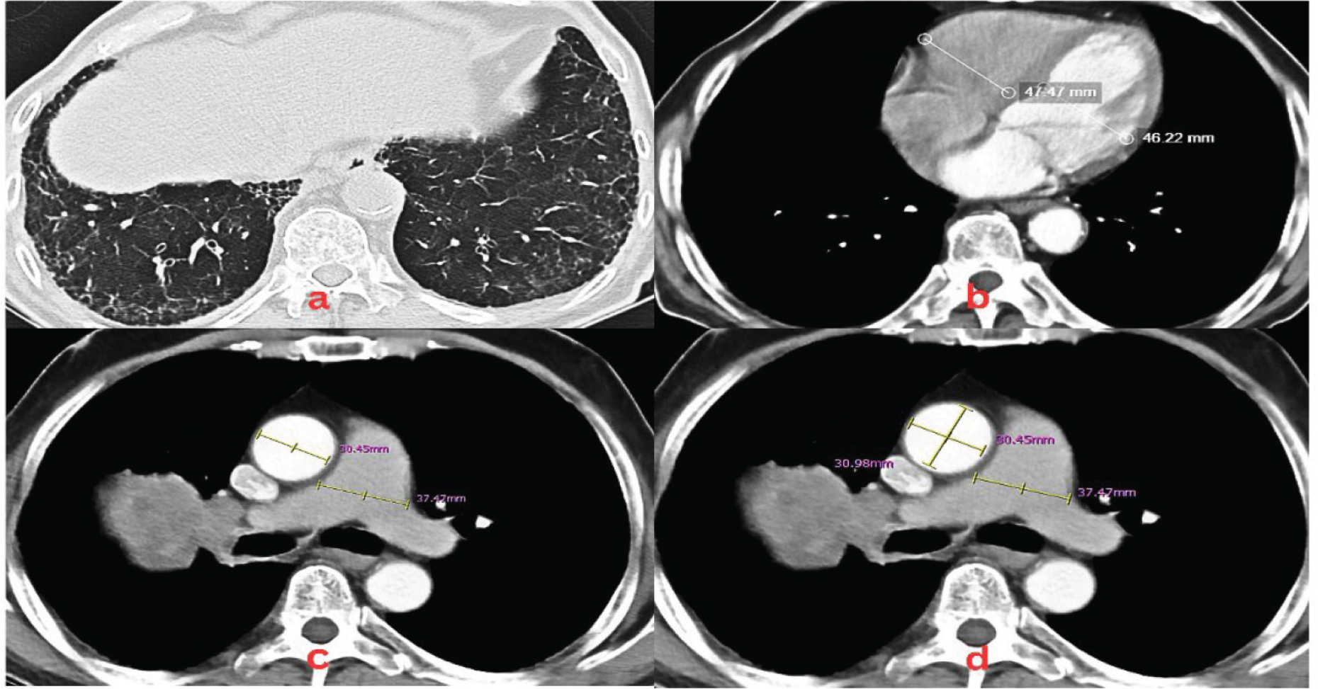
Figure 3.
Radiographic and tomographic features indicative of PH in a patient diagnosed with IPF. Echocardiography showed right heart dilatation (enlarged RA/RV) with flattening of the interventricular septum, while left heart dimensions were normal without LVH. Wall motion analysis revealed apical and apicoseptal hypokinesia with normokinetic remaining segments. Both LV systolic function (EF 46%) and RV contractility (TAPSE 1.6 cm) were decreased. Other findings included E/A <1, AoVTI 22 cm, mPA dilatation, mild MR, moderate TR, mild-to-moderate PR, and an estimated sPAP of 42 mmHg. (A) Axial lung window CT imaging of the thorax demonstrating reticular opacities associated with traction bronchiectasis, bronchiolectasis, and honeycombing are noted in the subpleural regions of the bilateral lower lobes. (B) RV/LV ratio 47.47 mm: 46.22 mm, resulting ratio 1.03 (C) Axial mediastinal window at the level of the bifurcation illustrating a ratio dilated pulmonary artery: ascending aorta (37.47 mm: 30.45 mm), resulting in a pathologic PA:A ratio of 1.23. An absolute PA diameter >30 mm can also serve as a standalone indicator for suspected PH. (D) Utilization of the average of two perpendicular measurements (30.98 + 30.45)/2 = 30.72 mm for the ascending aorta (AA) diameter to enhance reliability, yielding a pathologic PA:A ratio of 1.22. AoVTI, Ascending Aorta Velocity Time Integral; CT, computed tomography; EF, Ejection Fraction; IPF, idiopathic pulmonary fibrosis; LVH, Left Ventricular Hypertrophy; mPA, main pulmonary artery; MR, Mitral Regurgitation; PA, pulmonary artery; PH, pulmonary hypertension; PR, Pulmonic Regurgitation; sPAP, systolic pulmonary artery pressure; TAPSE, tricuspid annular plane systolic excursion; TR, Tricuspid Regurgitation.
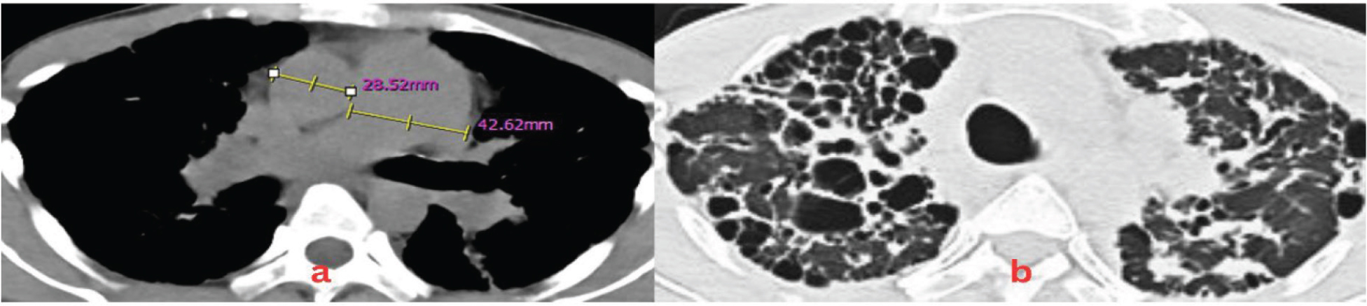
Figure 4.
Measurement of the mPA and ascending aorta (AA) diameters at the level of the pulmonary artery bifurcation on Thoracic HRCT in a patient with IPAF within the spectrum of CTD-ILD. Echocardiography revealed cardiac chamber dimensions with RA and RV dilatation. Global systolic function of the LV was adequate (EF 54%) with global normokinesia and a D-shaped left ventricle. Right ventricle contractility was preserved (TAPSE 1.8 cm). Valvular evaluation showed severe TR and moderate PR, indicating a high probability of PH. The definitive assessment was severe PH. (A) The mPA diameter was defined as the widest diameter perpendicular to its long axis, while the ascending AA was measured as the transverse diameter, resulting in a PA:A (40.21 mm: 27.85 mm) ratio of 1.44. An absolute PA diameter >30 mm can also serve as a standalone indicator for suspected PH (11). (B) Axial lung window illustrates honeycomb features, extensive traction bronchiectasis, ground-glass opacities, consolidation, and reticulation, predominantly in the right anterior region. CTD-ILD, Connective Tissue Disease-Associated Interstitial Lung Disease; HRCT, High-Resolution Computed Tomography; ILD, interstitial lung disease; IPAF, interstitial pneumonia with autoimmune features; mPA, main pulmonary artery; PA, pulmonary artery; PH, pulmonary hypertension; TAPSE, tricuspid annular plane systolic excursion.
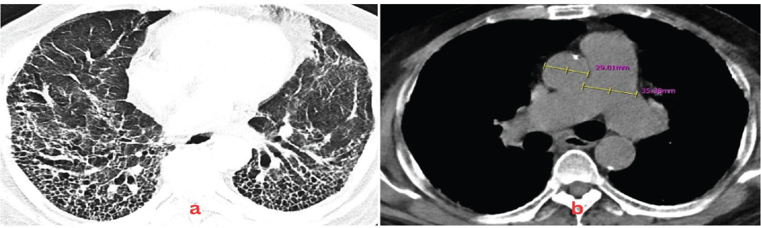
Figure 5.
Measurement of the mPA and ascending aorta (AA) diameters at the level of the pulmonary artery bifurcation on Thoracic HRCT in a patient with SLE - CTD ILD. Echocardiography revealed cardiac chamber dimensions with RA and RV dilatation. Global left ventricle (LV) contractility was within normal limits with an LVEF of 60% and global normokinesia, alongside grade I LV diastolic dysfunction. Right ventricle (RV) contractility was preserved (TAPSE 1.8 cm). Valvular evaluation showed severe TR and moderate PR, indicating a high probability of PH. The definitive assessment was severe PH. (A) Axial lung window reveals a loss of volume associated with traction bronchiectasis and exuberant honeycombing in the bilateral lower lobes, which was accompanied by reticulation and predominantly peripheral honeycombing in the bilateral upper lobes. (B) The mPA diameter was defined as the widest diameter perpendicular to its long axis, while the ascending AA was measured as the transverse diameter, resulting in a PA:A (35.38 mm: 29.01 mm) ratio of 1.22. An absolute PA diameter >30 mm can also serve as a standalone indicator for suspected PH. ILD, interstitial lung disease; LVEF, Left Ventricular Ejection Fraction; mPA, main pulmonary artery; PH, pulmonary hypertension; SLE, systemic lupus erythematosus; TAPSE, tricuspid annular plane systolic excursion.