
Introduction
Interstitial lung disease (ILD), particularly idiopathic pulmonary fibrosis (IPF), is a progressive and fatal pulmonary condition that is frequently complicated by pulmonary hypertension (PH). While the presence of PH significantly increases morbidity and mortality in patients with ILD, its early detection remains challenging. PH worsens functional status, increases the need for supplemental oxygen, and is associated with a marked reduction in survival (1–4). Early detection of PH-ILD is crucial for guiding diagnostic evaluation. However, a definitive diagnosis of PH is established by right heart catheterization (RHC), performed at rest, with a mean pulmonary artery pressure (mPAP) exceeding 20 mmHg. However, RHC is an invasive and costly procedure with limited availability. While transthoracic echocardiography (TTE) is the most commonly used non-invasive screening method, its accuracy is often limited in advanced ILD due to suboptimal acoustic windows (2–5).
Thoracic computed tomography (CT) is routinely performed in individuals with ILD to determine disease subtype and assess the degree of fibrotic involvement. This imaging modality enables the evaluation of a non-invasive radiologic marker: the pulmonary artery-to-ascending aorta diameter ratio (PA:A) ratio. It is important to emphasize that the PA:A ratio is not intended to replace TTE; instead, it serves as a pragmatic tool to strengthen the clinical indication for TTE in the ILD population (4–6). Furthermore, while vascular assessments are ideally conducted using contrast-enhanced imaging, the PA:A ratio remains a reliable and reproducible indicator even when measured on non-contrast (native) CT scans. Multiple studies have reported a significant association between elevated PA:A ratio values and the presence of PH (4, 5). Thresholds such as PA:A >0.9 or >1.0 are commonly cited as clinically meaningful indicators (4–6). In patients with IPF, a PA:A ratio above 0.9 has been identified as an independent predictor of increased (mPAP >20 mmHg) and is linked to poorer clinical outcomes (4, 5).
This ratio has also been incorporated into multivariable prediction models for identifying PH-ILD (2, 4, 6). This literature review aims to evaluate the scientific evidence regarding the diagnostic utility and predictive value of the PA:A ratio measured through thoracic CT as an effective screening indicator for detecting or predicting PH in patients with ILD. The findings of this review are expected to clarify the role of the PA:A ratio within the clinical algorithm for the early detection of PH-ILD (4, 5).
Definition
Following the 2022 ESC/ERS Guidelines for the diagnosis and treatment of PH, the hemodynamic definition of PH has been updated. A definitive diagnosis is established via RHC (7, 8), performed at rest, when the mPAP exceeds 20 mmHg (2). In the clinical context of ILD, which falls under WHO Group 3 (PH associated with lung diseases and/or hypoxia), the condition is further characterized by the following hemodynamic parameters: Pre-capillary PH: Defined as mPAP >20 mmHg, with a pulmonary capillary wedge pressure ≤15 mmHg and a pulmonary vascular resistance (PVR) >2 wood units (WU). Severe PH in Lung Disease: Specifically identified when the PVR is >5 WU, indicating a significant vascular component that contributes to poor clinical outcomes (2).
Pathophysiology
PH associated with ILD is classified under the World Health Organization (WHO) Group 3 category. The pathogenesis of PH-ILD is complex and multifactorial, involving the interplay between parenchymal changes and vasculocentric alterations, which are further exacerbated by comorbidities (Figure 1) (2). Fundamentally, the elevation in pulmonary pressure stems from:
Vascular Bed Reduction: Progressive fibrosis leads to the physical destruction and ablation of capillaries within the alveolar-capillary units. This resulting obliteration of the pulmonary capillary bed decreases the overall vascular cross-sectional area, consequently increasing resistance to blood flow (2).
Hypoxic Vasoconstriction: Chronic alveolar hypoxemia triggers hypoxic pulmonary vasoconstriction as a physiological response. This prolonged vasoconstriction causes narrowing of the pulmonary vessels, contributing significantly to the development of PH (2).
Vascular Remodeling: PH is characterized by degenerative changes and proliferation of pulmonary vascular cells, including intimal thickening and medial hypertrophy. Inflammatory mediators, such as Endothelin and Transforming Growth Factor-alpha (TGF-a), promote vasoconstriction and vascular smooth muscle proliferation, thereby exacerbating PH (2).
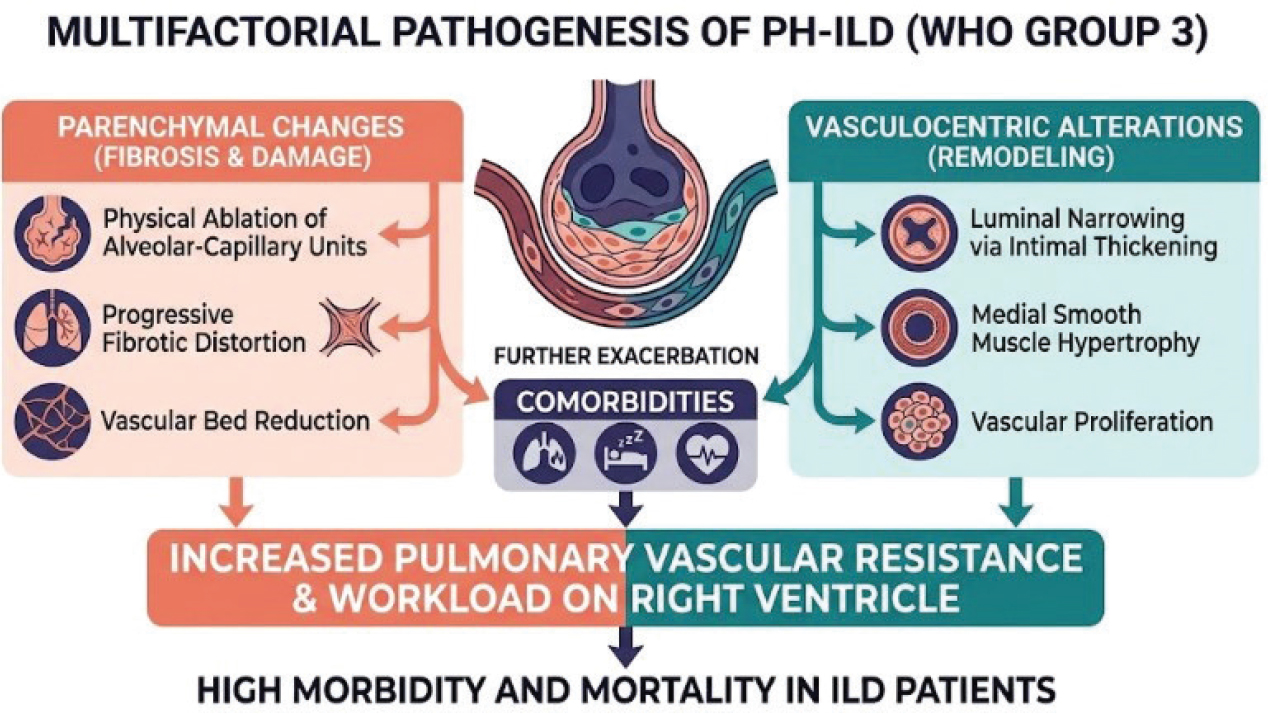
Figure 1.
Pathogenic factors associated with the development of PH in ILD [2]. Reproduced from Kattih et al. [2] under the terms of the Creative Commons Attribution (CC BY) License (https://creativecommons.org/licenses/by/4.0/). © 2025 by the authors. Licensee MDPI, Basel, Switzerland.
These mechanisms collectively lead to a structurally increased workload on the Right Ventricle (RV), contributing to the poor clinical outcomes observed in ILD patients (Figure 1) (2).
Other non-invasive screening modalities
While CT provides critical structural markers, other modalities are conventionally used to complement the screening of PH-ILD:
Echocardiography (TTE)
Transthoracic echocardiography is the most commonly used screening tool for estimating systolic pulmonary artery pressure (sPAP). However, its diagnostic accuracy is frequently hindered in advanced lung disease due to suboptimal acoustic windows. Secondary indicators, such as right ventricle (RV) dysfunction (e.g., reduced tricuspid annular plane systolic excursion [TAPSE]) and RV/RA dilation, can enhance the clinical suspicion of PH (2).
Pulmonary function testing and exercise capacity
A significant decline in the (diffusion capacity for carbon monoxide [DLCO] <40% predicted) is a strong physiological predictor of PH in ILD, reflecting extensive vascular obliteration. Additionally, an elevated forced vital capacity (FVC)/DLCO ratio and reduced distance in the (6-Minute Walk Test [6MWT] <350 m) are often incorporated into multivariable screening models (2).
Cardiac biomarkers
Elevated levels of (brain natriuretic peptide [BNP] >50 pg/mL) or N-terminal pro-B-type natriuretic peptide (NT-proBNP) (>300 pg/mL) serve as biochemical indicators of right ventricle pressure overload. While valuable for risk stratification, these markers are typically used alongside clinical and imaging findings to increase predictive accuracy (2).
Diagnostic
Indications for RHC
In the context of WHO Group 3 PH Pulmonary Hypertension Associated with Interstitial Lung Disease (PH-ILD), RHC remains the gold standard for definitive diagnosis (2). Following recent clinical guidelines, the emphasis has shifted from routine measurement to specific clinical indications. RHC should be prioritized when there is a high clinical suspicion of PH that is disproportionate to the underlying lung disease severity, or when precise hemodynamic data is required for lung transplant evaluation (4, 6). Additionally, RHC is essential to exclude other PH etiologies, such as Group 2 (Left Heart Disease) or Group 4 (Chronic Thromboembolic) PH, ensuring that patients receive the correct specialized management (2, 5).
According to the latest 2022 ESC/ERS guidelines and the 7th World Symposium on PH 2024 proceedings, RHC remains the gold standard for confirming PH in ILD patients, although its routine use is constrained by specific clinical indications. The primary justification for performing RHC is the potential for a change in patient management (7, 8). Key clinical situations where RHC is strongly indicated include pre-operative clearance, assistance in perioperative management, and phenotyping the disease to consider targeted PH-specific therapeutic interventions. Additionally, RHC is essential for patients being evaluated for lung transplantation, lung volume reduction therapy in relevant cases, or for those seeking enrollment in clinical trials (7, 8).
CT imaging markers
While the PA:A ratio is a primary focus, a comprehensive thoracic CT evaluation provides several indirect markers that strongly suggest underlying PH, even on non-ECG-gated scans (4). These cardiac-specific clues include:
Right Ventricle (RV) Strain: Morphological changes such as interventricular septum flattening or bowing toward the left ventricle, and hypertrophy of the RV free wall (5).
Chamber Enlargement: Dilatation of the Right Atrium (RA) and an increased RV/LV diameter ratio. It should be noted that while the PA:A ratio can be assessed on native scans, an accurate RV/LV ratio typically requires contrast-enhanced imaging for precise endocardial border definition (6).
Differential Etiology: CT can reveal indicators of alternative PH causes, such as left-sided heart enlargement (Group 2) or endoluminal webs and thrombi (Group 4) (4, 5).
Temporal Progression: Recent evidence highlights that the longitudinal increase in pulmonary artery diameter over time is a significantly stronger predictor of mortality than a single baseline measurement (4, 6).
Composite detection and threshold simplification
It is imperative to recognize that no non-invasive screening tool—including the PA:A ratio—can replace the definitive diagnosis provided by RHC (2, 4). For clinical utility, the various screening thresholds can be consolidated as follows:
PA Diameter >30 mm: A widely utilized initial marker of pulmonary artery enlargement (4).
PA:A ratio >0.9 or 1.0: These thresholds serve as critical cut-offs for risk stratification. A ratio >0.9 is a sensitive indicator for elevated mPAP (>20 mmHg) in IPF patients, while a ratio ≥1.0 is a more specific marker associated with significant survival decline and clinical deterioration (4, 6).
RV/LV ratio: A basal right ventricle (RV) to left ventricle (LV) diameter ratio greater than 1 may indicate the presence of PH, although this measurement generally requires contrast-enhanced CT pulmonary angiography (6).
By integrating these simplified CT-based metrics with clinical assessments, the PA:A ratio serves as an effective ‘gatekeeper’, identifying high-risk cohorts who require further invasive investigation and specialized care (Figure 2) (4, 5).
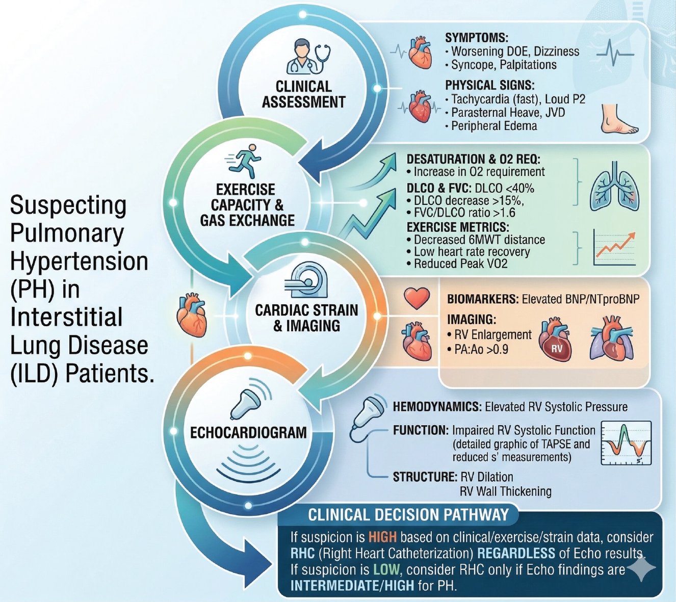
Figure 2.
Clinical, physiological, and imaging parameters indicative of pulmonary hypertension (PH) in individuals with interstitial lung disease (ILD). Patients at high risk are often identified by clinical manifestations such as worsening exertional dyspnea, syncope, and physical signs like an accentuated P2 heart sound or jugular venous congestion. From a physiological standpoint, a DLCO under 40%, an FVC/DLCO ratio surpassing 1.6, and diminished performance on the six-minute walk test (6MWT) are critical warning signs. These findings, when coupled with a CT-derived PA:A ratio over 0.9, elevated BNP or NT-proBNP levels, and echocardiographic evidence of right ventricular (RV) enlargement or low TAPSE, strongly indicate the necessity for gold-standard assessment through right heart catheterization (RHC) [2]. Reproduced from Kattih et al. [2] under the terms of the Creative Commons Attribution (CC BY) License (https://creativecommons.org/licenses/by/4.0/). © 2025 by the authors. Licensee MDPI, Basel, Switzerland.
Echocardiography (transthoracic echocardiography - TTE)
Transthoracic echocardiography is the most commonly used screening modality for PH. However, its accuracy in estimating pulmonary pressures is limited in the setting of advanced lung disease due to suboptimal acoustic windows, which can lead to misestimation of pressure values (9). Secondary (indirect) indicators of PH—such as right ventricle (RV) dysfunction (e.g., reduced TAPSE), RV/right atrium (RA) dilation, and RV wall thickening—can enhance diagnostic accuracy, especially in cases of severe PH (10). Measurement of tricuspid regurgitation velocity (TRV): TTE assesses the peak velocity of tricuspid regurgitation using continuous-wave Doppler. The right ventricle systolic pressure (RVSP), an estimate of the sPAP, is calculated using the Simplified Bernoulli Equation, which relates the tricuspid regurgitant gradient to the right atrium pressure (RAP): RVSP = sPAP = 4 x (TRV)2 + RAP. An elevated RVSP suggests a high likelihood of PH.
Pulmonary function testing and exercise capacity
DLCO: A markedly reduced DLCO (<40% of predicted) is a strong predictor of PH in ILD, reflecting vascular obliteration and heterogeneous perfusion characteristic of ILD-associated PH (2, 4, 6).
Ratio FVC/DLCO: An elevated FVC/DLCO ratio increases the suspicion for underlying PH (2, 4).
6MWT: Reduced walking distance (<350 m) and significant oxygen desaturation during the test (nadir SpO2 <88%) are common indicators of PH-ILD and are often incorporated into screening models (4, 6).
Technical steps for measuring the PA:A ratio
The vessel diameters are measured on axial (transverse) chest CT images at a specific anatomical level. Measurements must be performed on an axial slice taken at the level of the main pulmonary artery (mPA) bifurcation, the point at which the mPA divides into the right pulmonary artery and the left pulmonary artery (1, 3, 5). This bifurcation level serves as a consistent anatomical landmark, allowing for more accurate and comparable measurements across patients and between studies. Because this region is minimally affected by cardiac pulsation, aortic arch curvature, and variations in lung anatomy, the resulting diameter measurements are more reliable and reproducible. The PA:A ratio is calculated using a simple formula according to CT Scan thorax: Diameter of main pulmonary artery (PA)/Diameter of ascending aorta (AA) (Figure 3).
Proximal to the bifurcation: The diameter of the pulmonary artery should be measured proximal to the point where the mPA divides into the right and left pulmonary arteries (1, 3, 5). The measurement must capture the widest diameter of the vessel and should be taken perpendicular to the long axis of the mPA to ensure accuracy and consistency (Figure 4) (1, 5).
Measurement of the Ascending Aorta (AA): Location: The diameter of the ascending aorta is measured on the same CT slice used for the pulmonary artery measurement (1, 3, 5). Orientation: The AA diameter should be assessed as the transverse (inner-to-inner) diameter, obtained at a level adjacent to the mPA (Figure 5) (1, 5). Averaging Method: Some studies recommend using the average of two perpendicular diameter measurements of the AA to enhance reliability and reduce inter-observer variability (5).
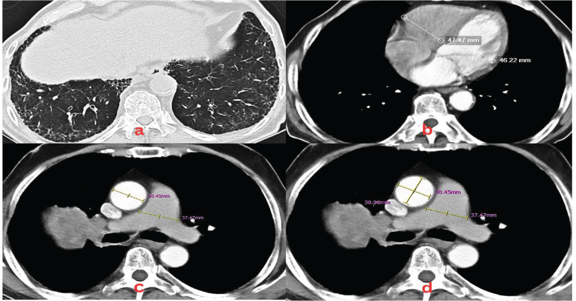
Figure 3.
Radiographic and tomographic features indicative of PH in a patient diagnosed with IPF. Echocardiography showed right heart dilatation (enlarged RA/RV) with flattening of the interventricular septum, while left heart dimensions were normal without LVH. Wall motion analysis revealed apical and apicoseptal hypokinesia with normokinetic remaining segments. Both LV systolic function (EF 46%) and RV contractility (TAPSE 1.6 cm) were decreased. Other findings included E/A <1, AoVTI 22 cm, mPA dilatation, mild MR, moderate TR, mild-to-moderate PR, and an estimated sPAP of 42 mmHg. (A) Axial lung window CT imaging of the thorax demonstrating reticular opacities associated with traction bronchiectasis, bronchiolectasis, and honeycombing are noted in the subpleural regions of the bilateral lower lobes. (B) RV/LV ratio 47.47 mm: 46.22 mm, resulting ratio 1.03 (C) Axial mediastinal window at the level of the bifurcation illustrating a ratio dilated pulmonary artery: ascending aorta (37.47 mm: 30.45 mm), resulting in a pathologic PA:A ratio of 1.23. An absolute PA diameter >30 mm can also serve as a standalone indicator for suspected PH. (D) Utilization of the average of two perpendicular measurements (30.98 + 30.45)/2 = 30.72 mm for the ascending aorta (AA) diameter to enhance reliability, yielding a pathologic PA:A ratio of 1.22. AoVTI, Ascending Aorta Velocity Time Integral; CT, computed tomography; EF, Ejection Fraction; IPF, idiopathic pulmonary fibrosis; LVH, Left Ventricular Hypertrophy; mPA, main pulmonary artery; MR, Mitral Regurgitation; PA, pulmonary artery; PH, pulmonary hypertension; PR, Pulmonic Regurgitation; sPAP, systolic pulmonary artery pressure; TAPSE, tricuspid annular plane systolic excursion; TR, Tricuspid Regurgitation.
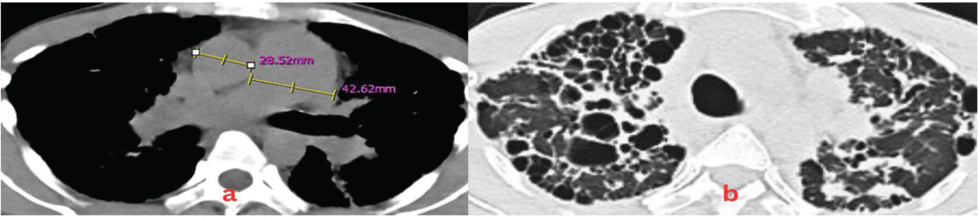
Figure 4.
Measurement of the mPA and ascending aorta (AA) diameters at the level of the pulmonary artery bifurcation on Thoracic HRCT in a patient with IPAF within the spectrum of CTD-ILD. Echocardiography revealed cardiac chamber dimensions with RA and RV dilatation. Global systolic function of the LV was adequate (EF 54%) with global normokinesia and a D-shaped left ventricle. Right ventricle contractility was preserved (TAPSE 1.8 cm). Valvular evaluation showed severe TR and moderate PR, indicating a high probability of PH. The definitive assessment was severe PH. (A) The mPA diameter was defined as the widest diameter perpendicular to its long axis, while the ascending AA was measured as the transverse diameter, resulting in a PA:A (40.21 mm: 27.85 mm) ratio of 1.44. An absolute PA diameter >30 mm can also serve as a standalone indicator for suspected PH (11). (B) Axial lung window illustrates honeycomb features, extensive traction bronchiectasis, ground-glass opacities, consolidation, and reticulation, predominantly in the right anterior region. CTD-ILD, Connective Tissue Disease-Associated Interstitial Lung Disease; HRCT, High-Resolution Computed Tomography; ILD, interstitial lung disease; IPAF, interstitial pneumonia with autoimmune features; mPA, main pulmonary artery; PA, pulmonary artery; PH, pulmonary hypertension; TAPSE, tricuspid annular plane systolic excursion.
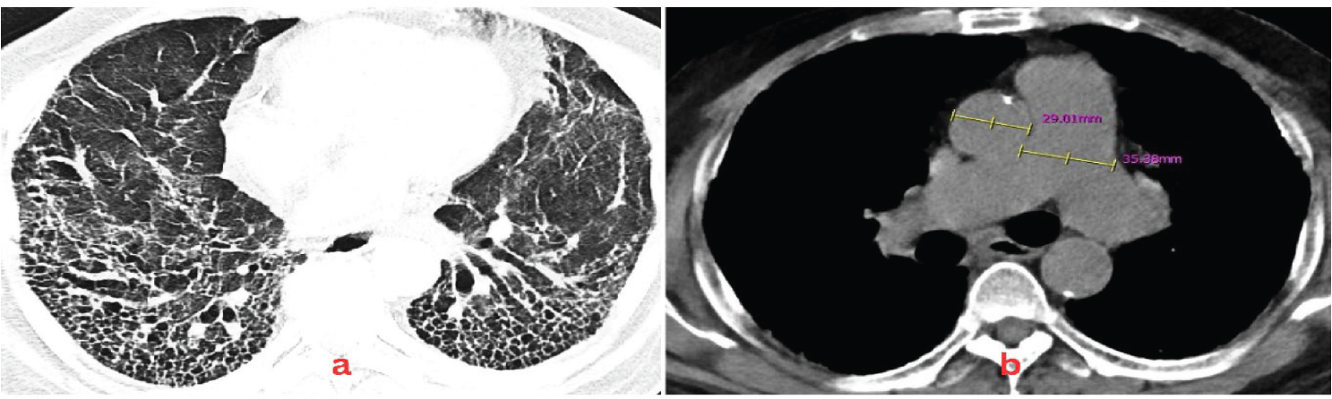
Figure 5.
Measurement of the mPA and ascending aorta (AA) diameters at the level of the pulmonary artery bifurcation on Thoracic HRCT in a patient with SLE - CTD ILD. Echocardiography revealed cardiac chamber dimensions with RA and RV dilatation. Global left ventricle (LV) contractility was within normal limits with an LVEF of 60% and global normokinesia, alongside grade I LV diastolic dysfunction. Right ventricle (RV) contractility was preserved (TAPSE 1.8 cm). Valvular evaluation showed severe TR and moderate PR, indicating a high probability of PH. The definitive assessment was severe PH. (A) Axial lung window reveals a loss of volume associated with traction bronchiectasis and exuberant honeycombing in the bilateral lower lobes, which was accompanied by reticulation and predominantly peripheral honeycombing in the bilateral upper lobes. (B) The mPA diameter was defined as the widest diameter perpendicular to its long axis, while the ascending AA was measured as the transverse diameter, resulting in a PA:A (35.38 mm: 29.01 mm) ratio of 1.22. An absolute PA diameter >30 mm can also serve as a standalone indicator for suspected PH. ILD, interstitial lung disease; LVEF, Left Ventricular Ejection Fraction; mPA, main pulmonary artery; PH, pulmonary hypertension; SLE, systemic lupus erythematosus; TAPSE, tricuspid annular plane systolic excursion.
Conclusion
Utilization of the pulmonary artery–to–aorta (PA:A) ratio on routine thoracic CT imaging offers a practical, highly reproducible, and non-invasive approach to screen for PH within the ILD cohort. This radiological marker effectively assists clinicians in risk stratification, identifying specific candidates who warrant further definitive but invasive screening through RHC. Furthermore, an increased PA:A ratio functions as a strong, standalone prognostic tool that correlates with adverse clinical outcomes, such as accelerated mortality rates and the urgent necessity for lung transplantation. When integrated into multi-parametric risk assessment frameworks alongside severe gas exchange impairment (decreased DLCO), diminished 6-min walk distance, and elevated cardiac biomarkers, the PA:A ratio comprehensively optimizes survival predictability and clinical management strategies for patients with ILD.
Acknowledgement
The authors declare that no specific funding, financial support, or non-author contributions were received for the preparation and publication of this manuscript.
Notes
[1] Contributed by Author’s Contributions
ABT: Conceptualization; Literature Search and Data Collection; Analysis and Interpretation of Data; Writing – Original Draft Preparation; Technical Visualization and Figure Creation; Final Approval of the Manuscript. FN: Expert Supervision and Validation (Pulmonology); Pathophysiology Framework Analysis; Critical Revision for Intellectual Content; Final Manuscript Editing and Submission Management. AAS: Clinical Supervision and Validation (Cardiology); Critical Review of Hemodynamic Data and Diagnostic Criteria; Methodology Oversight.
[2] Conflicts of interest Conflict of Interest
There is no conflict of interest.
[3] Ethics Approval
Ethics Committee approval was not required.
[4] Informed Consent Statement
No informed consent was necessary.