Have a personal or library account? Click to login
The importance of fibrobronchoscopy in the diagnosis of pulmonary diseases Cover

The importance of fibrobronchoscopy in the diagnosis of pulmonary diseases

By:  and    
Open Access
|Jul 2026

Figures & Tables

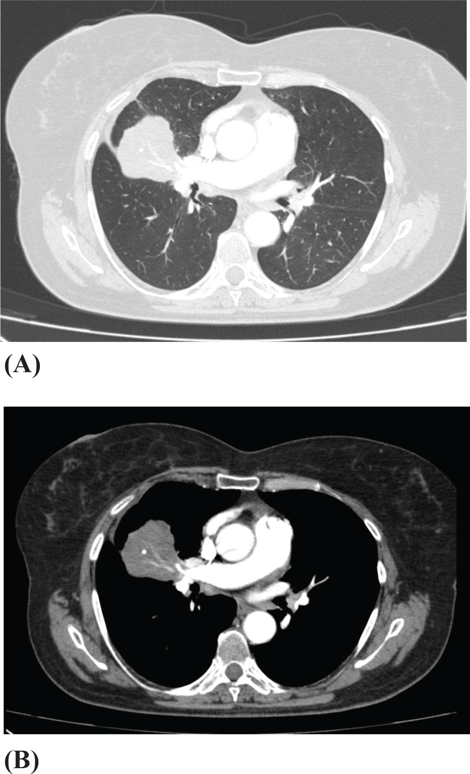
Figure 1.

Axial contrast-enhanced chest computed tomography (CT) in lung and mediastinal windows showing a persistent right upper lobe mass with adjacent post-obstructive atelectatic changes.
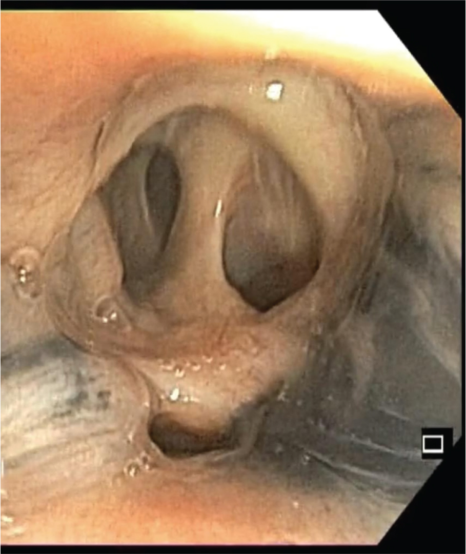
Figure 2.

Bronchoscopic view of anthracotic pigmentation with partial collapse of the middle lobe bronchus.
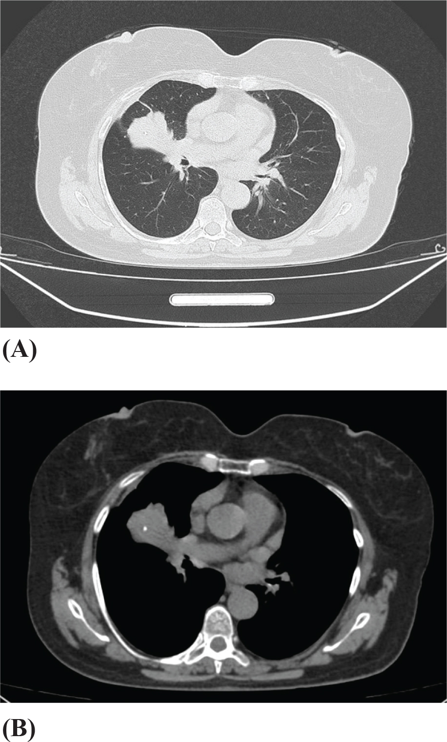
Figure 3.

Chest CT scan showing unchanged radiological findings after completion of tuberculosis treatment.
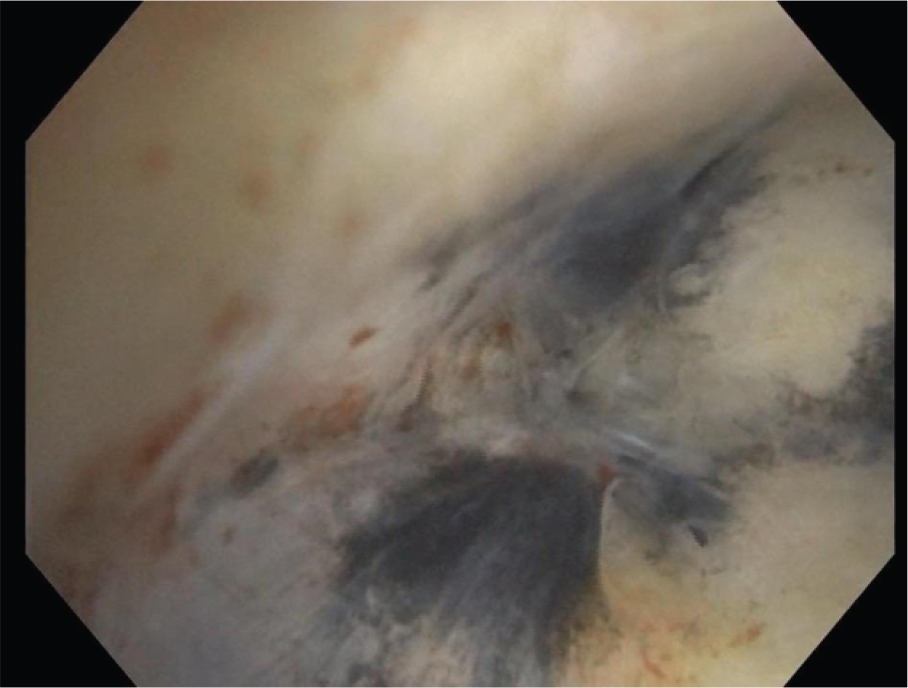
Figure 4.

Bronchoscopic image of a completely obstructed bronchus with no visible lumen.

DOI: https://doi.org/10.2478/pneum-2026-0011 | Journal eISSN: 2247-059X | Journal ISSN: 2067-2993
Language: English, Romanian
Page range: 77 - 82
Published on: Jul 31, 2026
Published by: Romanian Society of Pneumology
In partnership with: Paradigm Publishing Services
Publication frequency: Volume open

© 2026 Lavinia Cantea, Mihai Olteanu, published by Romanian Society of Pneumology
This work is licensed under the Creative Commons Attribution-NonCommercial-NoDerivatives 4.0 License.