
Introduction
Bronchoscopy is a minimally invasive and painless method for visualising the interior of the airways (larynx, trachea, bronchi). The first bronchoscopy using a flexible fibreoptic bronchoscope was performed in 1966 by Dr. Shigeto Ikeda (1); since then, bronchoscopy has remained an essential tool in the diagnosis and management of pulmonary pathologies (2).
The indications for flexible bronchoscopy are numerous but can be divided into three categories: diagnostic indications (such as persistent cough, wheezing, stridor or, in cases of the presence of abnormal radiological findings, hilar or mediastinal lymphadenopathy, infiltrates, atelectasis, pleural opacities, diffuse interstitial lung diseases, cancer staging), therapeutic indications (for the treatment of tracheobronchial stenoses, placement of stents or endobronchial prostheses, endobronchial tumour resections, foreign body removal, cryotherapy, laser therapy or bronchial aspirations) and special indications (bronchoscopically guided intubation, guidance of percutaneous tracheostomy, endobronchial ultrasound and local drug administration) (3,4).
Flexible bronchoscopes of various sizes are available, allowing for visualisation of the bronchial tree. They contain a fibreoptic system that transmits the image from the tip of the instrument to a video camera. The flexible fibreoptic bronchoscope contains a suction channel or an instrument channel (biopsy forceps, foreign body forceps, brushing brushes, electrode, electrocautery) (5).
Case presentation
We present the case of a 67-year-old female patient, from an urban area, who presented to our pulmonology department in October 2023, complaining of a febrile episode at home, dyspnoea on moderate exertion and marked physical asthenia, with symptoms that had begun suddenly 1 week prior to presentation. On the recommendation of her family physician, she underwent a chest X-ray, which revealed a mediastinal opacity of moderate intensity, homogeneous, triangular in shape, with the upper margin clearly demarcated by the horizontal fissure, approximately 8 cm in diameter, located in the right parahilar region. She was, therefore, referred to our hospital.
We note from the patient’s medical history that her father was diagnosed with pulmonary tuberculosis (TB) during the patient’s childhood, for which she received chemoprophylactic treatment at the time. From her personal medical history, we note that she is currently diagnosed with cardiovascular conditions (Grade 2 primary hypertension, mild mitral regurgitation, carotid atheromatosis), for which she is undergoing chronic treatment at home.
We note that the patient has not been exposed to respiratory toxins or irritants (as she is a teacher by profession), is a non-smoker and denies alcohol consumption.
On clinical examination, she appears unwell but is afebrile; auscultation of the respiratory system reveals diminished vesicular breath sounds in the lower half of the right lung field, without rales. The oxygen saturation (SaO2) is 95% on room air; auscultation of the cardiovascular system reveals rhythmic heart sounds; blood pressure is 160/90 mmHg, and the heart rate is 80 bpm; otherwise, the clinical examination is within normal limits.
As a preliminary diagnosis, based on the medical history, clinical examination and chest X-ray results, pneumonia was suspected. Laboratory tests indicated a complete blood count within normal limits, the presence of mild hepatocellular syndrome (aspartate aminotransferase [AST]: 43 U/L and alanine aminotransferase: 66 U/L), a non-specific inflammatory syndrome (erythrocyte sedimentation rate: 63 mm/hr) and mild hyperglycaemia (serum glucose: 133 mg/dL). Spirometry revealed mild restrictive ventilatory dysfunction (forced vital capacity: 73%). A sputum examination could not be performed because the patient did not present specific symptoms and was unable to expectorate.
As a supplementary investigation, we decided to perform a contrast-enhanced chest CT, which revealed an area of pulmonary consolidation, without an air bronchogram, with a heterogeneous structure and iodophilia, with areas of necrosis and subcentimetre calcification, occupying almost the entire middle lobe, encasing the middle lobar bronchus and leading to a reduction in lung volume (Figure 1A and 1B). Given the imaging findings, a suspicion of a bronchopulmonary neoplasm arose and a bronchoscopy was required to establish a definitive diagnosis. Bronchoscopy revealed extensive anthracotic patches in the right upper lobe and left upper lobe bronchi, as well as in the right lower lobe bronchus. The middle lobe bronchus was found to be subtotally compressed, with its opening covered by anthracotic patches (Figure 2). No proliferative elements were found. Bronchoalveolar lavage for cytology and bronchial lavage for M. tuberculosis [microscopy (M), cultures (C) and GeneXpert] were performed. The bronchoscopic appearance suggests middle lobe atelectasis due to changes in bronchial patency, a finding suggestive of sequelae of pulmonary TB. Bronchoalveolar lavage results revealed no atypical or malignant cells, but M. tuberculosis was identified in bronchial aspirate cultures with 10 BAAR-positive colonies (M-, C+ 10 BAAR-positive colonies, with sensitivity to Isoniazid and Rifampicin, very low GeneXpert). Thus, based on the results, the diagnosis was established as ‘Secondary pulmonary TB, nodular infiltrative pattern in the middle lobe, bronchial lavage examination – M, C+ 10 BAAR at T0, DOT initiated on 28/02/2024 and HIV negative.’ DOT was initiated according to the National Tuberculosis Prevention, Surveillance and Control Program, at maximum doses, with a 7/7 administration schedule as follows: the first 2 months with Isoniazid 300 mg/day, Rifampicin 600 mg/day, Pyrazinamide 2000 mg/day and Ethambutol 1600 mg/day, followed by a 6-month period of Isoniazid 300 mg/day and Rifampicin 600 mg/day; additionally, a gastric protector, a liver protector and vitamin B6 were administered throughout the treatment. The patient demonstrated good tolerance and adherence to treatment, with favourable clinical and biological outcomes.
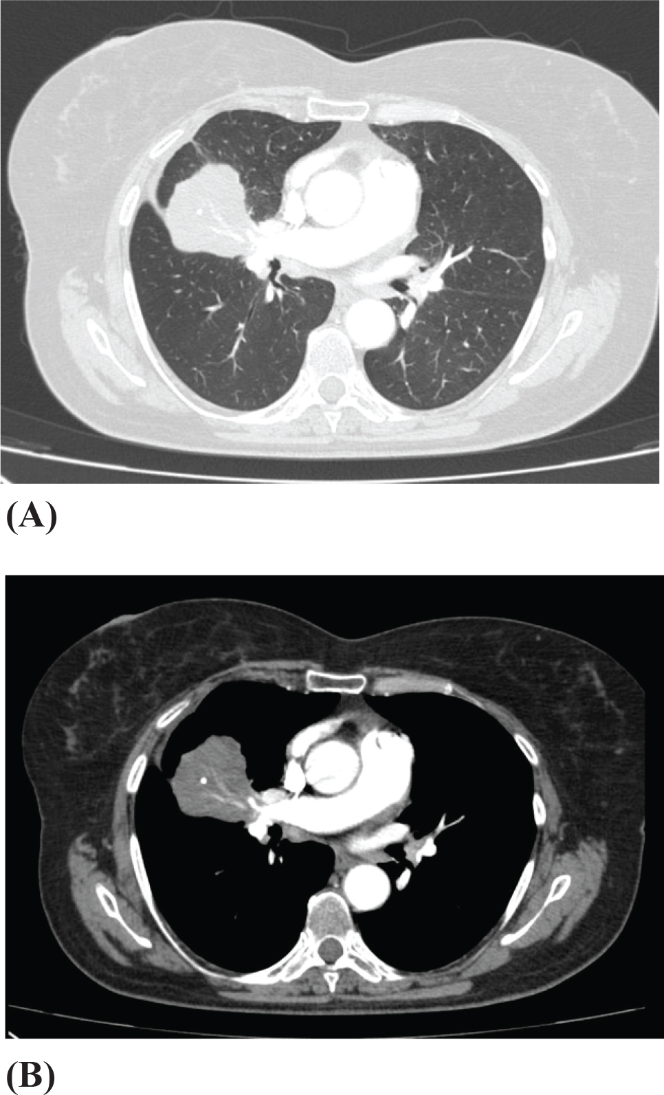
Figure 1.
Axial contrast-enhanced chest computed tomography (CT) in lung and mediastinal windows showing a persistent right upper lobe mass with adjacent post-obstructive atelectatic changes.
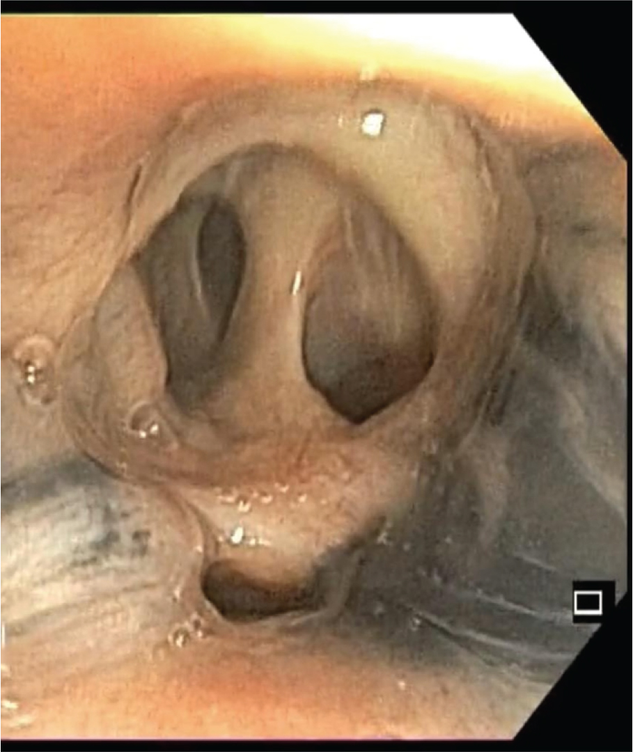
Figure 2.
Bronchoscopic view of anthracotic pigmentation with partial collapse of the middle lobe bronchus.
After 1 month of treatment, the imaging response remained stable.
At the end of treatment, the patient was in good general condition with no subjective complaints, and laboratory tests revealed mild hepatic cytolysis (AST: 54 U/L) and mild hypochromic microcytic anaemia (haemoglobin: 11.9 mg/dL). It was decided to repeat the CT scan with contrast. The same imaging findings were observed, showing the same area of pulmonary consolidation, without air bronchogram, with heterogeneous structure and iodophilia, with areas of necrosis and subcentimetre calcification, occupying almost the entire middle lobe, encasing the middle lobar bronchus and leading to reduced lung volume (Figure 3A and 3B).
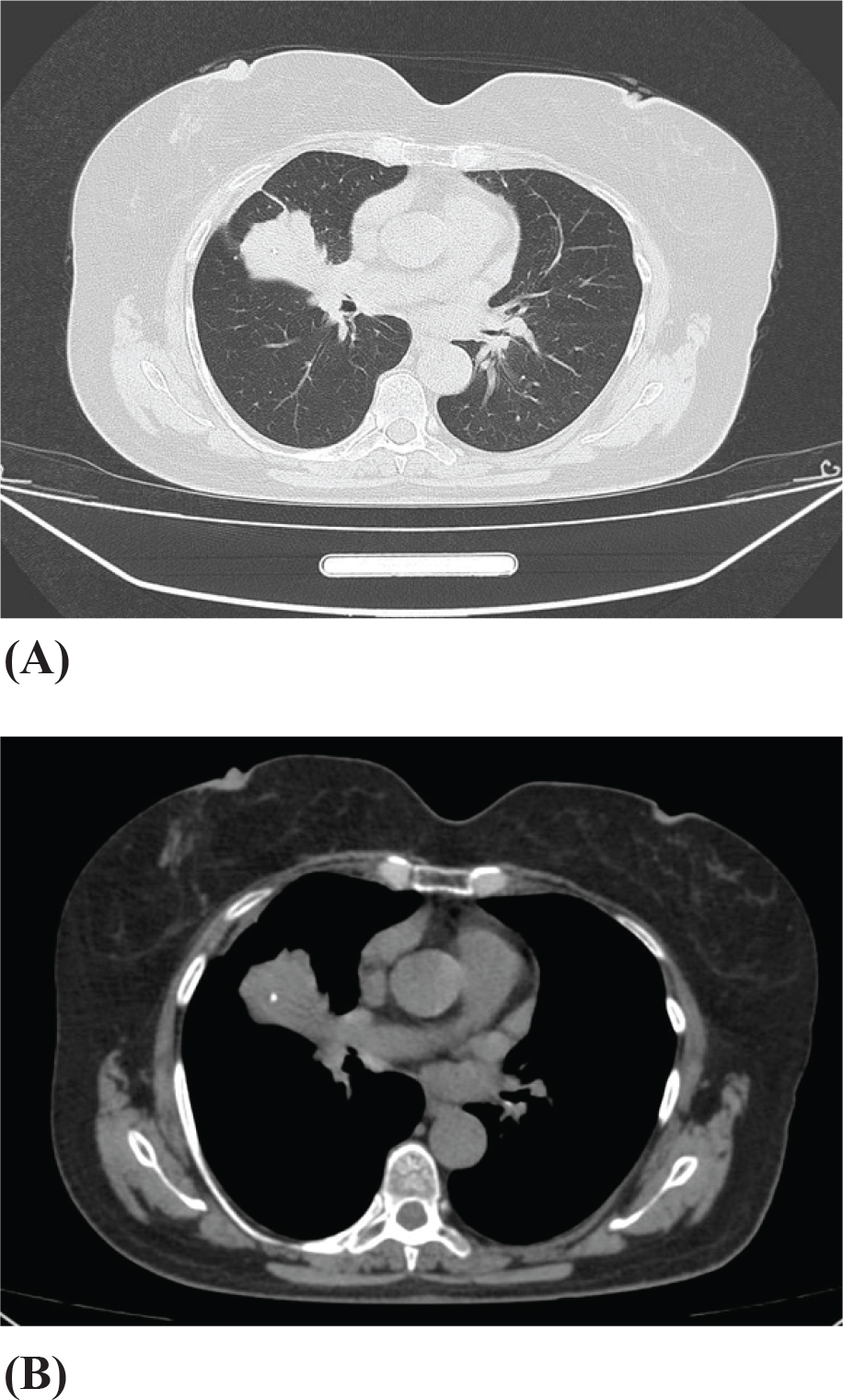
Figure 3.
Chest CT scan showing unchanged radiological findings after completion of tuberculosis treatment.
Given the persistence of the imaging findings, suspicion of a pulmonary neoplasm arises again, hence a repeat bronchoscopy is decided upon. Bronchoscopic images reveal the same anthracotic patches in the right and left upper lobar bronchi, as well as in the middle lobar bronchus, but, this time, the middle lobe bronchus is found to be completely collapsed; an attempt is made to open the lumen, revealing complete collapse with a ‘finger in glove’ appearance (Figure 4). Based on the endoscopic findings, the diagnosis of middle lobe atelectasis due to bronchial static changes was confirmed. No malignant cells were found in the bronchoalveolar lavage, and the bronchial aspirate was negative for M. tuberculosis both microscopically and in cultures.
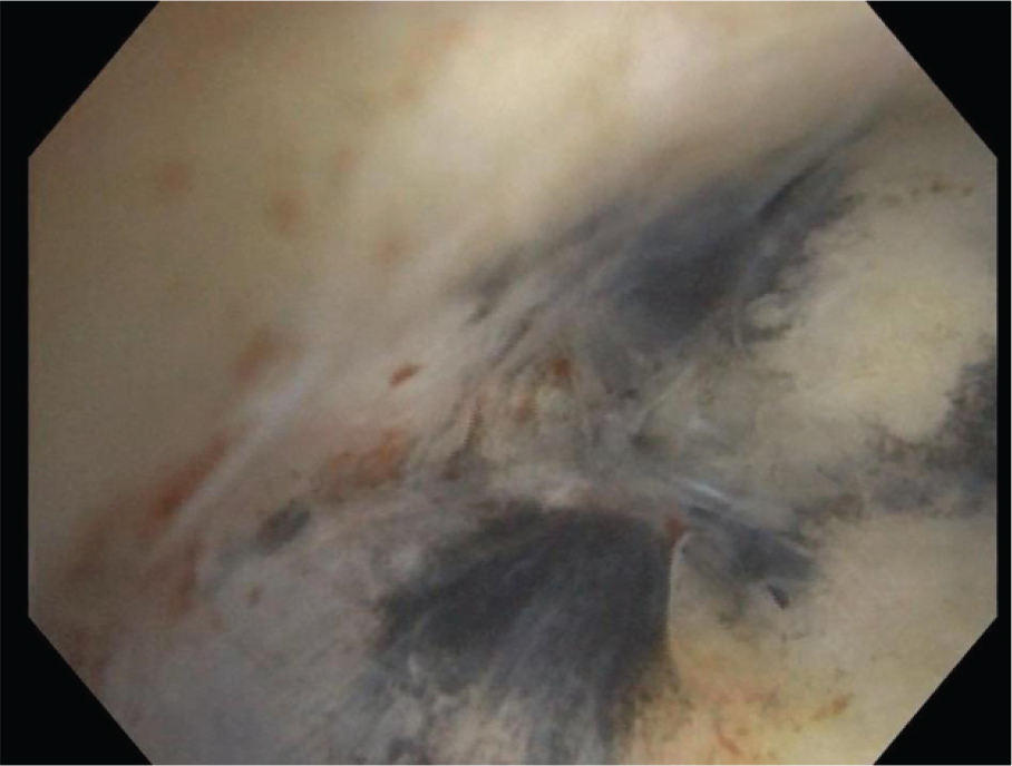
Figure 4.
Bronchoscopic image of a completely obstructed bronchus with no visible lumen.
Discussion
We present a case in which the diagnosis was confirmed via bronchoscopy, demonstrating the importance of using this method. Our 67-year-old patient underwent chemoprophylaxis during childhood following direct contact with her father, who had been diagnosed with pulmonary TB and had no respiratory symptoms until her presentation at our clinic.
Given the high prevalence of TB in Romania, similar radiological findings, as well as the similarity between bacillary infiltration syndrome and neoplastic infiltration syndrome, the two conditions are often confused, leading to a delay in diagnosis and the initiation of targeted treatment (6). At the time of admission, the radiological image raised suspicion of a neoplastic process; hence, a bronchoscopy was performed. Thus, upon visualisation of the bronchi, stenosis of the middle lobe bronchus was observed, leading to the conclusion of middle lobe atelectasis due to changes in bronchial patency. Bronchoalveolar lavage for cytology ruled out a neoplastic process, and following bronchial lavage, the diagnosis of pulmonary TB was confirmed. Two mechanisms underlying the onset of TB are known: the first is through the reactivation of endogenous foci of primary infection against the backdrop of an impaired immune system, and the second is through exogenous infection from a bacilliferous source. According to studies, the most common mechanism in endemic countries is reinfection with a strain of M. tuberculosis different from the original strain (7). The delayed onset of symptoms, more than 50 years after the end of chemoprophylactic treatment, suggests, in our case, an exogenous infection rather than endogenous reactivation, which, according to studies, occurs, on average, between 8 months and 57 months after the end of initial treatment (7). Although the patient had a favourable course throughout the period of anti-TB treatment, the radiological findings remained stable, as it is known that atelectasis can occur as a complication of endobronchial TB, which may be permanent despite complete treatment (8). Surgical resection was proposed, a procedure the patient decided to postpone; however, it will become necessary, especially if other complications arise, such as worsening dyspnoea, development of bronchiectasis or haemoptysis. For these reasons, the patient will continue to be monitored clinically, with imaging, and via bronchoscopy annually.
Endobronchial lesions in pulmonary TB are diverse and closely related to disease progression, the activity of the infection and the inflammatory response (9). According to the Chung Lee classification, seven endobronchial patterns have been described (caseous, oedematous-hyperaemic, fibrostenotic, tumoural, granular, ulcerative and non-specific bronchitic) (10). In our case, bronchoscopy allowed us to visualise anthracotic patches and stenosis of the middle lobar bronchus, a finding suggestive of the fibrostenotic form of endobronchial TB (11–13).
At the same time, this case highlights the importance of considering TB in the differential diagnosis of malignant lung diseases, especially in areas with high prevalence.
Although fibrobronchoscopy is a minimally invasive method, there are risks and complications that may occur post-procedure – particularly when a biopsy is performed (14,15), the most common complications being bleeding and pneumothorax and less commonly bronchospasm, hypoxaemia or hypotension (16).
Unlike surgical diagnostic methods (mediastinoscopy, thoracoscopy or CT-guided percutaneous biopsy), which require rigorous preoperative preparation, a longer procedure time, higher costs and a longer patient recovery time, bronchoscopy allows, in a short time, using ultra-thin instruments, the exploration of the airways, the evaluation of endobronchial lesions and the collection of samples for microbiological or histopathological examinations, with complications reported in 1.1% of cases and a mortality risk of 0.04% (17,2).
Conclusions
Given the modernisation of bronchoscopic instruments in recent years, bronchoscopy has become an extremely useful tool in the management of pulmonary diseases, particularly due to the minimal risk associated with the procedure, as well as the safe and efficient approach to specimen collection, which is essential for establishing a diagnosis.
Notes
[1] Contributed by Author’s contribution
The author participated in the conception, study design, data collection, analysis and interpretation, manuscript writing, reviewing and approval of the final version for publication.
[2] Conflicts of interest Conflict of interest
There is no conflict of interest.
[3] Ethics approval
Ethics Committee approval was not required.
[4] Informed consent statement
Informed consent was obtained from all subjects involved in the study.