
Table 1.
Similarities and differences in ICON and ESID criteria for CVID diagnosis (adaptation from Lee, Theodore K et al. “State-of-the-art diagnostic evaluation of common variable immunodeficiency.” Annals of allergy, asthma & immunology : official publication of the American College of Allergy, Asthma, & Immunology vol. 127,1 (2021): 19-27. doi:10.1016/j.anai.2021.03.005)
| CVID ICON criteria | CVID ESID criteria |
|---|---|
5 criteria for CVID diagnosis:
| ICON criteria 1 - 4 plus
|
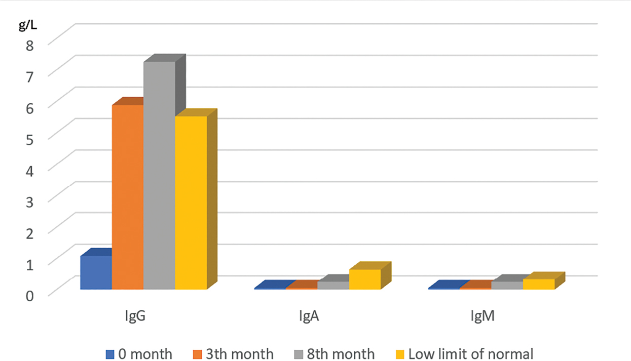
Figure 1.
The patient’s immunograms (mg/L) at 0, 3, and 8 months.
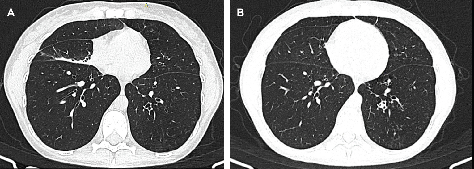
Figure 2.
Chest CT image (A) at the first presentation: the appearance of ‘middle lobe syndrome’. (B) After 8 months of treatment: re-expansion of the partial atelectasis of the middle lobe.