
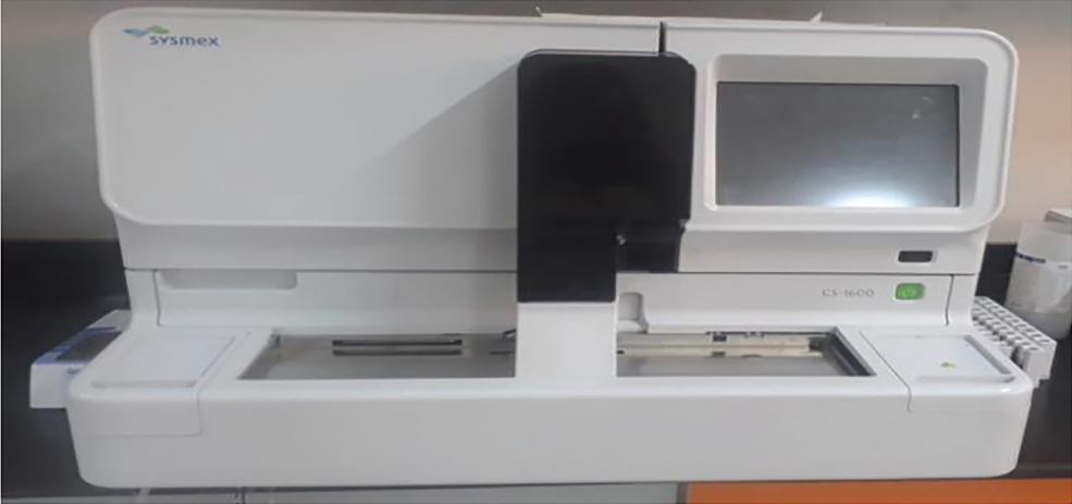
Figure 1.
CS-1600, automated hemostasis analyzer (Sysmex, Wakinohama, Japan).
Table 1.
Baseline demographic and clinical characteristics of the studied patients.
| Variable | Summary statistics (n = 65) n (%) |
|---|---|
| Age: Mean ± SD | 45.83 ± 16.23 |
| Gender: | |
| Female | 37 (56.9) |
| Male | 28 (43.1) |
| BMI: Mean kg/m2 ± SD | 22.11 ± 4.39 |
| Smoking: | |
| Non-smokers | 37 (56.9) |
| Current | 13 (20) |
| Ex-smoker | 15 (23.1) |
| Comorbidities: | |
| Yes | 41 (63.1) |
| No | 24 (36.9) |
| Comorbidities: | |
| Hypertension | 15 (23.1) |
| Diabetes Mellitus | 10 (15.4) |
| CVD | 32 (49.2) |
Table 2.
Clinical, laboratory, and radiological parameters of the studied patients.
| Variable | Summary statistics (n = 65) n (%) |
|---|---|
| CBC: mean ± SD | |
| WBCs 109/L | 11.66 ± 5.04 |
| Neutrophils | 8.88 ± 4.49 |
| Lymphocyte | 1.54 ± 0.69 |
| Eosinophils | 0.04 ± 0.07 |
| Platelet | 317.55 ± 103.87 |
| Haemoglobin | 12.34 ± 2.07 |
| ESR | 41.31 ± 20.62 |
| Albumin g/dL | 3.22 ± 0.49 |
| Fibrinogen mg/dL | 432.17 ± 63.3 |
| FEV1% | 55.69 ± 15.63 |
| Respiratory failure | |
| Yes | 20 (30.8) |
| No | 45 (69.2) |
| Number of affected lobes | |
| 0–2 | 35 (53.8) |
| ≥3 | 30 (46.2) |
| Radiologic appearance | |
| Cylindrical | 21 (32.3) |
| Cystic | 39 (60) |
| Varicose | 5 (7.7) |
| Pseudomonas colonisation | |
| No | 33 (50.8) |
| Yes | 32 (49.2) |
| Previous hospital admission | |
| No | 22 (33.8) |
| Yes | 43 (66.2) |
| Number of follow-up exacerbations | |
| 0–2 | 28 (43.1) |
| ≥3 | 37 (56.9) |
| MRC dyspnoea scale | |
| 0–2 | 18 (27.7) |
| ≥3 | 47 (72.3) |
| Number of previous exacerbations | |
| 0–2 | 26 (40) |
| ≥3 | 39 (60) |
| BSI score: mean ± SD | 9.29 ± 4.46 |
| Severity of bronchiectasis according to BSI score Mild | 14 (21.5) |
| Moderate | 13 (20) |
| Severe | 38 (58.5) |
| FACED score: mean ± SD | 3.6 ± 1.75 |
| Severity of bronchiectasis according to FACED score Mild | 21 (32.3) |
| Moderate | 11 (16.9) |
| Severe | 33 (50.8) |
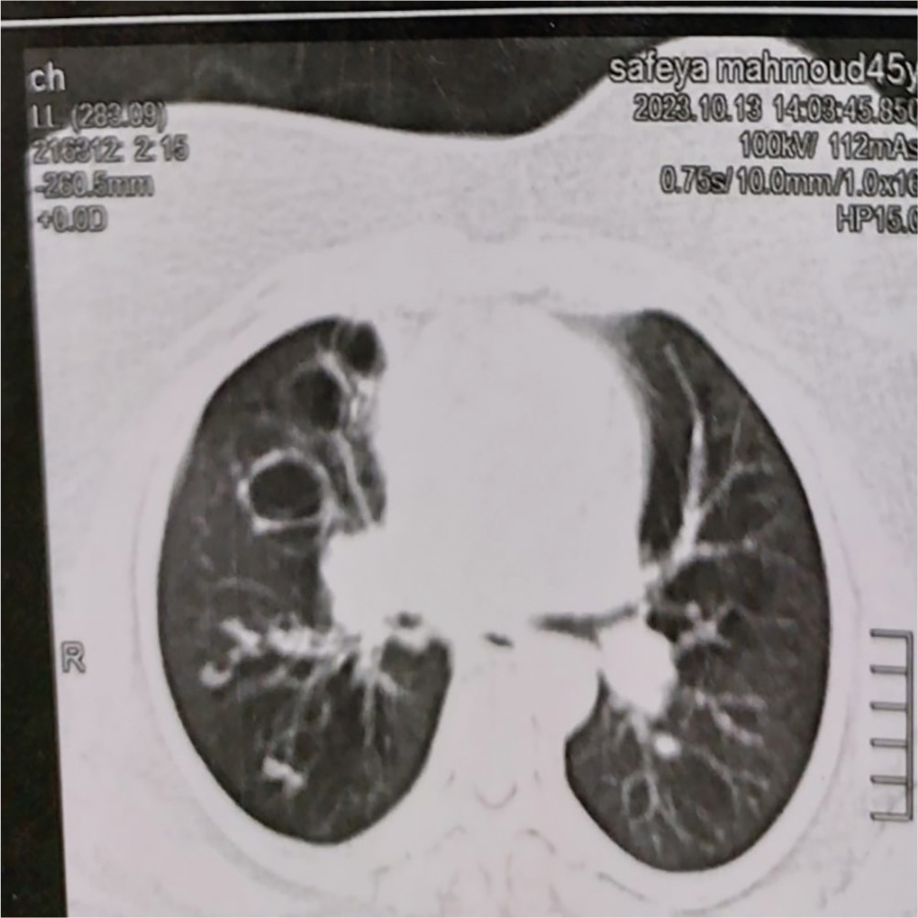
Figure 2.
Cystic bronchiectasis.
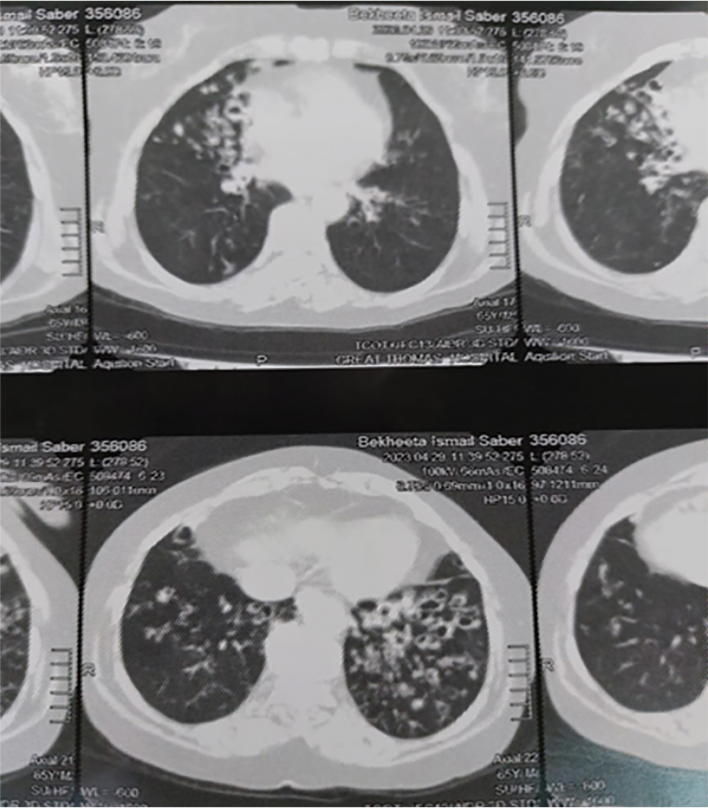
Figure 3.
Cylindrical bronchiectasis.
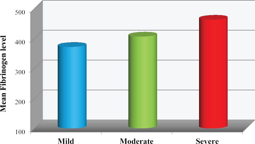
Figure 4.
Relation between serum fibrinogen level and BSI score severity. BSI, bronchiectasis severity index.
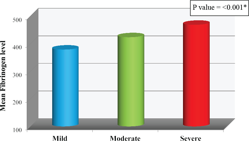
Figure 5.
Relation between serum fibrinogen level and FACED score severity.
Table 3.
Correlation between BSI, FACED scores, and laboratory findings
| Variable | BSI score | FACED score | ||
|---|---|---|---|---|
| Correlation coefficient | P-value | Correlation coefficient | P-value | |
| CBC: WBCs 109/L | r = 0.29 | 0.02* | r = 0.26 | 0.04* |
| Neutrophils | r = 0.26 | 0.04* | r = 0.24 | 0.051 |
| Lymphocyte | r = 0.23 | 0.06 | r = 0.09 | 0.48 |
| Eosinophils | r = 0.24 | 0.06 | r = 0.1 | 0.41 |
| Platelet | r = 0.16 | 0.21 | r = 0.24 | 0.06 |
| Haemoglobin | r = 0.2 | 0.11 | r = 0.11 | 0.37 |
| ESR | r = 0.34 | 0.01* | r = 0.32 | 0.01* |
| Albumin g/dL | r = –0.16 | 0.2 | r = –0.15 | 0.24 |
| Fibrinogen mg/dL | r = 0.64 | <0.001* | r = 0.61 | <0.001* |
Table 4.
Univariate and multivariate linear regression analyses of factors associated with BSI and FACED scores
| Variable | BSI score | FACED score | ||||||
|---|---|---|---|---|---|---|---|---|
| Univariate analysis | Multivariable analysis | Univariate analysis | Multivariable analysis | |||||
| Unadjusted B (95% CI) | P-value | Adjusted B (95% CI) | P-value | Unadjusted B (95% CI) | P-value | Adjusted B (95% CI) | P-value | |
| Age (years) | 0.06 (–0.01:0.13) | 0.07 | 0.02 (–0.01:0.05) | 0.17 | ||||
| Smoking | ||||||||
| Non-smokers | 3.24 (0.5:5.97) | 0.02* | –0.35 (–2.78:2.08) | 0.77 | 1.75 (0.72:2.77) | 0.001* | 0.6 (–0.41:1.61) | 0.42 |
| Current | 3.15 (0.56:5.74) | 0.02* | 2.31 (0.33:4.29) | 0.02* | 1.32 (0.35:2.3) | 0.01* | 1.1 (0.29:1.92) | 0.01* |
| Ex-smoker | ||||||||
| Comorbidities | ||||||||
| Yes | –0.59 (–2.9:1.71) | 0.61 | –0.2 (–1.11:0.72) | 0.67 | ||||
| No | ||||||||
| CBC: WBCs 109/L | 0.26 (0.04:0.47) | 0.02* | 0.64 (0.01:1.27) | 0.048* | 0.09 (0.003:0.17) | 0.04* | 0.03 (–0.03:0.11) | 0.28 |
| Neutrophils | 0.25 (0.01:0.5) | 0.04* | –0.63 (–1.34:0.08) | 0.08 | 0.1 (0:0.19) | 0.051 | ||
| Lymphocyte | 1.51 (–0.07:3.09) | 0.06 | 0.23 (–0.42:0.87) | 0.48 | ||||
| Eosinophils | 16.27 (–0.44:32.98) | 0.06 | 2.84 (–3.97:9.64) | 0.41 | ||||
| Platelet | 0.01 (0:0.02) | 0.21 | 0 (0:0.01) | 0.06 | ||||
| Haemoglobin | 0.44 (–0.1:0.97) | 0.11 | 0.1 (–0.12:0.31) | 0.37 | ||||
| ESR | 0.07 (0.02:0.12) | 0.01* | 0.03 (–0.01:0.08) | 0.13 | 0.03 (0.01:0.05) | 0.01* | 0.01 (–0.01:0.03) | 0.39 |
| Albumin g/dL | -1.47 (–3.76:0.81) | 0.2 | –0.54 (–1.45:0.37) | 0.24 | ||||
| Fibrinogen mg/dL | 0.04 (0.03:0.06) | <0.001* | 0.04 (0.03:0.06) | <0.001* | 0.02 (0.01:0.02) | <0.001* | 0.01 (0:01.02) | <0.001* |
Table 5.
Univariate and multivariate logistic regression analyses of factors associated with follow-up exacerbation among the studied patients
| Variable | Univariate analysis | Multivariable analysis | ||
|---|---|---|---|---|
| OR (95% CI) | P-value | OR (95% CI) | P-value | |
| Age (years) | 1 (0.97:1.03) | 0.89 | ||
| BMI kg/m2 | 0.97 (0.86:1.08) | 0.57 | ||
| Smoking | ||||
| Non-smokers | 1 | 0.03* | 1 | 0.16 |
| Current | 6.47 (1.26:33.34) | 0.36 | 10.17 (0.4:258.41) | 0.84 |
| Ex-smoker | 1.77 (0.52:5.97) | 1.25 (0.14:11.09) | ||
| Comorbidities | ||||
| Yes | 0.91 (0.33:2.53) 1 | 0.86 | ||
| No | 1 | |||
| Fibrinogen mg/dL | 1.04 (1.02:1.06) | < 0.001* | 1.03 (1.001:1.06) | 0.046* |
| FEV1% | 0.94 (0.9:0.97) | 0.001* | 1.01 (0.94:1.1) | 0.75 |
| Respiratory failure | ||||
| Yes | 1 | 0.2 | ||
| No | 2.01 (0.69:5.85) | |||
| Number of affected lobes | ||||
| 0–2 | 1 | 0.15 | ||
| ≥3 | 2.12 (0.77:5.8) | |||
| Radiologic appearance | ||||
| Cylindrical | 1 | 0.02* | 1 | 0.43 |
| Cystic | 4 (1.3:12.33) | 0.09 | 0.38 (0.03:4.31) | 0.98 |
| Varicose | 8 (0.75:85.73) | 1.06 (0.02:57.05) | ||
| Pseudomonas colonisation | ||||
| No | 1 | 0.001* | 1 | 0.91 |
| Yes | 6.25 (2.09:18.74) | 1.13 (0.13:9.97) | ||
| Previous hospital admission | ||||
| No | 1 | < 0.001* | 1 | 0.11 |
| Yes | 14.85 (4.07:54.16) | 6.14 (0.66:56.8) | ||
| MRC dyspnoea scale | ||||
| 0–2 | 1 | 0.001* | 1 | 0.32 |
| ≥3 | 8.25 (2.31:29.52) | 3.85 (0.27:55.13) | ||
| Previous exacerbations: | ||||
| 0–2 | 1 | < 0.001* | 1 | 0.04* |
| ≥3 | 19.2 (5.38:68.56) | 6.75 (1.11:41.26) | ||
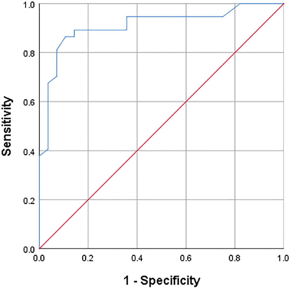
Figure 6.
ROC curve of serum fibrinogen level in predicting the occurrence of high-frequency exacerbations. ROC, receiver operating characteristic.
Table 6.
Cut-off point of serum fibrinogen level in predicting the occurrence of high-frequency exacerbations.
| Area under curve | P–value | 95% CI | Cut-off point | Sensitivity | Specificity |
|---|---|---|---|---|---|
| 0.91 | <0.001* | 0.84:99 | 416.5 | 87% | 89% |