
Introduction
Boron neutron capture therapy (BNCT) has long been proposed as a novel form of radiotherapy that, in theory, could be an ideal treatment for many types of cancer due to the selective accumulation of boron in tumor tissues [1, 2]. In this therapy, a 10B-containing agent, which has a high thermal neutron capture cross-section and can selectively target tumors, is first administered to the patient. Subsequently, the tumor site is irradiated with epithermal neutrons. These neutrons become thermalized in tissue and then react with 10B, producing short-range alpha particles (range ~9 μm) and lithium recoil nuclei (range ~5 μm), which kill tumor cells within the cell scale.
For a long time, BNCT has relied on reactor-based neutron sources (RB-BNCT) [3]. However, RB-based BNCT is complex, costly, and difficult to implement in a hospital setting, which has greatly hindered the development of BNCT. In recent years, accelerator-based BNCT (AB-BNCT) has gained increasing attention due to its low cost and ease of control [4, 5], and many countries are vigorously advancing this technology [6].
The most common targets for BNCT are beryllium and lithium [7,8,9], via the 9Be(p,n)9B and 7Li(p,n)7Be reactions, respectively. The Be target offers low heat dissipation due to its high melting point and high thermal conductivity; however, its neutron yield is low, necessitating an increase in incident proton energy (above 8 MeV) to produce more neutrons. The high proton energy also leads to higher neutron energies, which increases the complexity and manufacturing cost of the beam shaping assembly (BSA) [2, 10,11,12]. In contrast, the Li target is widely used in BNCT because of its high neutron yield and an energy spectrum that is easier to moderate. Nevertheless, Li has a low melting point (180°C) and poor thermal conductivity, and it is prone to oxidation. Therefore, careful design is required to ensure the lifetime of the Li target [13].
When a proton penetrates the target material, it loses energy through collisions with atomic electrons. The collision probability increases as the proton energy decreases, so the ion loses most of its initial kinetic energy just before reaching the end of its range, forming a high energy-loss peak known as the Bragg peak. Beyond the Bragg peak, the energy loss approaches zero. If the target is too thin, the neutron yield is insufficient. Conversely, if the target is too thick, the Bragg peak will be partially or even completely contained within the target, making heat dissipation difficult. Most neutrons generated by proton-target interactions are fast neutrons (>10 keV). An epithermal neutron beam (0.5 eV – 10 keV) can only be obtained after moderation by the BSA. Therefore, the choice of target thickness and the design of the BSA are both key factors for the success of BNCT.
This work is based on an existing 3.5 MeV proton accelerator. The proton current is 10 mA. Considering the relationship between target thickness and neutron yield, the optimal thickness of the Li target is selected. The IAEA standards [14] are used as the criteria for BSA design, and various neutron-moderating materials as well as different gamma-filter materials are evaluated [3].
Model and method
Figure 1 shows a cross-sectional view of the BSA described herein. The system was designed to bombard a Li target using a 3.5 MeV proton beam. The assembly mainly consists of a moderator, a reflector, a primary collimator, a photon filter layer, and a thermal neutron filter layer. The BSA outlet has a diameter of 15 cm, and the collimation length is 20 cm. Both the primary collimator, which aims to direct all neutrons forward, and the reflector, which is used to reflect neutrons back into the beam, are made of lead. The thermal neutron filter layer is made of 6Li with a thickness of 0.1 cm, which reduces damage to shallow tissue by filtering out thermal neutrons.
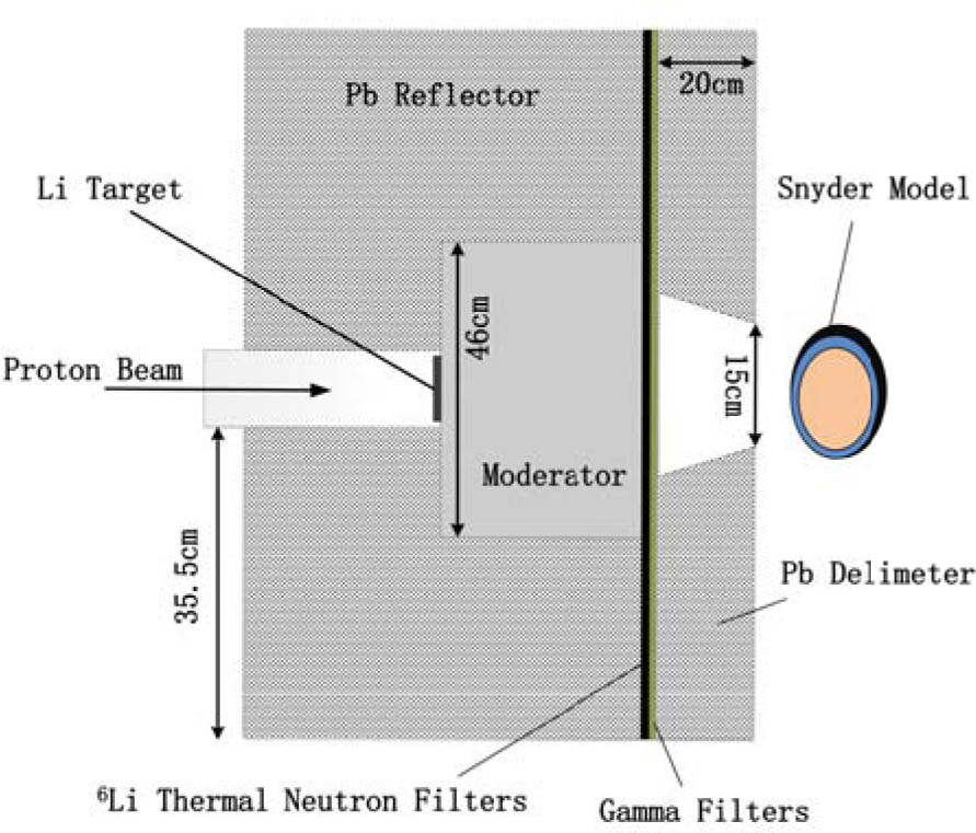
Fig. 1.
The cross-sectional view of a beam shaping assembly for Geant4 simulation.
Li target thickness selection
In selecting the actual target thickness, we should not only ensure a high neutron yield but also avoid making the target so thick that it contains the proton Bragg peak and increases the complexity of target heat dissipation. The target thickness was optimized.
Moderator and thickness selection
The ideal neutron energy for BNCT is the epithermal range (0.5 eV – 10 keV). Typically, moderator materials are selected to have large cross-sections for fast neutrons (>10 keV) and thermal neutrons (<0.5 eV), while having smaller cross-sections for epithermal neutrons [3, 15]. 19F has a low first excited state at 109.9 keV and a second excited state at 197.1 keV, which results in a relatively high cross-section for inelastic scattering and enables rapid moderation of neutrons with energies above 100 keV. Therefore, materials containing 19F can serve as ideal moderators. In this paper, materials with different 19F concentrations, such as MgF2, Fluental (composed of 69% AlF3, 30% Al, and 1% 7LiF), AlF3, and 7LiF, are selected as candidate moderators to compare their effectiveness on fast neutrons.
Lead shielding material selection
Gamma rays are generated when neutrons enter the moderators and reflectors. Therefore, in BSA design, high-atomic-number materials are typically used to filter gamma rays from the beam to protect patients from excessive gamma radiation. The most commonly used shielding materials are Pb and Bi. This paper compares the shielding effectiveness of these two materials as well as their influence on the neutron energy spectrum.
The IAEA standards [14] can only provide a preliminary assessment of the neutron energy spectrum; the dose distribution inside the phantom needs to be completed through further dose calculation. For BNCT, there are mainly four dose components: (1) boron dose, (2) thermal neutron dose, (3) fast neutron dose, and (4) photon dose. The total dose is the sum of each component multiplied by its corresponding biological effectiveness factor.
where Da is the total dose delivered by BNCT, CFB is the compound biological effectiveness factor for the boron dose, which is 1.3 in normal tissues and 3.8 in tumors; RBE and RBEf are the compound biological effectiveness factors for the thermal neutron dose and fast neutron dose, respectively, both of which are always 3.2 in both normal tissues and tumors; and RBEγ is the compound biological effectiveness factor for the photon dose, which equals 1 in both normal tissues and tumors.Several parameters are commonly used to evaluate dose distribution, including the maximum dose rate received by normal tissues (advantage depth dose rate, ADDR), the advantage depth (AD) at which the tumor dose rate equals ADDR, and the therapeutic gain (TG), defined as the ratio of the average tumor dose rate before AD to ADDR. Irradiation time is also a crucial parameter. It is determined by ADDR and the maximum tolerated dose of normal tissues (12.5 Gy-eq), and is also influenced by the maximum tolerated dose of skin (11 Gy-eq). The smaller the ADDR, and the larger the AD and TG, the better the therapeutic effect. Furthermore, the more dose the tumor receives within a shorter time, the better the outcome.
Currently, the modified Snyder ellipsoid model is commonly used in theoretical calculations. This model uses ellipsoids to define the boundaries of different tissues. To distinguish it from mesh-based models, it is often referred to as an analytical model. The original model consisted of two ellipsoids, dividing the head into two parts: the brain and the skull. The modified model further adds a 0.5 cm thick skin layer. The brain ellipsoid has an eccentricity of 1 cm in the Z-direction. Each interface satisfies the following equations (i.e., Eqs. (2) to (4)):
To reflect the effect of the moderator thickness on the dose distribution within the model, a modified Snyder model is placed at the exit of the BSA. The ratio of boron concentration in tumor to that in normal tissue is assumed to be 3:1, with a boron concentration of 30 ppm in the tumor tissue. Figure 1 shows a geometrical cross-sectional view of the modified Snyder head model. The right ellipsoidal structure represents the modified Snyder head model.
Results and discussion
Li target thickness selection
Figure 2 shows the relationship between neutron yield and lithium target thickness for a 3.5 MeV proton beam. It can be seen that as the target thickness increases, the neutron yield initially increases. After the thickness reaches a certain value (approximately 300 μm), the neutron yield reaches its maximum and no longer increases with further thickening. Notably, the maximum neutron yield is achieved before the Bragg peak, which ensures a high neutron yield while avoiding most of the energy deposition inside the target.
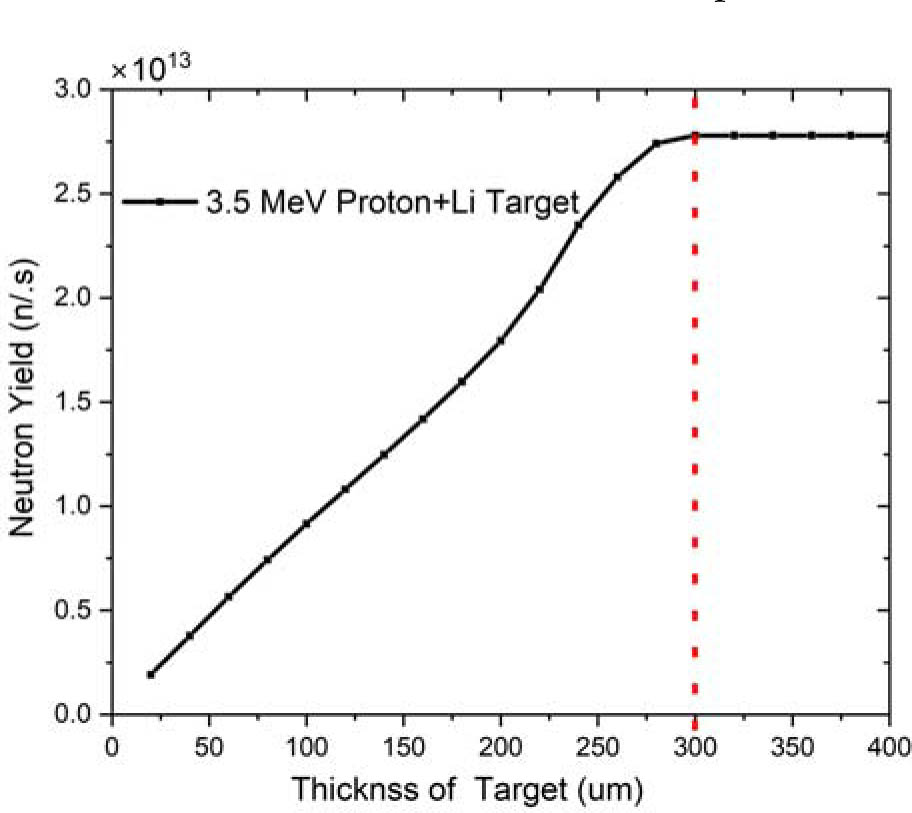
Fig. 2.
The relationship curve between neutron yield and target thickness for a 3.5 MeV proton beam bombarding a lithium target.
Figure 3 shows the relationship between the neutron distribution per unit arc angle and the Li target thickness. It can be seen that neutrons per unit arc angle are mainly distributed within a small angle; the neutron yield reaches a maximum at 40 degrees and then gradually decreases slightly toward 90 degrees. In BSA design, a higher neutron yield is desirable. Therefore, according to Fig. 3, utilizing the forward-directed neutrons is an effective approach to obtain a higher neutron yield in the BSA design.
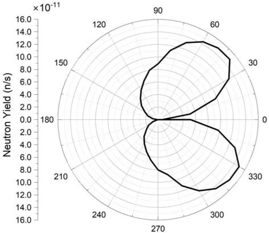
Fig. 3.
Neutron angular distribution with a Li target thickness of 290 μm.
Moderator and thickness selection
Figure 4 shows the variation of the neutron energy spectrum at the BSA outlet when MgF2, Fluental, AlF3, and 7LiF are each used alone as the moderating material with thicknesses ranging from 22 cm to 30 cm. It can be seen that 7LiF and MgF2 exhibit the best moderating ability at the same thickness, slowing neutrons down to approx. 10 keV. AlF3 performs worse than 7LiF and MgF2; although the neutron energy is also moderated to around 10 keV, a large number of fast neutrons (E >10 keV) remain. Fluental has the poorest moderating effect, with most neutrons remaining unmoderated in the fast energy range. The reasons for the different moderating abilities of these four materials are analyzed, which are mainly attributed to their different 19F contents: MgF2 (63.2%), 7LiF (73.2%), Fluental (47.6%), and AlF3 (41.3%). The better performance of AlF3 compared to Fluental at the same thickness is due to its higher 27Al content. 27Al also has a very low first excited state (843.8 keV), which allows inelastic scattering to readily occur with neutrons above 843.8 keV, reducing the neutron energy to below 843.8 keV before further interaction with 19F.
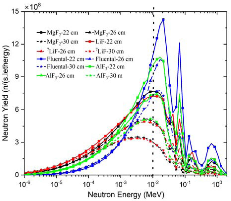
Fig. 4.
The relationship curve between the neutron energy spectrum at the BSA exit and the thickness of the moderated material (MgF2, 7LiF, Fluental, and AlF3) with a range of 22–30 cm.
The dose parameters corresponding to different 7LiF thicknesses in the BSA are shown in Table 1, while those for different MgF2 thicknesses are presented in Table 2. It can be observed from Table 1 that as the thickness of 7LiF increases, the depth of the maximum dose rate in normal tissue gradually shifts from the skin surface to deeper locations, reaching its maximum when the 7LiF thickness is 30 cm. At this thickness, the maximum depth reaches 2.4 cm, and the advantage depth also increases to a maximum of 9.2 cm. Further increasing the 7LiF thickness does not change the ADDR or the advantage depth. Additionally, increasing the irradiation time leaves the maximum tumor dose almost unchanged. Therefore, when 7LiF is used as the sole moderator, a thickness of 30 cm is considered the most suitable.
Table 1.
The dose evaluation parameters of 7LiF in the thickness range of 22–36 cm
| 7LiF thickness (cm) | ADDR (Gy-eq) | Depth of ADDR (cm) | Max dose rate of tumor (Gy-eq) | Advantage depth (cm) | Therapeutic gain (TG) | Treatment time (min) | Max dose rate of tumor (Gy-eq) |
|---|---|---|---|---|---|---|---|
| 22 | 61.1 | 0 | 157 | 8.2 | 1.95 | 12.3 | 33.0 |
| 24 | 42.8 | 0.4 | 135 | 8.8 | 2.21 | 17.5 | 40.5 |
| 26 | 33.2 | 0.4 | 115 | 9.0 | 2.46 | 22.6 | 43.7 |
| 28 | 25.9 | 2 | 99 | 9.2 | 2.61 | 29.0 | 48.5 |
| 30 | 21.7 | 2.4 | 85 | 9.2 | 2.66 | 34.6 | 49.3 |
| 32 | 18.3 | 2.4 | 73 | 9.2 | 2.68 | 41.4 | 50.4 |
| 34 | 15.5 | 2.4 | 63 | 9.2 | 2.70 | 48.6 | 51.0 |
| 36 | 13.2 | 2.4 | 54 | 9.2 | 2.72 | 57.0 | 51.3 |
Table 2.
The dose evaluation parameters of MgF2 in the thickness range of 22–36 cm
| MgF2 thickness (cm) | ADDR (Gy-eq) | Depth of ADDR (cm) | Max dose rate of tumor (Gy-eq) | Advantage depth (cm) | Therapeutic gain (TG) | Treatment time (min) | Max dose rate of tumor (Gy-eq) |
|---|---|---|---|---|---|---|---|
| 22 | 64.1 | 0 | 152 | 8.0 | 1.81 | 12.0 | 32.4 |
| 24 | 47.6 | 0 | 131 | 8.4 | 2.00 | 16.2 | 35.3 |
| 26 | 34.2 | 0 | 109 | 8.8 | 2.28 | 22.2 | 40.6 |
| 28 | 25.8 | 2 | 95 | 9.2 | 2.50 | 29.4 | 46.6 |
| 30 | 20.9 | 2 | 80 | 9.2 | 2.63 | 36.0 | 48.3 |
| 32 | 17.8 | 2.4 | 69 | 9.2 | 2.63 | 42.6 | 48.8 |
| 34 | 15.1 | 2.4 | 59 | 9.2 | 2.63 | 49.8 | 49.3 |
| 36 | 12.9 | 2.4 | 51 | 9.2 | 2.63 | 58.2 | 49.3 |
It can be observed that as the thickness of MgF2 increases, the depth of the maximum dose rate in normal tissue gradually shifts from the skin surface to deeper locations, reaching its maximum when the MgF2 thickness is 32 cm. At this thickness, the maximum depth reaches 2.4 cm, and the advantage depth also increases to a maximum of 9.2 cm. Further increasing the MgF2 thickness does not change the ADDR or the advantage depth. Moreover, increasing the irradiation time leaves the maximum tumor dose almost unchanged. Therefore, when MgF2 is used as the sole moderator, a thickness of 32 cm is considered the most suitable.
It can be seen from Tables 1 and 2 that the moderating effect of 7LiF is better than that of MgF2 at the same thickness. Consequently, in this paper, 30 cm of 7LiF was selected as the BSA moderator material.
Lead shielding material selection
Figure 5 and Table 3 show the neutron energy spectra at the BSA exit with Pb and Bi used as gamma filters, respectively. It can be seen that their filtering effects are almost equivalent. Considering that the density of Bi is lower (9.79 g/cmZ), a 0.8 cm thick Bi layer was ultimately selected as the gamma filter.
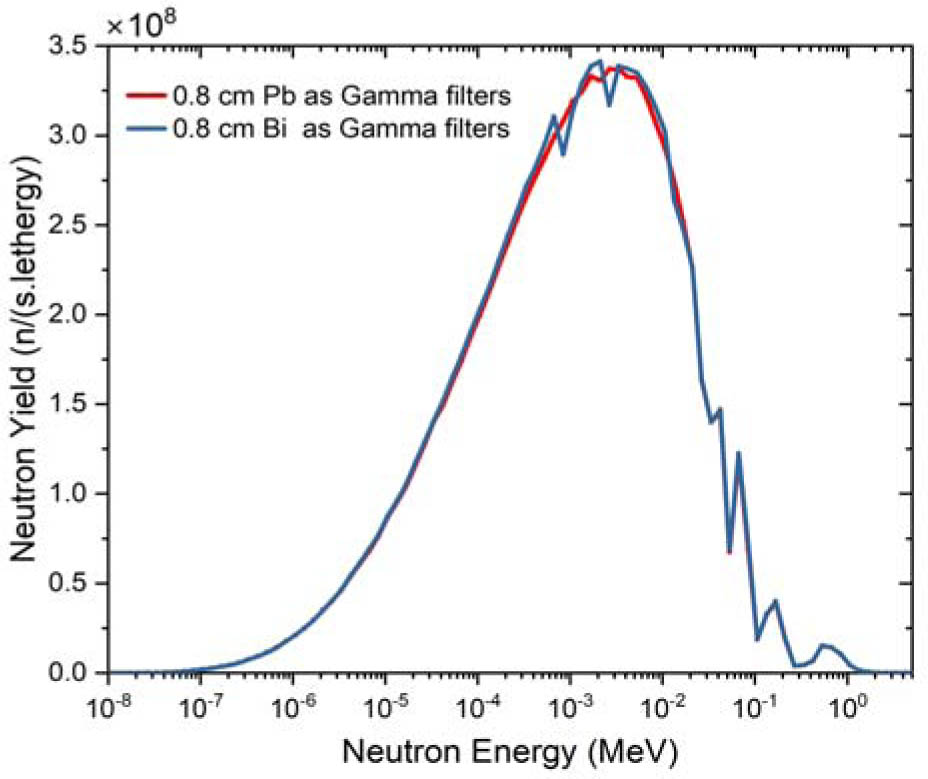
Fig. 5.
Neutron energy spectrum at the BSA exit obtained with 0.8 cm Pb and 0.8 cm Bi as gamma filters, respectively, where the thickness of 7LiF is 30 cm.
Table 3.
The IAEA parameters [14] obtained by using 0.8 cm thick Pb and Bi as gamma filters, respectively, where the thickness of 7LiF is 30 cm
| Parameter | φepi (n/cm−2·s−1) | φth/φepi | Df/φepi (Gy-cm2/n) | Dγ/φepi (Gy-cm2/n) | Current/flux |
|---|---|---|---|---|---|
| IAEA advised | >1.00 × 10⁹ | <0.05 | <2.00 × 10⁻¹³ | <2.00 × 10⁻¹³ | >0.70 |
| Pb 0.8 cm | 1.82 × 10⁹ | 0.005 | 4.22 × 10⁻¹⁴ | 3.31 × 10⁻¹⁴ | 0.71 |
| Bi 0.8 cm | 1.84 × 10⁹ | 0.005 | 4.21 × 10⁻¹⁴ | 3.30 × 10⁻¹⁴ | 0.71 |
Based on the above results and discussion, the final moderator material is 30 cm of 7LiF, the gamma filter is a 0.8 cm thick Bi layer, and the depth-dose curve in the Snyder head model is shown in Fig. 6. Considering that in actual treatment the maximum dose to the skin should be less than 11 Gy-eq and the maximum dose to normal tissue less than 12.5 Gy-eq, both tolerance limits were taken into account in this paper. The irradiation time and related dose parameters were calculated for these two cases, and they yielded the same results. The maximum dose rate to normal tissue is 22 Gy/h, the advantage depth is 9.2 cm, the therapeutic gain is 2.64, and the neutron irradiation time is 34.1 min. The maximum dose that the tumor can receive is 48.6 Gy-eq.
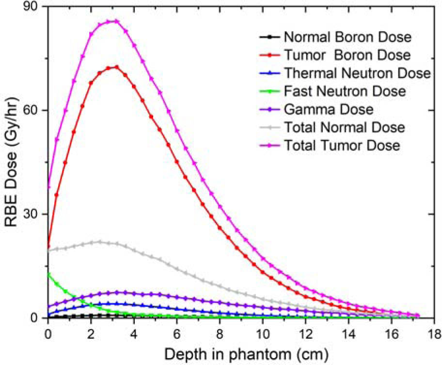
Fig. 6.
The depth-dose curve of the final BSA.
Conclusion
Based on a 3.5 MeV proton accelerator, the corresponding BSA design and parameter evaluation were carried out. The BSA designed in this paper effectively moderates the primary neutron energy spectrum generated by the 3.5 MeV proton bombardment of a lithium target, meeting the IAEA requirements. Furthermore, dose distribution calculations at the exit of the Snyder phantom show that the safe dose limits for both normal tissue and skin are simultaneously satisfied. Under this condition, the maximum tumor dose reaches 48.6 Gy-eq with an irradiation time of 34.1 min.
Acknowledgment
This work was supported by the Institutional Fund of the Engineering and Technical College of Chengdu University of Technology under Grant C122025002 and the Research Center of Leshan Nuclear Technology Application Engineering Technology under Grant 7813HK0025007.
Notes
[1] Contributed by Author contributions. All authors contributed to the study conception and design. Material preparation, data collection and analysis were performed by Hong Huang, Yucheng Yan and Tao Fu. The first draft of the manuscript was written by Hong Huang and all authors commented on previous versions of the manuscript. All authors read and approved the final manuscript.