
Table 1.
Hydrodynamic diameter and zeta potential of Aeroxide P25 TiO2 NPs in water, EMEM, F12 and DMEM media
| Medium | Hydrodynamic diameter (nm) | Polydispersity index (PDI) | Zeta potential 7.4 (mV) |
|---|---|---|---|
| Water | 356 ± 1 | 0.457 ± 0.04 | –15.3 ± 0.5 |
| EMEM | 246 ± 1 | 0.193 ± 0.07 | –25.5 ± 0.6 |
| F12 | 295 ± 3 | 0.220 ± 0.05 | –28.7 ± 2.5 |
| DMEM | 300 ± 2 | 0.280 ± 0.07 | –27.5 ± 0.2 |
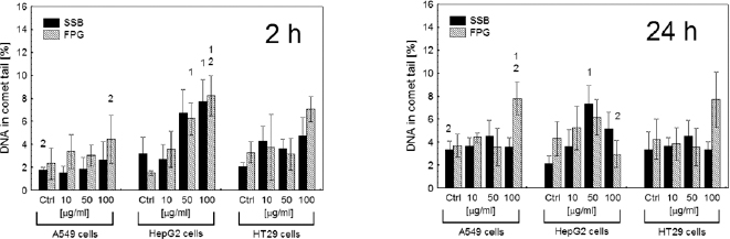
Fig. 1.
SSB and base damage recognized by FPG in A549, HepG2 or HT29 cells treated with Aeroxide P25 TiO2 NP for 2 h (left panel) and 24 h (right panel) at concentrations indicated. Numbers (1, 2) denote results of Tukey’s test post hoc analysis, indicating a significant difference of the means from the control when NP concentration was considered (1) or a significant difference of the means between the same NP concentration, but different treatment times (2).
Table 2.
Frequency of micronuclei and mean number of γ-H2AX foci per nucleus after 2 h and 24 h treatment with TiO2 NP concentrations indicated, and with the effect of X-irradiation with 2 Gy shown for comparison
| Micronuclei frequency | |||||||
|---|---|---|---|---|---|---|---|
| Treatment | Dose | Cell line/time of treatment | |||||
| A549 | HepG2 | HT29 | |||||
| 2 h | 24 h | 2 h | 24 h | 2 h | 24 h | ||
| Control | 22.9 ± 9.5 | 31.5 ± 2.0 | 34.6 ± 3.4 | 26.3 ± 7.8 | 5.9 ± 3.2 | 7.5 ± 1.8 | |
| X-ray | 2 Gy | 80.1 ± 5.2a | 155.2 ± 7.7a | 45.5 ±0.3a | |||
| TiO2 NP 21 nm | 10 μg/mL | 33.2 ± 6.9 | 26.8 ± 7.5 | 22.3 ± 8.8 | 27.0 ± 9.5 | 6.8 ± 1.0 | 8.5 ± 6.6 |
| 50 μg/mL | 22.5 ± 9.6 | 25.2 ± 3.7 | 24.6 ± 4.5 | 23.0 ± 4.4 | 5.5 ± 0.9 | 7.8 ± 1.6 | |
| 100 μg/mL | 25.5 ± 5.8 | 30.0 ± 8.2 | 24.5 ± 12.5 | 23.5 ± 6.4 | 6.1 ± 2.8 | 10.0 ± 7.2 | |
| The mean number of γ-H2AX foci per nucleus | |||||||
| Treatment | Dose | Cell line/time of treatment | |||||
| A549 | HepG2 | HT29 | |||||
| 2 h | 24 h | 2 h | 24 h | 2 h | 24 h | ||
| Control | 10.8 ± 5.7 | 10.7 ± 3.0 | 23.6 ± 10.8 | 7.2 ± 2.1 | 25.5 ± 10.1 | 26.4 ± 10.1 | |
| X-ray | 2 Gy | 78.2 ± 10.2a | 87.2 ± 10.0” | 87.9 ± 3.4a | |||
| TiO2 NP 21 nm | 10 μg/mL | 11.2 ± 6.8 | 10.5 ± 3.1 | 21.1 ± 6.0 | 9.7 ± 4.0 | 25.4 ± 10.0 | 25.0 ± 9.4 |
| 50 μg/mL | 10.3 ± 3.5 | 7.8 ± 1.5 | 19.3 ± 5.6 | 11.4 ± 2.7 | 26.6 ± 6.8 | 23.7 ± 8.8 | |
| 100 μg/mL | 10.6 ± 4.1 | 6.4 ± 2.5 | 18.3 ± 8.0 | 9.5 ± 3.2 | 28.0 ± 5.0 | 24.5 ± 10.5 | |
Table 3.
The extent of apoptosis after 2 h and 24 h treatment with TiO2 NP at indicated concentrations
| Treatment | Dose | Cell line/time of treatment | |||||
|---|---|---|---|---|---|---|---|
| A549 | HepG2 | HT29 | |||||
| 2h | 24 h | 2h | 24 h | 2h | 24 h | ||
| Control | Viable | 99.16 ± 0.14 | 98.36 ± 0.16 | 97.63 ± 0.29 | 96.34 ± 0.43 | 98.03 ± 0.55 | 97.04 ± 0.69 |
| Early apoptosis | 0.54 ± 0.21 | 1.34 ± 0.26 | 2.20 ± 0.30 | 3.22 ± 0.29 | 1.80 ± 0.56 | 2.61 ± 0-43 | |
| Late apoptosis and necrosis | 0.30 ± 0.10 | 0.30 ± 0.08 | 0.17 ± 0.15 | 0.44 ± 0.17 | 0.17 ± 0.06 | 0.35 ± 0.09 | |
| Staurosporine/24 hb | Viable | 32.48 ± 8.88a | 23.70 ± 5.33a | 20.20 ± 3.20a | |||
| Early apoptosis | 7.98 ± 2.33a | 48.20 ± 3.22a | 58.03 ± 4.98a | ||||
| Late apoptosis and necrosis | 59.54 ± 3.99a | 28.10 ± 2.56a | 21.77 ± 2.37a | ||||
| TiO2 NP 10 mg/mL | Viable | 93.07 ± 0.81a | 93.30 ± 1.20a | 93.20 ± 0.26a | 92.93 ± 0.64a | 93.13 ± 0.25a | 92.50 ± 1.46a |
| Early apoptosis | 5.10 ± 0.44a | 4.70 ± 0.75a | 5.03 ± 1.03 | 4.53 ± 0.40a | 4.43 ± 0.75 | 4.17 ± 0.79 | |
| Late apoptosis and necrosis | 1.83 ± 0.40a | 2.00 ± 0.46a | 1.77 ± 0.81 | 2.53 ± 0.26a | 2.43 ± 0.50a | 3.33 ± 0.81a | |
| TiO2 NP 50 mg/mL | Viable | 92.10 ± 0.80a | 93.93 ± 1.75a | 93.97 ± 0.65a | 91.13 ± 1.37a | 93.17 ± 0.78a | 91.83 ± 1.60a |
| Early apoptosis | 5.97 ± 0.76a | 3.83 ± 1.33a | 3.97 ± 0.61 | 6.00 ± 1.15a | 3.70 ± 0.60 | 5.17 ± 1.40a | |
| Late apoptosis and necrosis | 1.93 ± 0.15a | 2.23 ±0.42a | 2.07 ±0.15a | 2.87 ± 0.35a | 3.13 ±0.25a | 3.00 ± 0.26a | |
| TiO2NP 100 mg/mL | Viable | 93.63 ± 1.20a | 91.87 ± 1.10a | 93.73 ± 0.64a | 90.87 ± 0.96a | 92.23 ± 1.46a | 90.57 ± 0.23a |
| Early apoptosis | 3.97 ± 0.75a | 5.90 ± 1.47a | 4.27 ± 0.40a | 6.33 ± 0.91a | 4.70 ± 0.79a | 6.17 ± 0.65a | |
| Late apoptosis and necrosis | 2.40 ± 0.46a | 2.23 ± 0.40a | 2.00 ± 0.26a | 2.80 ± 0.26a | 3.07 ± 0.81a | 3.27 ± 0.47a | |
Table 4.
Caspase 3 activity after 2 h and 24 h treatment with TiO2 NP at indicated concentrations
| Treatment | Cell line/time of treatment | |||||
|---|---|---|---|---|---|---|
| A549 | HepG2 | HT29 | ||||
| 2 h | 24 h | 2 h | 24 h | 2 h | 24 h | |
| Control | 0.12 ±0.05 | 0.05 ±0.01 | 1.02 ±0.03 | 1.42 ±0.02 | 0.26 ±0.02 | 0.42 ±0.03 |
| TiO2 NP 10 mg/mL | 0.10 ± 0.02 | 0.06 ± 0.01 | 0.98 ± 0.02 | 1.43 ± 0.09 | 0.31 ± 0.01 | 0.41 ± 0.06 |
| TiO2 NP 50 mg/mL | 0.13 ± 0.02 | 0.07 ± 0.01 | 1.08 ± 0.07 | 1.37 ± 0.05 | 0.51 ± 0.05a | 0.63 ±0.05a |
| TiO2 NP 100 mg/mL | 0.11 ± 0.01 | 0.07 ± 0.01 | 1.39 ± 0.06a | 1.39 ± 0.03 | 0.37 ± 0.02a | 0.55 ± 0.04a |
| Positive control: Jurkat cells treated with 1 mM staurosporine for 24 h : 39.89 ± 3.82a | ||||||
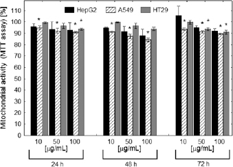
Fig. 2.
Mitochondrial activity (MTT assay) of cells treated with Aeroxide P25 TiO2 NP for 24 h and measured 72 h after treatment expressed as a percent of the control (untreated cells). The asterisk denotes a statistically significant difference from the control. Two-way ANOVA with Tukey’s HDS post hoc comparison. P < 0.05.
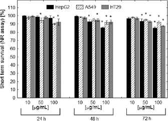
Fig. 3.
Short-term survival of cells treated with Aeroxide P25 TiO2 NP for 24 h, as measured by neutral red assay. The asterisk denotes a statistically significant difference from the control. Two-way ANOVA with Tukey’s HDS post hoc comparison. P < 0.05.
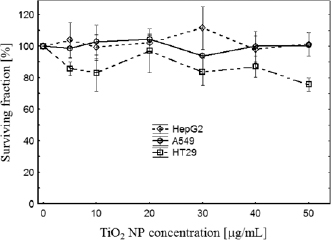
Fig. 4.
Cloning ability determination of A549, HT29 and HepG2 cells growing in the presence of Aeroxide P25 TiO2 NP.
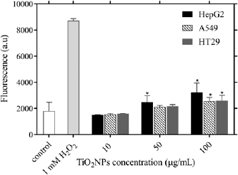
Fig. 5.
Generation of ROS in HepG2, A549 and HT29 cells incubated with Aeroxide P25 TiO2 NP. H2O2 in a concentration of 1 mM served as a positive control. The data provided are in the form mean ± SD. n denotes the number of experiments; n = 3. The asterisk denotes statistically significant difference from unexposed control, P < 0.05.