Fig. 1.
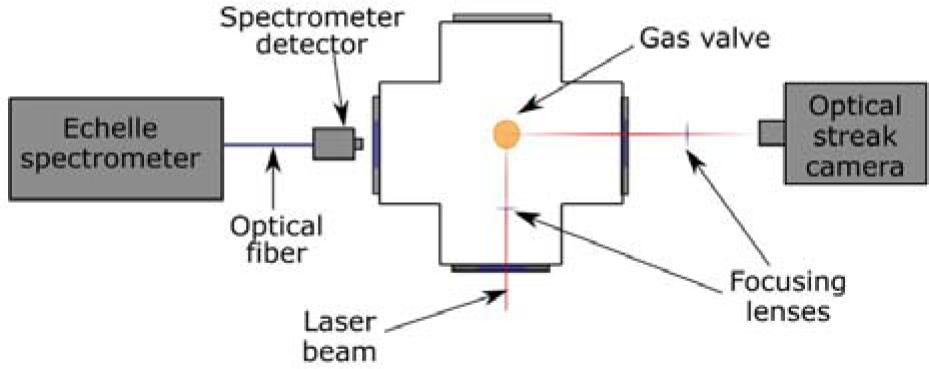
Fig. 2.
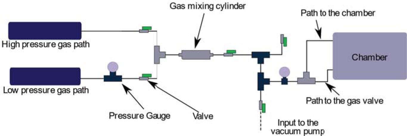
Fig. 3.
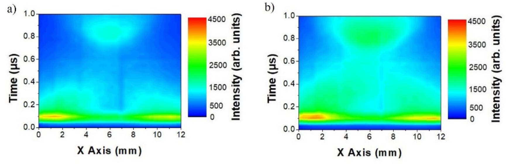
Fig. 4.
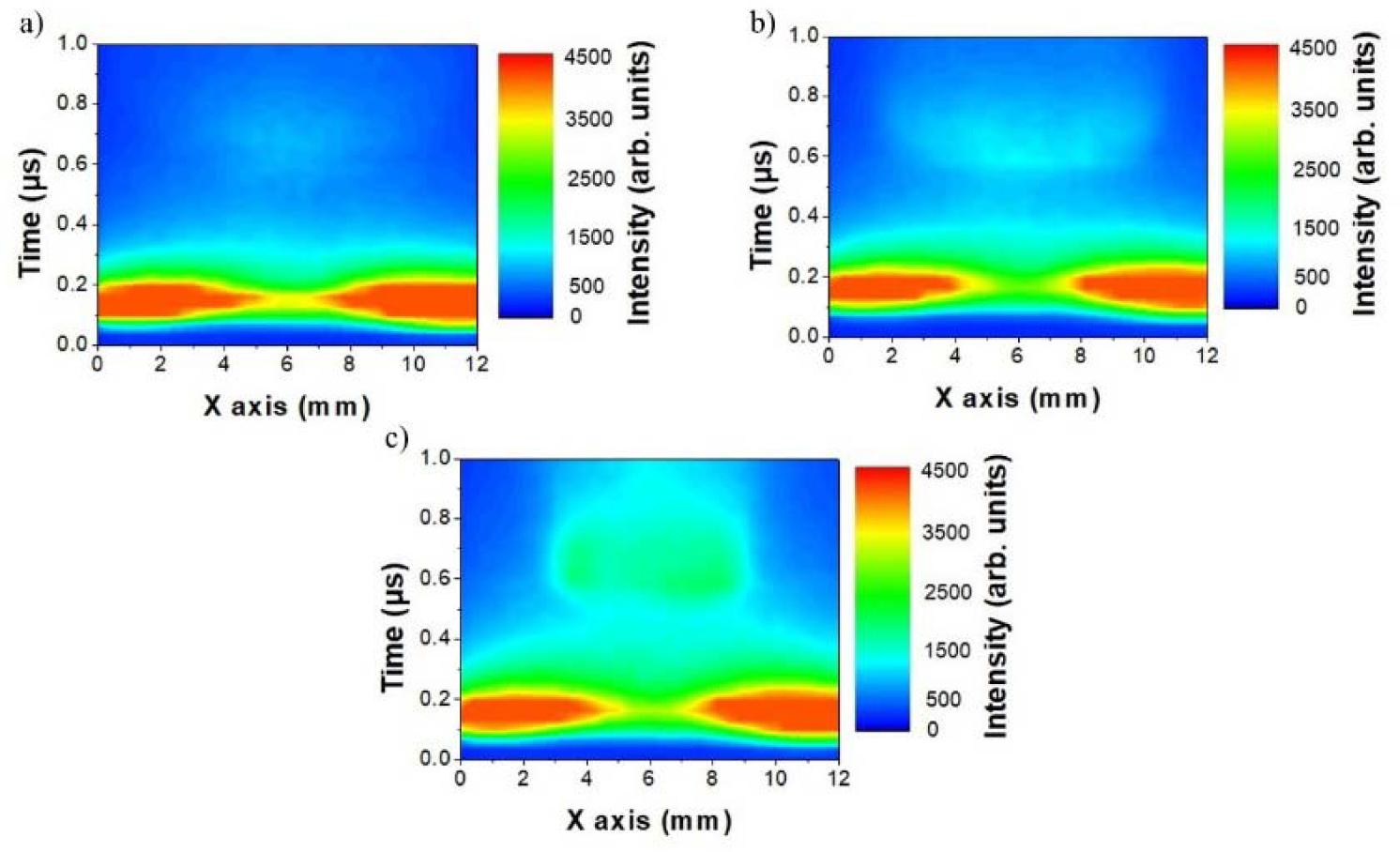
Fig. 5.
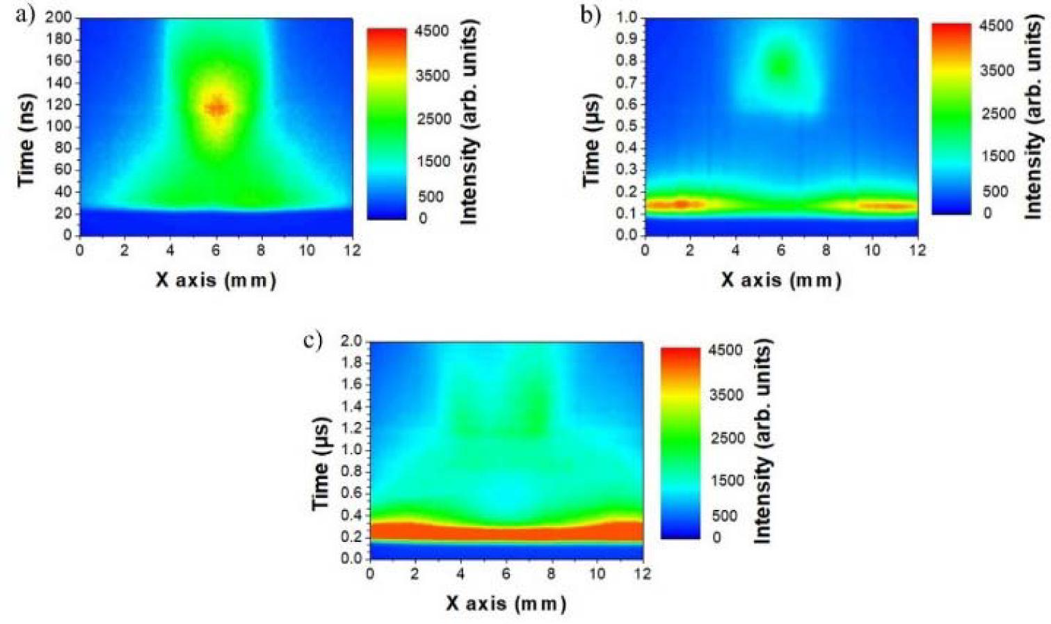
Fig. 6.
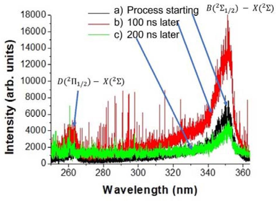
Fig. 7.
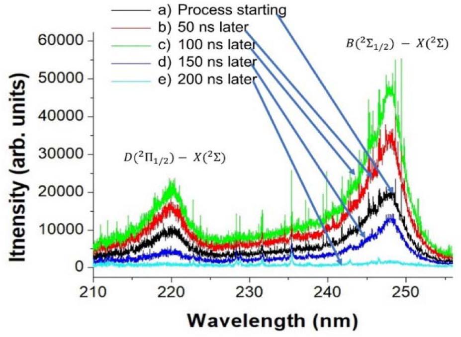
Fig. 8.
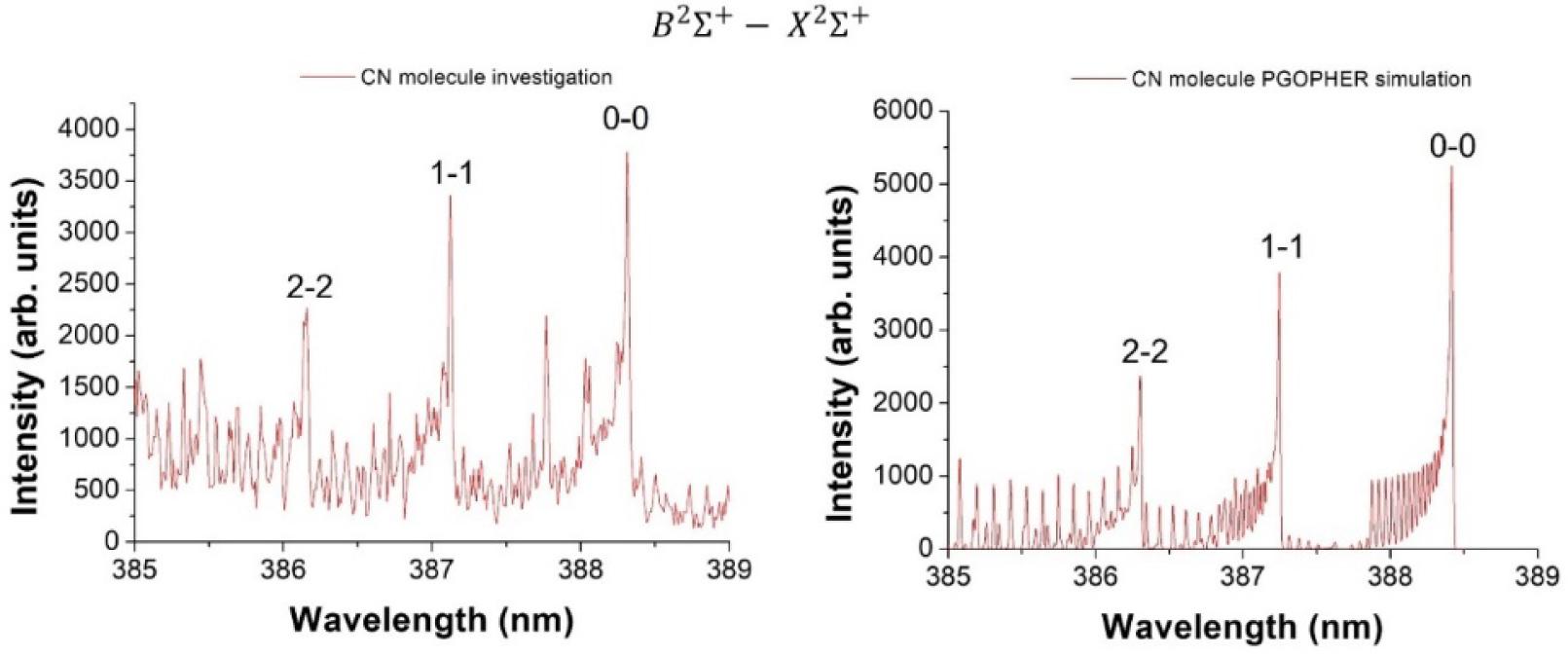
Fig. 9.

© 2023 Mateusz Majszyk, Andrzej Bartnik, Wojciech Skrzeczanowski, Tomasz Fok, Łukasz Węgrzyński, Mirosław Szczurek, Henryk Fiedorowicz, published by Institute of Nuclear Chemistry and Technology
This work is licensed under the Creative Commons Attribution-NonCommercial-NoDerivatives 4.0 License.