
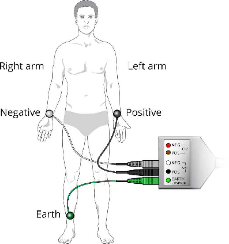
Fig. 1.
The placement of the ECG electrodes with PowerLab, a physiological signal acquisition instruments from ADInstruments Inc. [30].
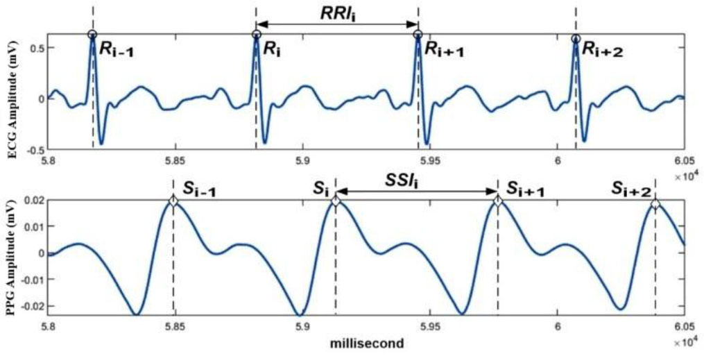
Fig. 2.
Examples of ECG and PPG signals.
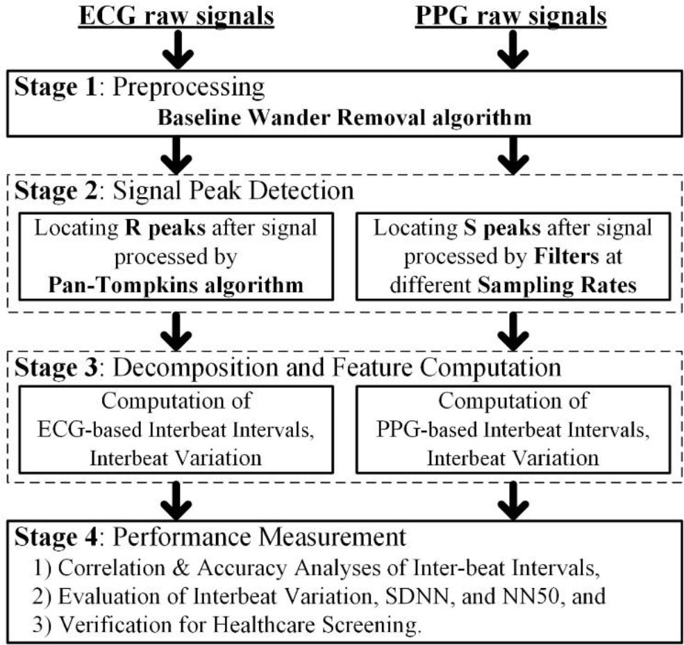
Fig. 3.
Signal processing flow chart for ECG and PPG.
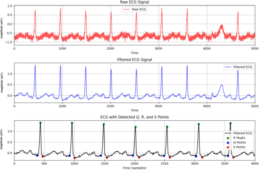
Fig. 4.
The process of extracting QRS from raw signals.
Table 1.
A descriptive listing of PPG signal processing methods.
| No. | Filter name | Description |
|---|---|---|
| F01 | Butterworth | The passband frequency is from 1 Hz to 5 Hz, and the filter order is 2. |
| F02 | Bessel | |
| F03 | Chebyshev | |
| F04 | Elliptic | |
| F11 | Savitzky-Golay | The polynomial order is 3, and the window size 401. |
| F12 | Average | The passband frequency is from 1 Hz to 5 Hz. |
| F13 | Periodic moving | After normalizing each cardiac cycle with the same number of sampled signals, the ith sampled signals of each of the 5 cycles are averaged, where 1 ≤ i ≤ the normalized cycle length. |
| F21 | Daubechies | Use the inverse DWT of Daubechies db6 and Coiflet C3 to reconstruct the selected band from 1 Hz to 5 Hz. |
| F22 | Coiflet C3 | |
| F23 | Morlet (CWT) | Apply the inverse WT of Morlet to reconstruct the selected band from 1 Hz to 5 Hz. |
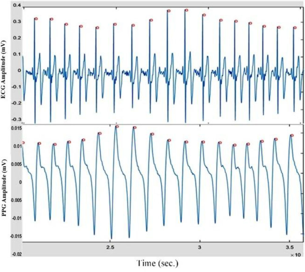
Fig. 5.
Detected ECG and PPG peaks.
Table 2.
Average absolute errors between the RRIV/ECG SDNN/ECG, NN50 at 1000 Hz and the SSIV/PPG SDNN/PPG NN50 at different sampling rates.
| Filter/Sample rates | Phase | Parameter | 1000 [Hz] | 500 [Hz] | 250 [Hz] | 200 [Hz] | 125 [Hz] | 100 [Hz] | 50 [Hz] | 40 [Hz] | 25 [Hz] | 20 [Hz] |
|---|---|---|---|---|---|---|---|---|---|---|---|---|
| F01 | R | SSIV [%] | 0.6 | 0.6 | 1.2 | 1.3 | 1.3 | 1.4 | 1.8 | 2.2 | 3.4 | 4.4 |
| F11 | R | SSIV [%] | 1.4 | 1.5 | 2.1 | 2.2 | 2.3 | 2.3 | 2.8 | 3 | 4.1 | 7.3 |
| F23 | R | SSIV [%] | 2 | 2.2 | 3 | 2.5 | 2.9 | 2.8 | 3.2 | 4.7 | 4.6 | 5.8 |
| F01 | B | SSIV [%] | 0.9 | 1.1 | 1.2 | 1.1 | 1.2 | 1.2 | 1.8 | 2 | 2.9 | 3.8 |
| F11 | B | SSIV [%] | 2.5 | 2.3 | 2.3 | 2.5 | 2.6 | 2.7 | 3.1 | 3.3 | 4.2 | 14.4 |
| F23 | B | SSIV [%] | 2.6 | 2.7 | 2.9 | 2.5 | 2.9 | 3 | 3.4 | 5 | 4.3 | 5.6 |
| F01 | R | SDNN [ms] | 0.6 | 0.6 | 0.6 | 0.6 | 0.8 | 0.9 | 2.1 | 3.1 | 6.5 | 9.5 |
| F11 | R | SDNN [ms] | 1.3 | 1.4 | 1.5 | 1.6 | 1.7 | 1.9 | 3.2 | 4 | 7.3 | 12.4 |
| F23 | R | SDNN [ms] | 2.7 | 2.6 | 2.8 | 2.1 | 2.7 | 2.3 | 3.4 | 6.9 | 7.5 | 10.8 |
| F01 | B | SDNN [ms] | 1.2 | 1.2 | 1.3 | 1.3 | 1.3 | 1.4 | 2.2 | 2.7 | 5 | 7.2 |
| F11 | B | SDNN [ms] | 2.2 | 2.2 | 2.2 | 2.4 | 2.4 | 2.6 | 3.3 | 3.9 | 6.3 | 15.3 |
| F23 | B | SDNN [ms] | 2.7 | 2.7 | 2.9 | 2.4 | 2.7 | 2.6 | 3.3 | 6.1 | 5.6 | 8.1 |
| F01 | R | NN50 [%] | 0.8 | 0.9 | 0.8 | 1 | 1 | 1.3 | 1.7 | 1.7 | 3.6 | 3.1 |
| F11 | R | NN50 [%] | 1.7 | 1.6 | 1.8 | 1.6 | 1.9 | 2 | 3.5 | 2.2 | 5.1 | 4.3 |
| F23 | R | NN50 [%] | 1.5 | 1.5 | 1.5 | 1.6 | 1.6 | 1.8 | 2.2 | 2.1 | 4.1 | 3.3 |
| F01 | B | NN50 [%] | 0.8 | 0.8 | 0.8 | 0.9 | 0.9 | 1.1 | 2.2 | 1.8 | 3.6 | 3.1 |
| F11 | B | NN50 [%] | 2.4 | 2.4 | 2.8 | 2.3 | 2.7 | 2.3 | 4.9 | 2.8 | 5.9 | 5.3 |
| F23 | B | NN50 [%] | 1.6 | 1.6 | 1.7 | 1.6 | 1.7 | 1.9 | 3.1 | 2.5 | 4.8 | 4.1 |
Table 3.
RRIV assessment range of normal cardiac autonomic function in each age group based on ECG [38].
| Age [y] | 20 ∼ 29 | 30 ∼ 39 | 40 ∼ 49 | 50 ∼ 59 | 60 ∼ 69 |
|---|---|---|---|---|---|
| Rest [ms] | 12 ∼ 46 | 6 ∼ 32 | 6 ∼ 36 | 5 ∼ 23 | 7 ∼ 19 |
| Breath [ms] | 19 ∼ 62 | 9 ∼ 54 | 14 ∼ 48 | 11 ∼ 59 | 8 ∼ 28 |
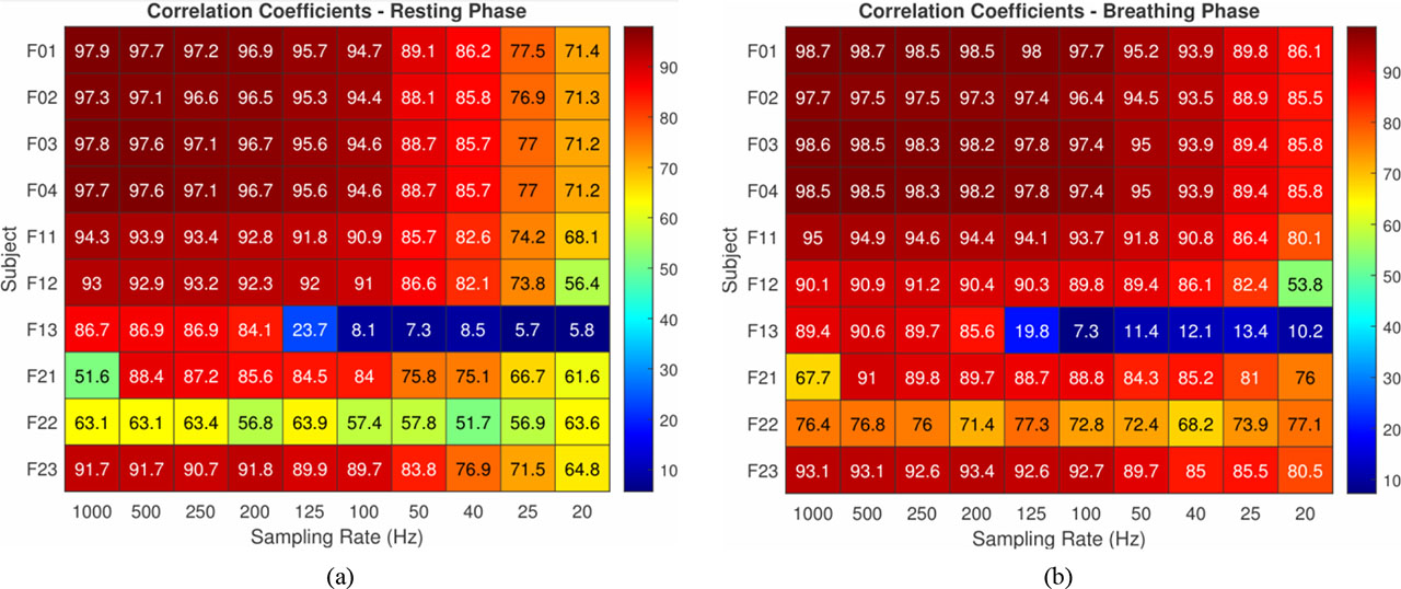
Fig. 6.
HeatMap of correlation coefficients between the RRI at 1 kHz and the SSIs at various rates in resting (a), breathing phase (b).
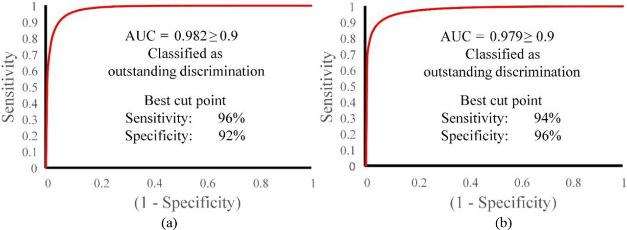
Fig. 7.
Evaluation of the best SSIV assessment range of normal cardiac autonomic function with the ROC curve, (a) – the resting phase; (b) – the deep breathing phase.
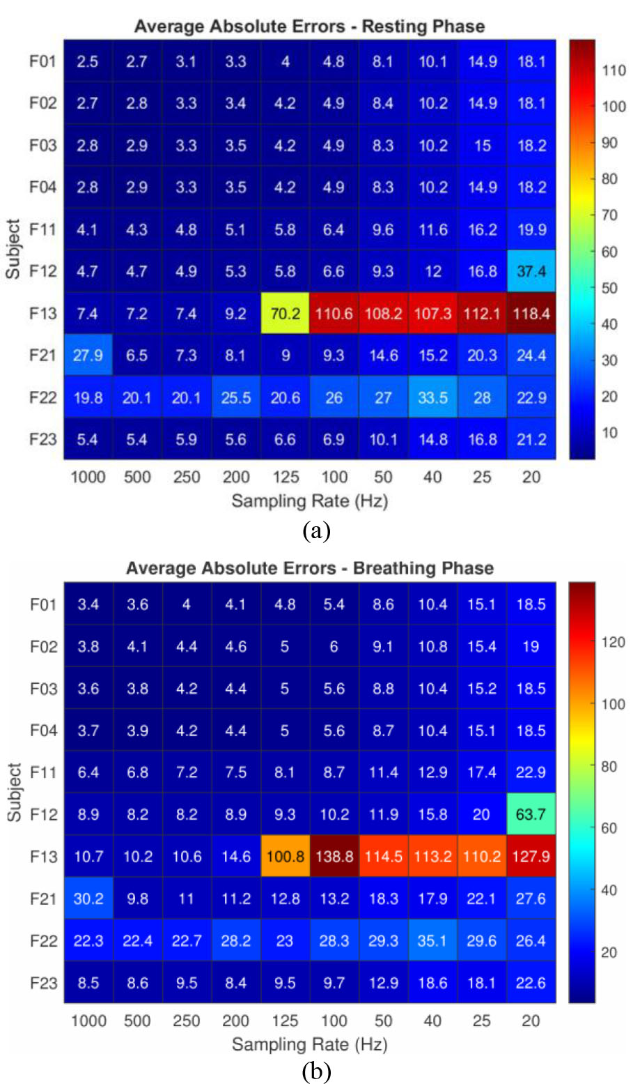
Fig. 8.
HeatMap of average absolute errors between the RRI at 1000 Hz and the SSIs at different sampling rates in resting (a) and breathing phases (b).
Table 4.
Confusion matrix between RRIV and SSIV in the resting and deep breathing phases.
| Phase | SSIV | RRIV Abnormal [ms] | RRIV Normal [ms] |
|---|---|---|---|
| Resting | Abnormal | 44 | 2 |
| Normal | 8 | 190 | |
| Deep breathing | Abnormal | 44 | 3 |
| Normal | 7 | 190 |
Table 5.
The sensitivity and specificity of SSIV in different assessment ranges.
| Phase | Rate [%] | Out+2 | Out+1 | RRIV source | In-1 | In-2 | In-3 | In-4 |
|---|---|---|---|---|---|---|---|---|
| Rest | Sensitivity | 36.54 | 63.46 | 84.62 | 96.15 | 96.15 | 100.00 | 100.00 |
| Specificity | 98.96 | 98.96 | 98.96 | 91.67 | 79.69 | 70.83 | 59.90 | |
| Breath | Sensitivity | 54.90 | 70.59 | 86.27 | 94.12 | 94.12 | 96.08 | 96.08 |
| Specificity | 99.48 | 99.48 | 98.45 | 95.85 | 91.19 | 84.97 | 78.24 |