
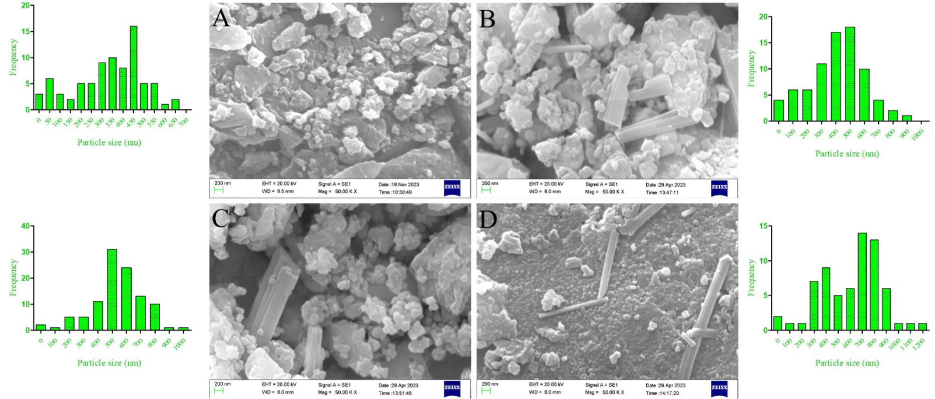
Fig. 1.
Representative SEM imaging results for (A) ZIF, (B) PROZIF, (C) SAFROZIF, and (D) CURCUZIF nanocarriers and their size distribution
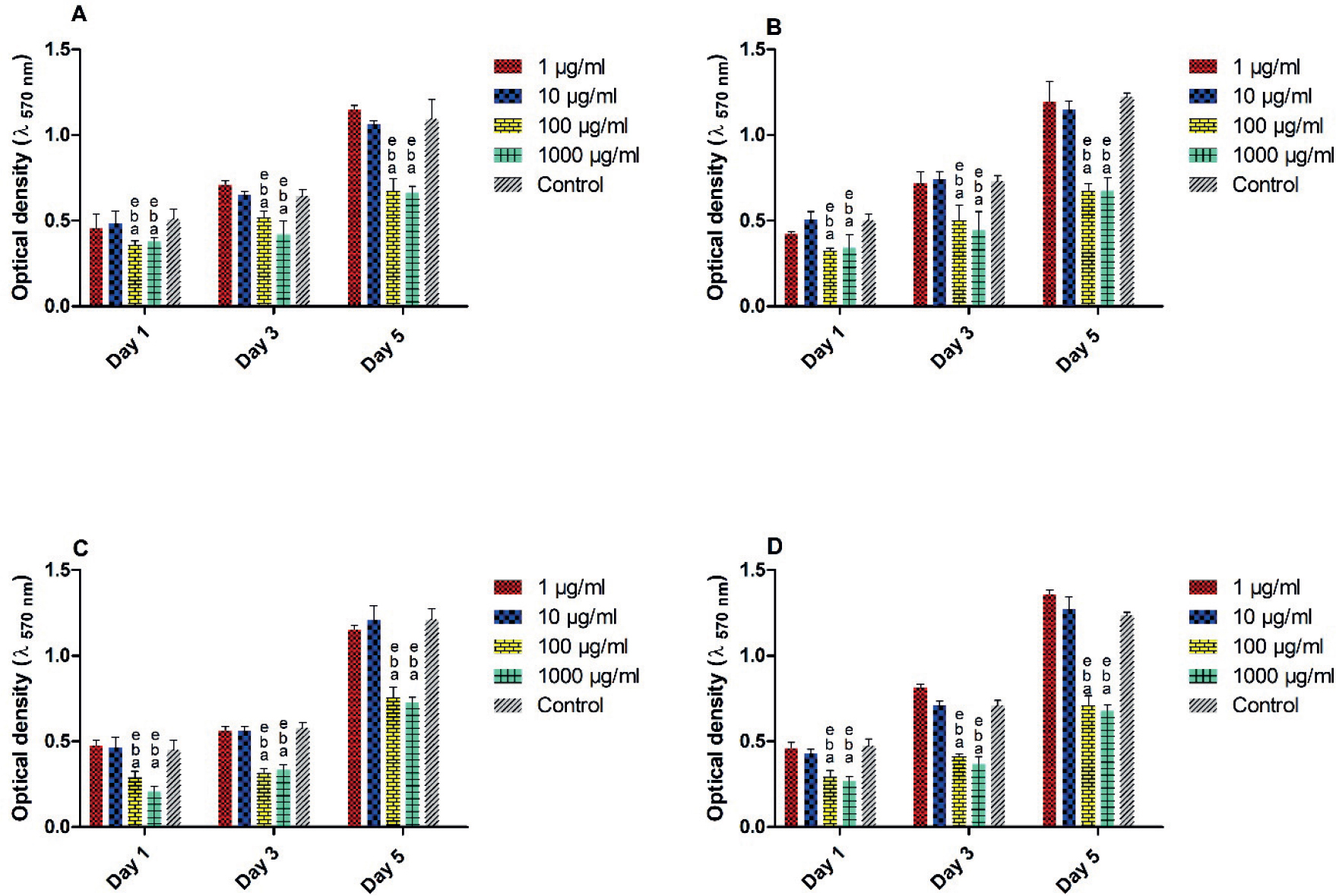
Fig. 2.
MTT assay with L929 cells cultured with different concentrations of (A) PROZIF, (B) SAFROZIF, (C) CURCUZIF, and (D) ZIF nanoparticles, a, b, and e show p-values < 0.05 relative to 1 μg/mL, 10 μg/mL, and control groups, respectively
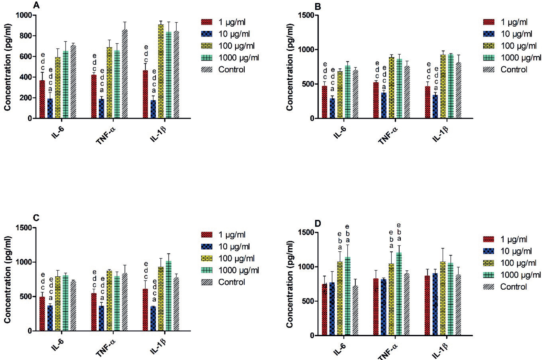
Fig. 3.
Histograms comparing the anti-inflammatory activity of (A) PROZIF, (B) SAFROZIF, (C) CURCUZIF, and (D) ZIF nanoparticles at different concentrations, a, b, c, and d show p-value < 0.05 relative to 1 μg/mL, 10 μg/mL, 100 μg/mL, and 1000 μg/mL concentrations, respectively
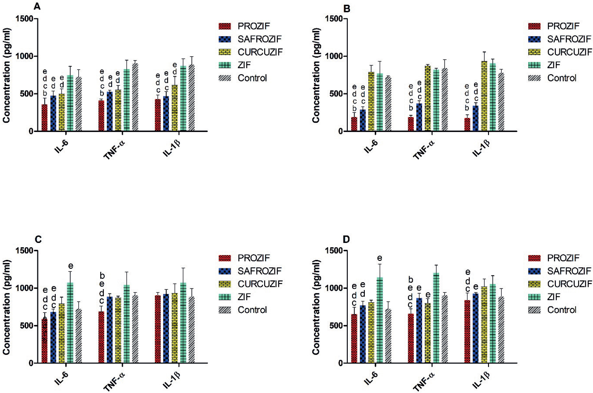
Fig. 4.
Histograms comparing the anti-inflammatory activity of ZIF, PROZIF, SAFROZIF, and CURCUZIF at (A) 1 μg/mL, (B) 10 μg/mL, (C) 100 μg/mL, and (D) 1000 μg/mL concentrations
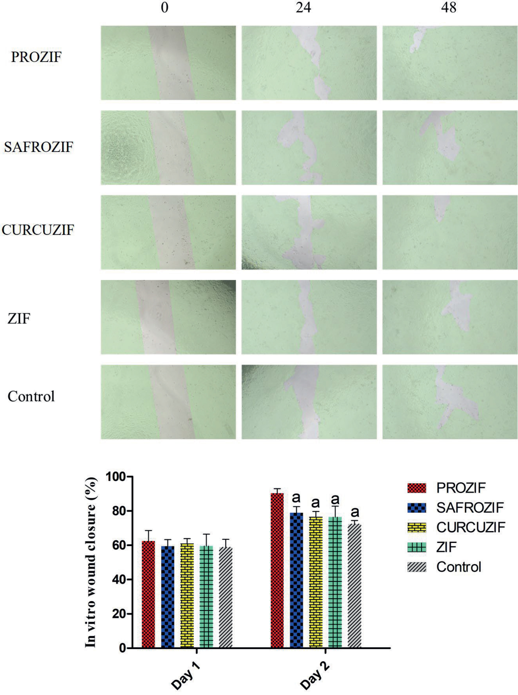
Fig. 5.
Cell migration assay with L929 cells cultured with different nanocarriers on day 0 (left panel), day 1 (middle panel), and day 2 (right panel), a shows p-value < 0.05 relative to PROZIF group
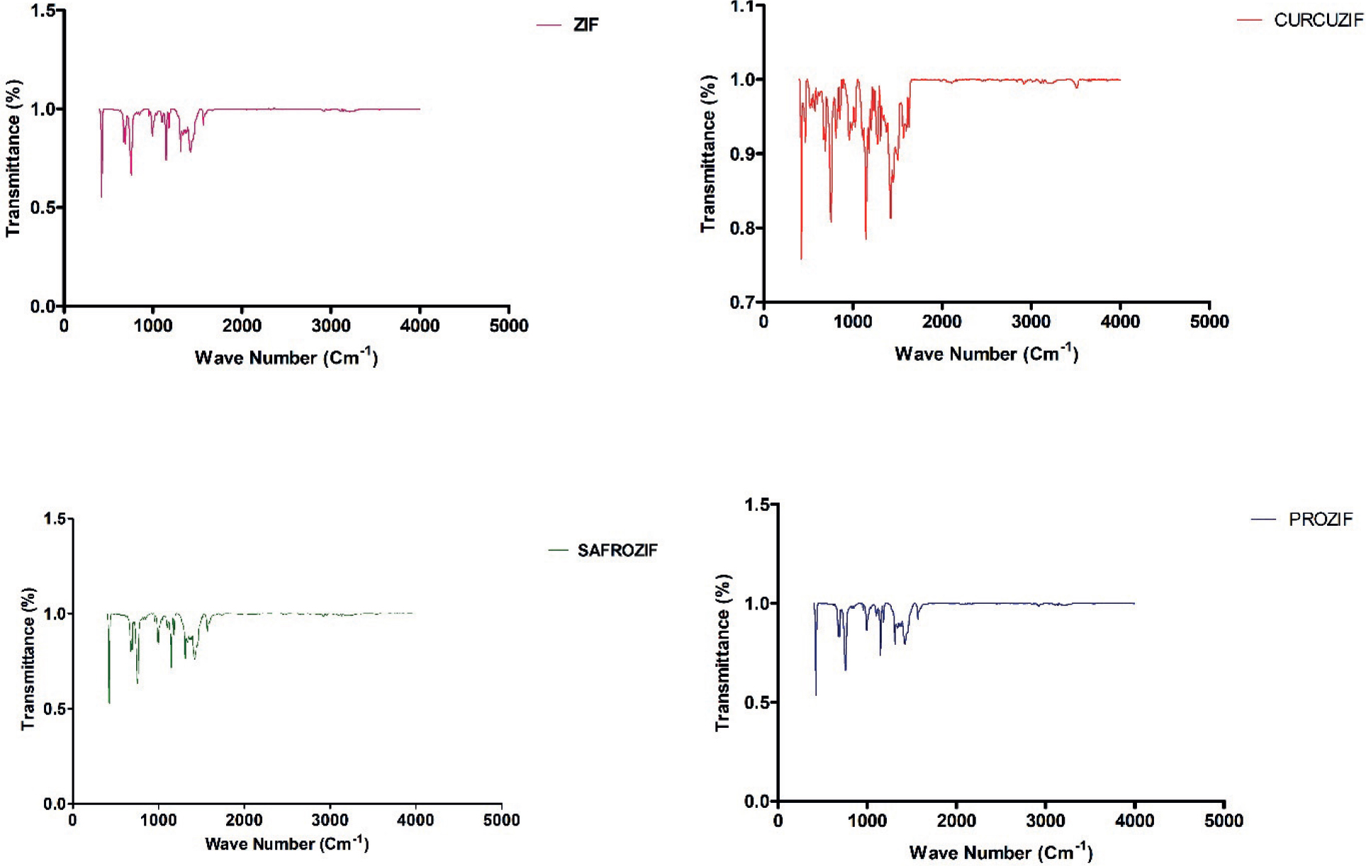
Fig. 6.
FTIR spectra of ZIF, PROZIF, SAFROZIF, and CURCUZIF nanoparticles
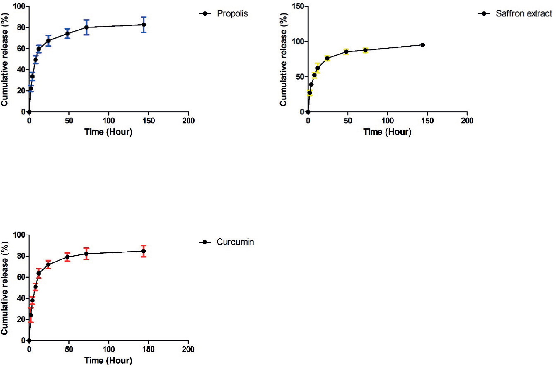
Fig. 7.
Cumulative release profile of propolis, saffron extract, and curcumin from PROZIF, SAFROZIF, and CURCUZIF during the course of 7 days in PBS