Fig. 1

Fig. 2
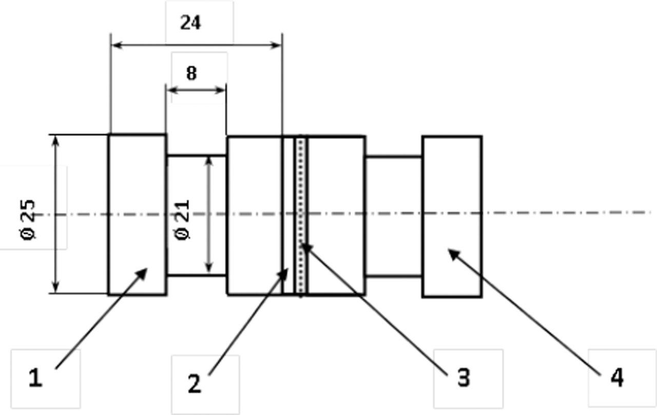
Fig. 3
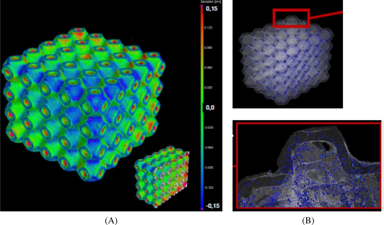
Fig. 4

Fig. 5
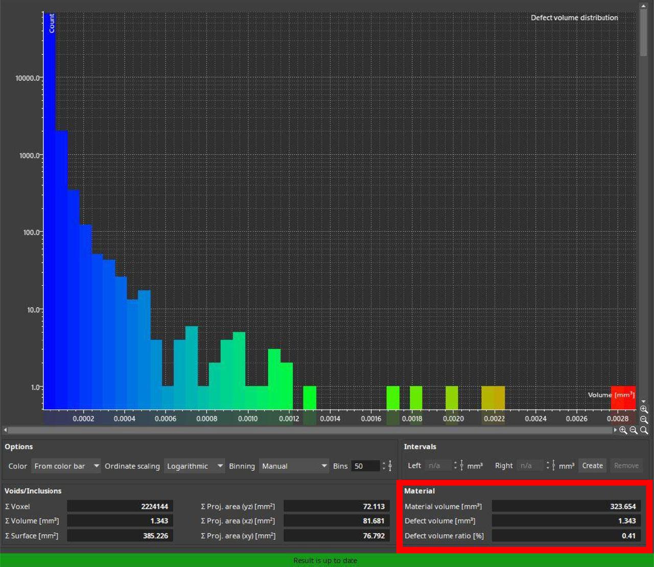
Fig. 6
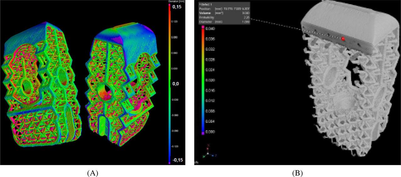
Fig. 7
Fig. 8
Fig. 9
Fig. 10

Chemical composition of Ti6Al4V titanium alloy (powder) according to ISO 5832-3
| Element | Wt.% of element |
|---|---|
| Fe | <0.3 |
| N | <0.05 |
| O | <0.2 |
| Al | 5.5–6.75 |
| C | <0.08 |
| V | 3.5–4.5 |
| H | <0.015 |
| Ti | Balance |

| Element | Wt.% of element |
|---|---|
| Fe | <0.3 |
| N | <0.05 |
| O | <0.2 |
| Al | 5.5–6.75 |
| C | <0.08 |
| V | 3.5–4.5 |
| H | <0.015 |
| Ti | Balance |
© 2023 Albina Kadyroldina, Darya Alontseva, Sergey Voinarovych, Leszek Łatka, Oleksandr Kyslytsia, Bagdat Azamatov, Aleksandr Khozhanov, Nadezhda Prokhorenkova, Almira Zhilkashinova, Svitlana Burburska, published by Wroclaw University of Science and Technology
This work is licensed under the Creative Commons Attribution-NonCommercial-NoDerivatives 4.0 License.