
Table 1
Chemical composition of the examined steel [%wt]
| C | Mn | Al | V | Nb | B | P | S | Ce | La | Nd | Fe |
|---|---|---|---|---|---|---|---|---|---|---|---|
| 0.47 | 20.1 | 3.0 | – | 0.10 | 0.003 | <0.01 | 0.003 | 0.011 | 0.004 | 0.004 | rest |
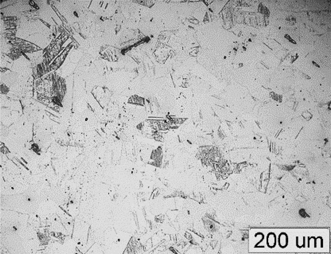
Fig. 1
Microstructure of the high-manganese MnAlNb steel after solution annealing at 1,100°C/2 h: austenite with annealing twins; the average grain diameter – 125 μm; light microscopy
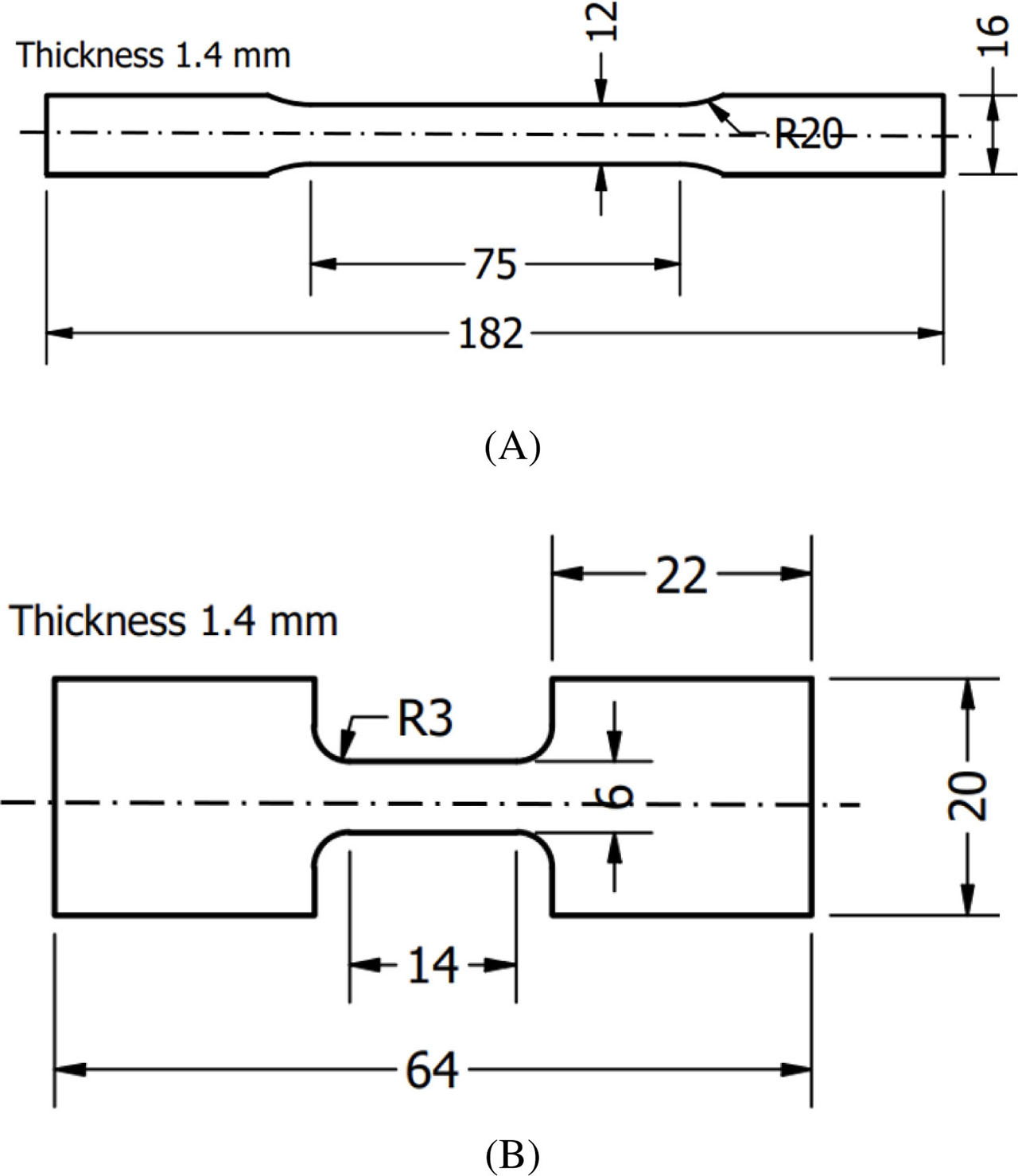
Fig. 2
Geometric features of specimens used in the tensile tests at strain rates of: (A) 0.001–0.1 s−1 and (B) 0.5 s−1
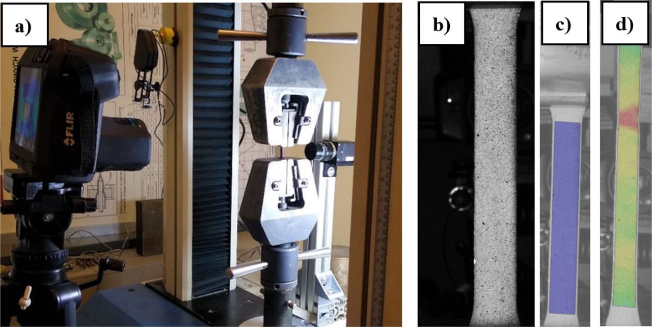
Fig. 3
The simultaneous measurement of temperature and strain during deformation: (A) experimental set-up for the mechanical testing, (B) black–white pattern for DIC measurement, (C) evaluation area in GOM Correlate, (D) example of the strain distribution image. DIC, Digital Image Correlation

Fig. 4
Regions selected for the microstructure analysis
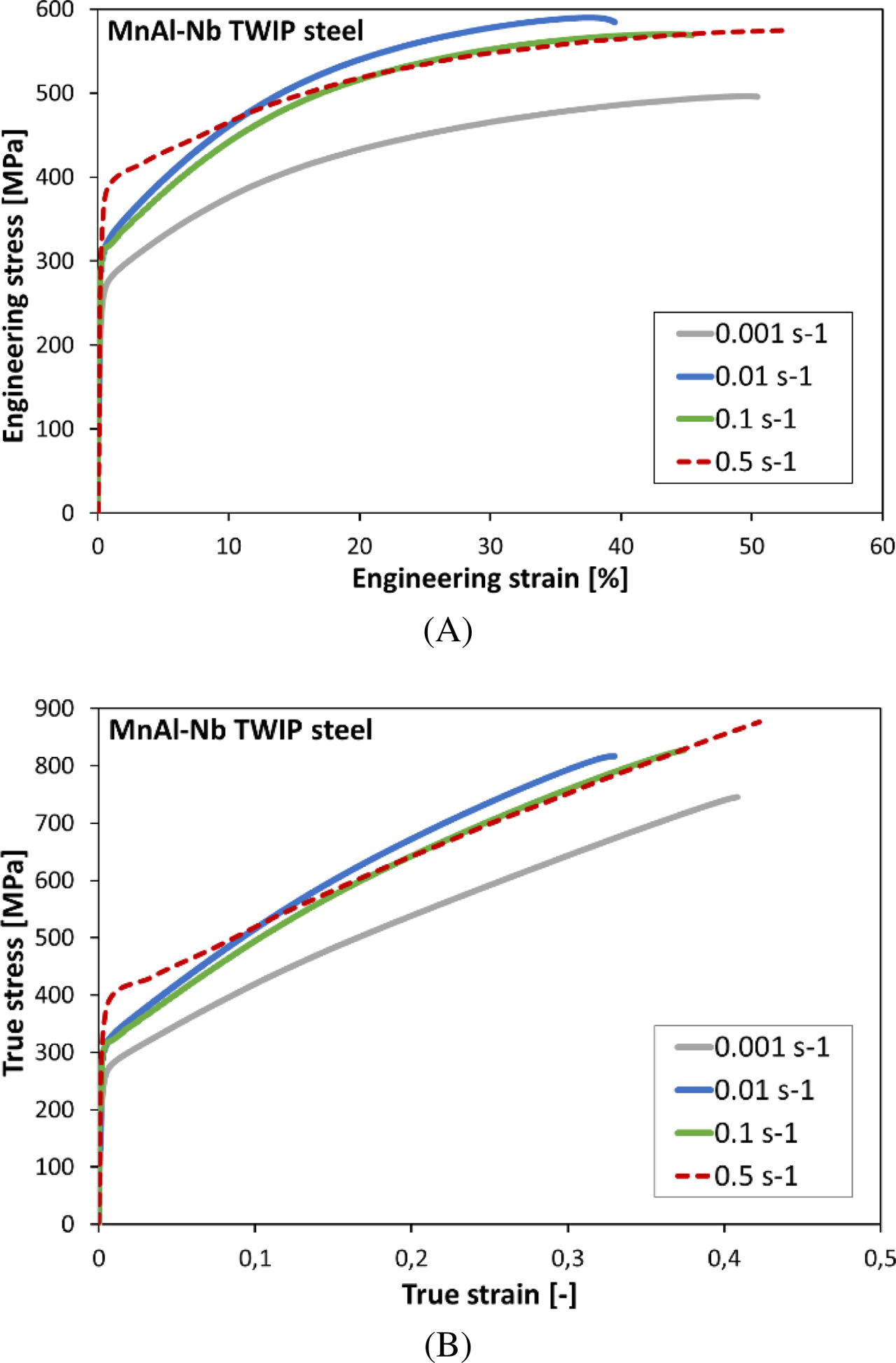
Fig. 5
The engineering (A) and true (B) stress–strain curves obtained for the MnAlNb steel from the tensile tests conducted at various strain rates
Table 2
Values of UTS, total elongation and temperature of MnAlNb steel for different strain rates
| Strain rate (s−1) | Ultimate tensile strength, UTS (MPa) | Total elongation (%) | Average temperature in the necked region (°C) |
|---|---|---|---|
| 0.001 | 496 | 50 | 42 |
| 0.01 | 590 | 39 | 66 |
| 0.1 | 570 | 45 | 79 |
| 0.5 | 575 | 52 | 104 |
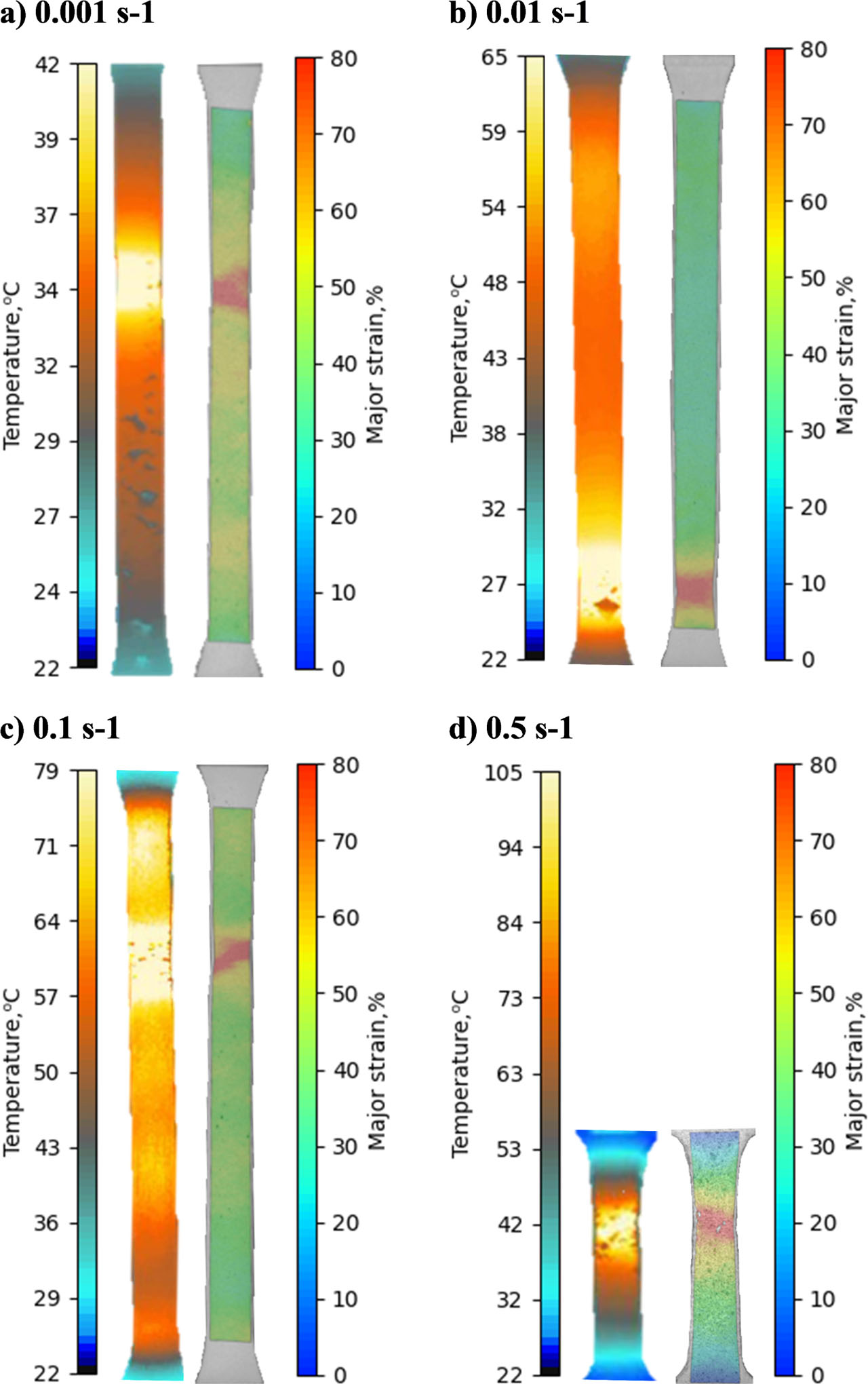
Fig. 6
Thermograms and DIC strain distribution maps at the moment just before fracture. DIC, digital image correlation
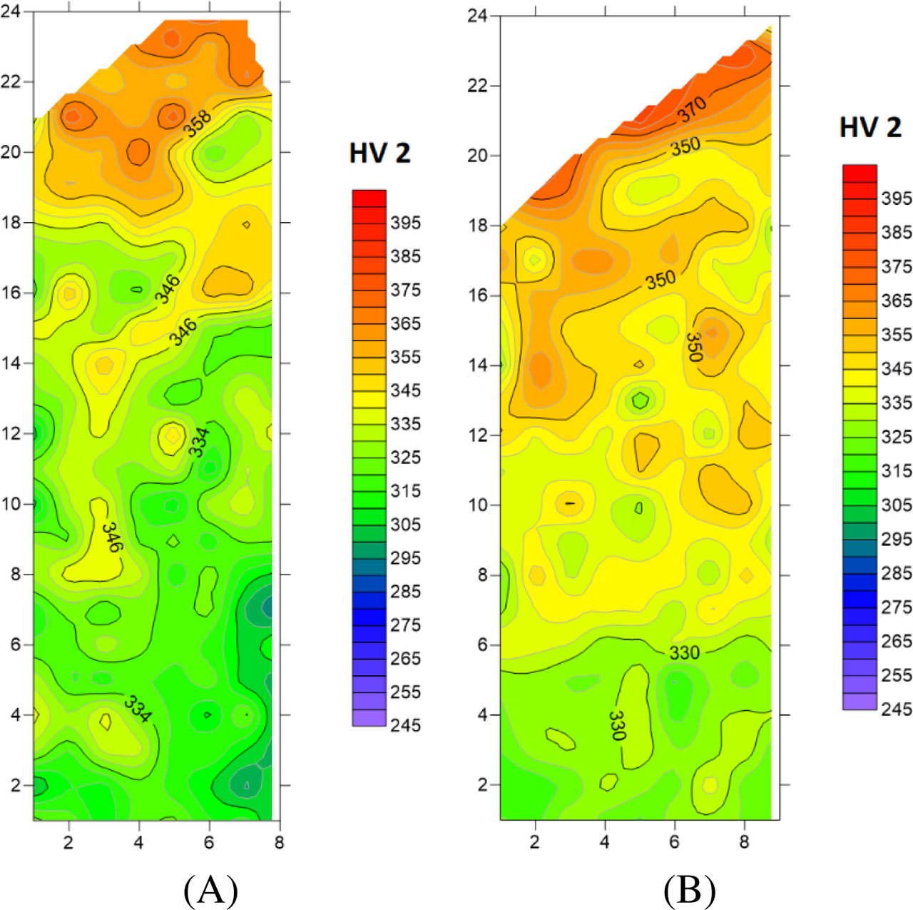
Fig. 7
The hardness distribution in one piece of the fractured specimen: (A) 0.001 s−1 and (B) 0.1 s−1
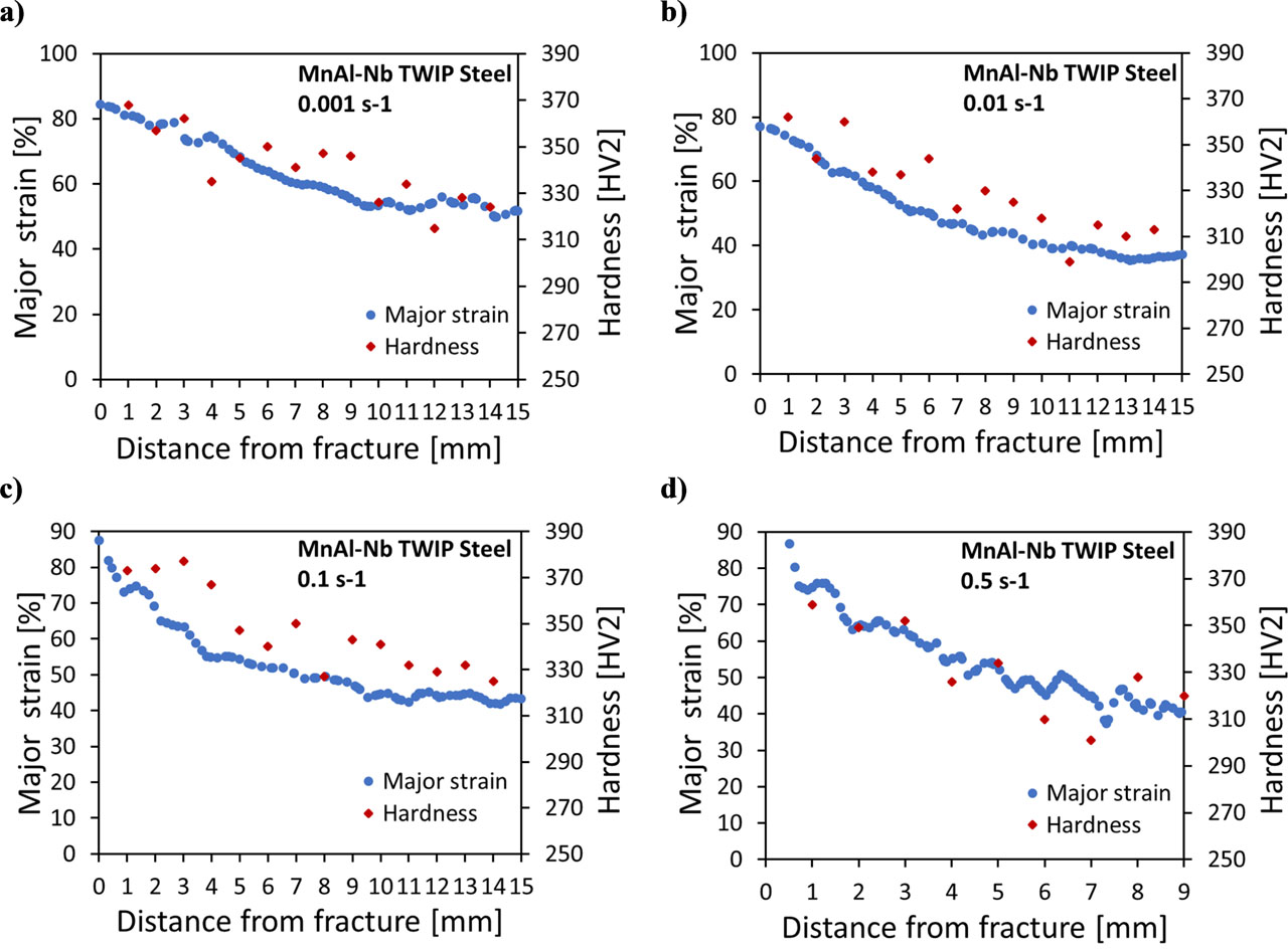
Fig. 8
Dependence of the major strain and hardness value on a distance from the fracture: a) 0.001 s−1, b) 0.01 s−1, c) 0.1 s−1, d) 0.5 s−1
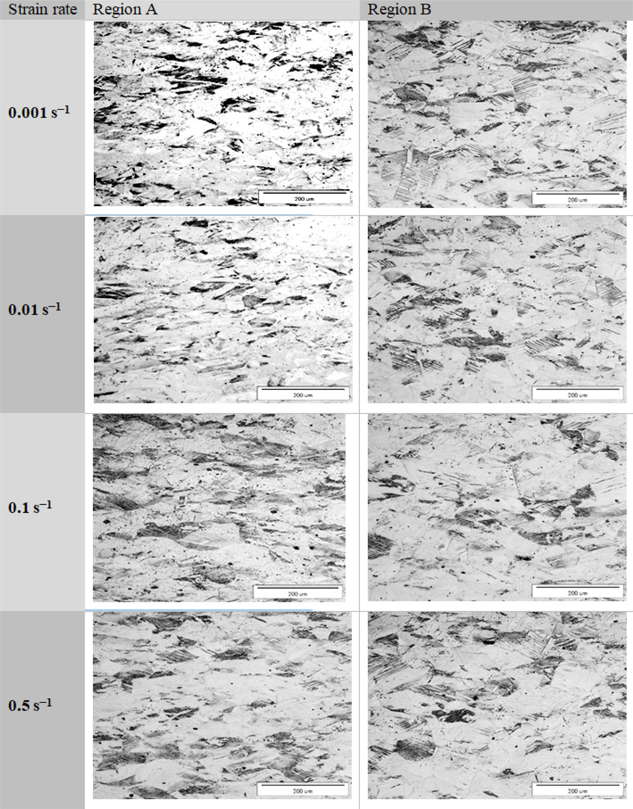
Fig. 9
Microstructure of MnAl-Nb steel deformed at various strain rates; longitudinal sections taken in regions A and regions B; light microscopy
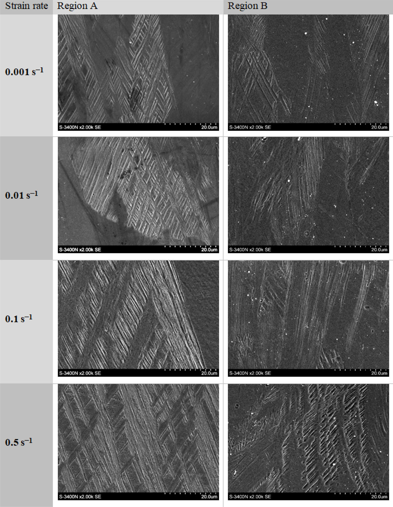
Fig. 10
Microstructure of MnAlNb steel deformed at various strain rates in the regions A and B, SEM. SEM, scanning electron microscopy