
Table 1
Hydrogel groups, reagents concentration, and synthesis process
| Groups | NIPAM (mmol) | MBA (mmol) | APS (mmol) | NMB (mmol) | Mol % | Magnetic stirring |
|---|---|---|---|---|---|---|
| H1.A* | 2.21 | 0.135 | 0.078 | 0.097 | 87.7×5.4×3.1×3.8 | Yes |
| H2.A* | 2.26 | 0.239 | 0.074 | 0.101 | 84.5×8.9×2.8×3.8 | Yes |
| H3.A* | 2.22 | 0.446 | 0.059 | 0.076 | 79.3×15.9×2.1×2.7 | Yes |
| H1.0 | 2.21 | 0.132 | 0.076 | 0.097 | 87.8×5.2×3.2×3.8 | No |
| H2.0 | 2.22 | 0.235 | 0.075 | 0.102 | 84.4×8.9×2.8×3.9 | No |
| H3.0 | 2.23 | 0.443 | 0.057 | 0.074 | 79.5×15.8×2.3×2.6 | No |
| H4.0 | 2.22 | 0.443 | 0.076 | 0.075 | 78.9×15.7×2.8×2.6 | No |
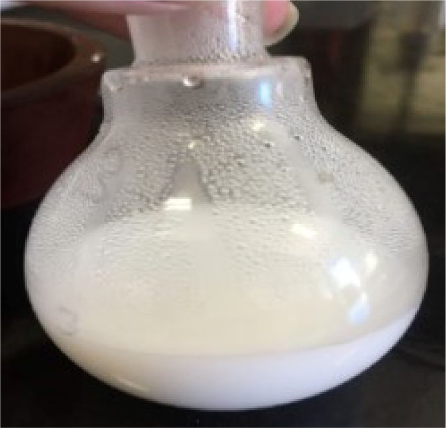
Fig. 1
Sample H1.0 after gelation
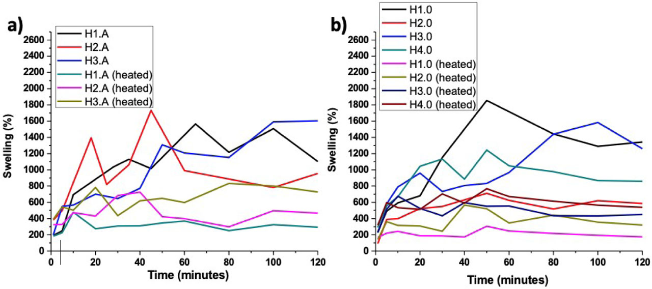
Fig. 2
Swelling ratios of the hydrogels at room temperature and 37°C in (a) stirred samples and (b) unstirred samples
Table 2
NMR 1H Spectra results
| Groups | Chemical dislocation (ppm) | Integration ratio/number of hydrogens involved |
|---|---|---|
| H1.0 | 1.22 | 6.04 / 6 |
| 1.66 | 1.89 / 2 | |
| 2.09 | 1.17 / 1 | |
| 3.98 | 1.00 / 1 | |
| H2.0 | 1.08 | 6.00 / 6 |
| 1.64 | 2.19 / 2 | |
| 1.96 | 1.17 / 1 | |
| 3.83 | 1.16 / 1 | |
| H3.0 | 1.22 | 5.93 / 6 |
| 1.78 | 1.98 / 2 | |
| 2.10 | 1.33 / 1 | |
| 3.97 | 1.00 / 1 | |
| H4.0 | 1.21 | 6.00 / 6 |
| 1.68 | 2.32 / 2 | |
| 2.11 | 1.12 / 1 | |
| 3.98 | 0.85 / 1 |
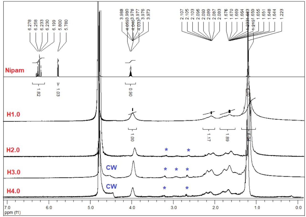
Fig. 3
1H NMR spectra of Nipam, H1.0, H2.0, H3.0, and H4.0 (500 MHz, D2O).
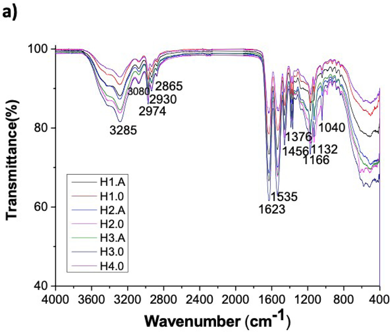
Fig. 4
FTIR-ATR spectra of all hydrogel samples
Table 3
Semiquantitative chemical composition (wt%) of the samples using EDS analysis
| Groups | Carbon | Oxygen | Nitrogen | Platinum |
|---|---|---|---|---|
| H1.A | 77.70 | 8.09 | 12.24 | 1.97 |
| H1.0 | 72.51 | 4.86 | 3.93 | 18.70 |
| H2.A | 74.76 | 4.00 | 0.05 | 21.20 |
| H2.0 | 80.19 | 10.90 | 5.42 | 3.48 |
| H3.A | 75.76 | 6.02 | 0 | 18.21 |
| H3.0 | 73.65 | 7.03 | 0 | 19.32 |
| H4.0 | 65.41 | 7.81 | 0 | 26.78 |
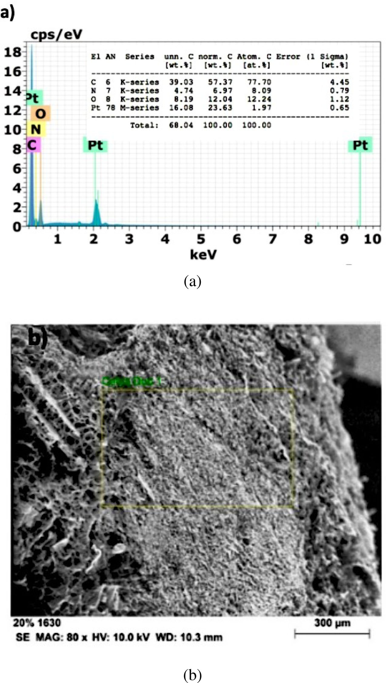
Fig. 5
(a) EDS spectrum of hydrogel H1.A and (b) image of the region analyzed by EDS
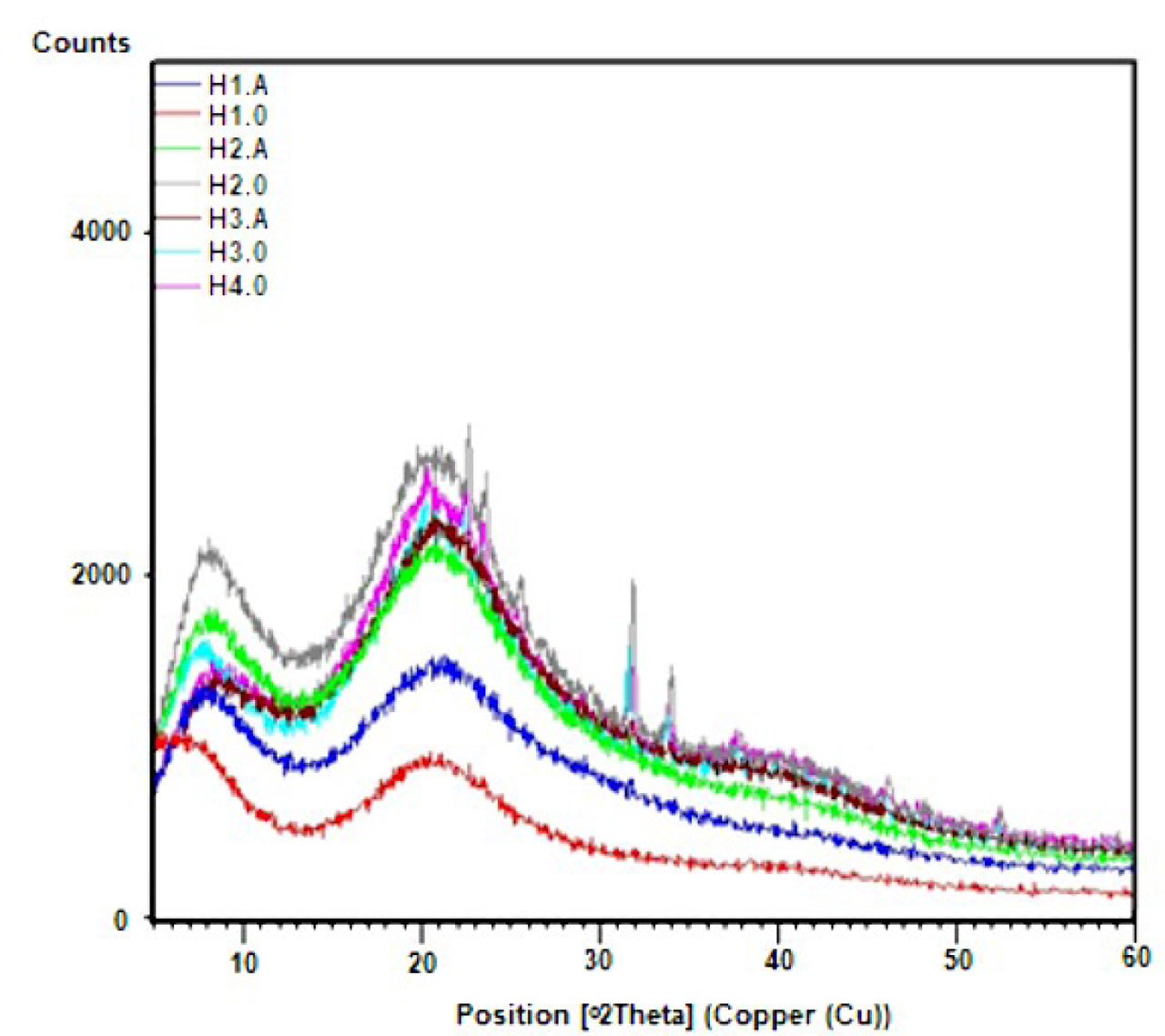
Fig. 6
XRD patterns of the hydrogels.
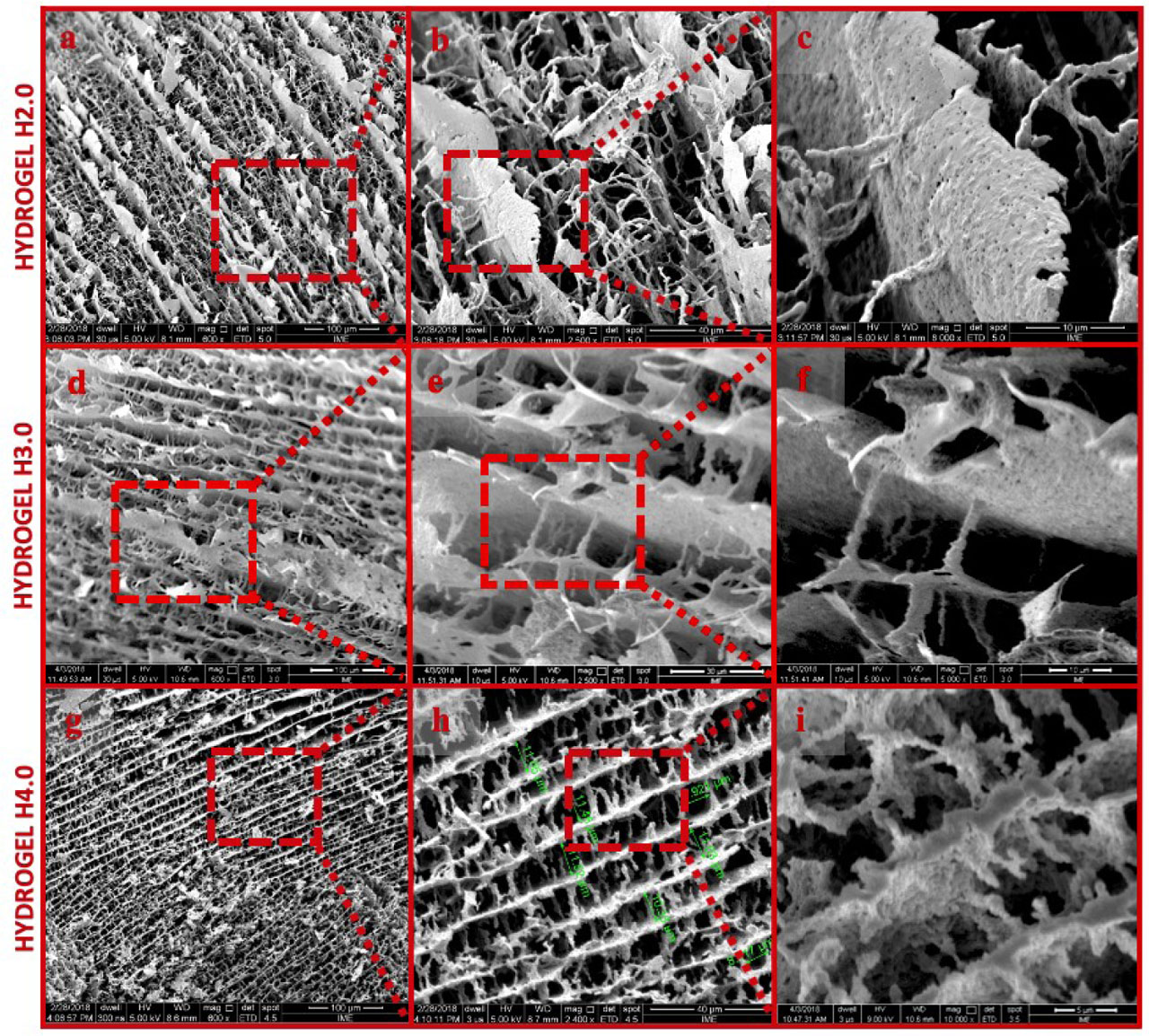
Fig. 7
SEM morphologies of the samples of H2.0, H3.0, and H4.0. Magnification of the sample of H2.0: (a) 600 ×; (b) 2,500 ×; (c) 8,000 ×. Magnification of the sample of H3.0: (d) 600 ×; (e) 2,500 ×; (f) 5,000 ×. Magnification of the sample of H4.0: (g) 600 ×; (h) 2,400 ×; (i) 10,000 ×
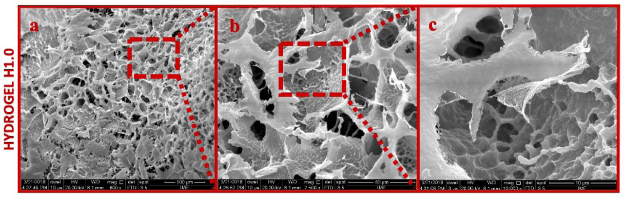
Fig. 8
Morphologies of the sample of H1.0. Magnifications: (a) 600 ×; (b) 2,500 ×; (c) 10,000 ×.
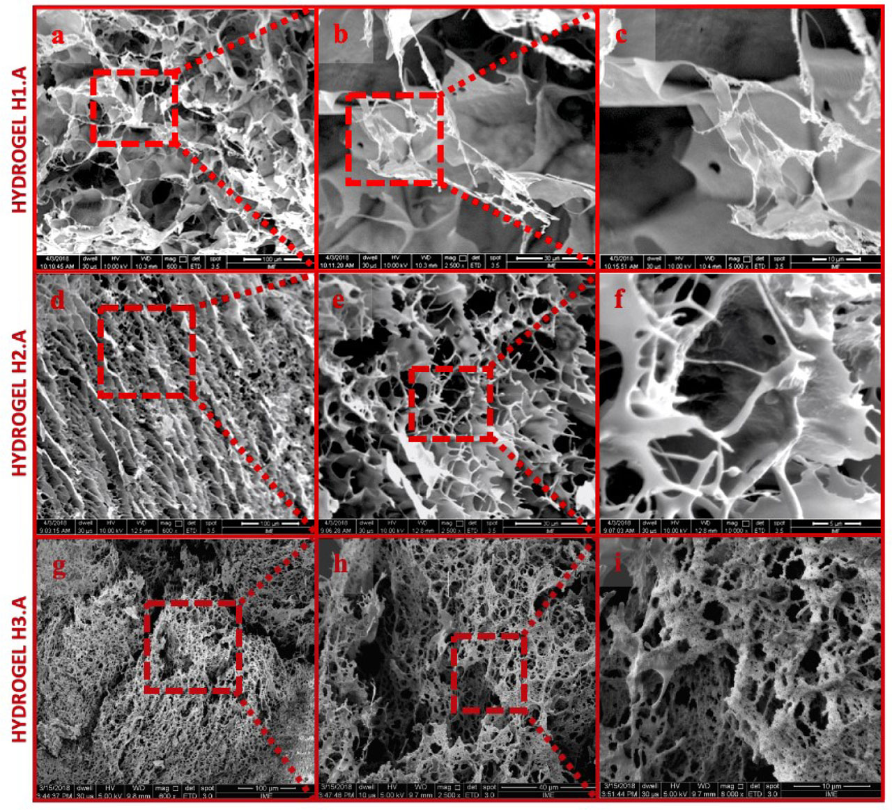
Fig. 9
Morphologies of the samples of H1.A, H2.A, and H3.A. Magnification of the sample of H1.A: (a) 600 ×; (b) 2,500 ×; (c) 5,000 ×. Magnification of the sample of H2.A: (d) 600 ×; (e) 2,500 ×; (f) 10,000 ×. Magnification of the sample of H3.A: (g) 600 ×; (h) 2,500 ×; (i) 8,000 ×
Table 4
Thermogravimetric data of the hydrogels
| Sample | 1st event | 2nd event | 3rd event | |||||
|---|---|---|---|---|---|---|---|---|
| Tpeak (°C) | Residue | Tmax (°C) | Tpeak (°C) | Residue | Tmax (°C) | Tpeak (°C) | Residue | |
| H1.A | 74 | 4% | 265 | 291 | 20% | 349 | 381 | 61% |
| H1.0 | 54 | 5% | 262 | 286 | 18% | 360 | 397 | 71% |
| H2.A | 104 | 7% | 342 | 389 | 67% | - | - | - |
| H2.0 | 56 | 6% | 271 | 307 | 20% | 378 | 412 | 69% |
| H3.A | 111 | 6% | 346 | 381 | 62% | - | - | - |
| H3.0 | 61 | 4% | 259 | 279 | 21% | 350 | 383 | 66% |
| H4.0 | 65 | 4% | 260 | 281 | 23% | 351 | 376 | 65% |
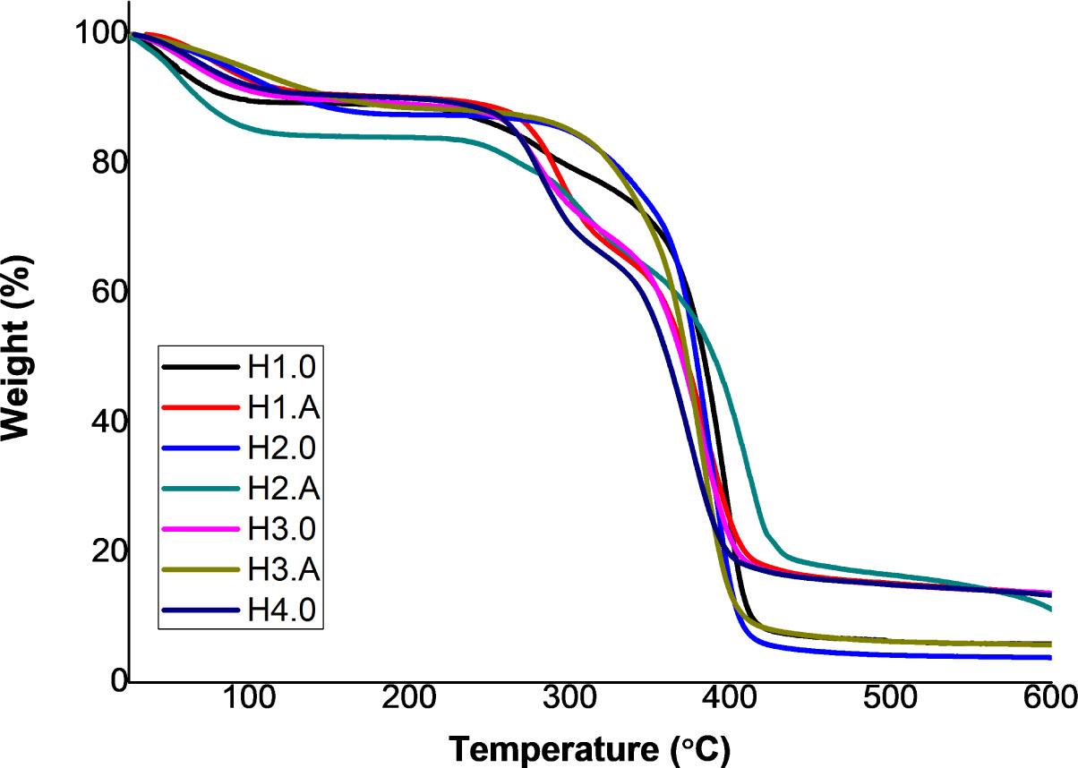
Fig. 10
PNIPAAm hydrogels’ thermogravimetric analysis (TGA)