
Introduction
The Pasteurella genus belongs to the Pasteurellaceae family, which comprises a large and diverse group of Gram-negative microorganisms (8). Pasteurellaceae Pohl 1981 is classified within the Proteobacteria phylum, Gammaproteobacteria class and Pasteurellales order, and currently consists of 17 genera and over 60 species of microorganisms. These have been isolated from animals and humans (11). Comparative genomic and phylogenetic analyses of Pasteurellaceae have revealed that many members of this highly diverse family have been insufficiently classified. Indeed, numerous species within the Pasteurellaceae family have already been reclassified, including Mannheimia haemolytica, Bibersteinia trehalosi, Actinobacillus ureae, Avibacterium gallinarum, Avibacterium volantium, Avibacterium avium and Gallibacterium anatis (8, 11). Advances in microbiological diagnostics mean that the taxonomy of the Pasteurellaceae family continues to undergo significant changes and revisions.
Pasteurella multocida is a commensal or opportunistic pathogen found in the upper respiratory tract of most domestic and wild animals. This bacterium has been isolated from chickens, turkeys, wild birds, cattle and American bison, pigs, rabbits, dogs, domestic cats and large wild felids such as tigers, leopards, cougars and lions, as well as from chimpanzees, marine mammals including seals, sea lions and walruses, and even from Komodo dragons (15, 31). It was first described by Italian botanist Vittore Trevisan in 1887. He proposed the genus name Pasteurella in honour of the famous French microbiologist and chemist Louis Pasteur. The researcher first isolated the bacterium in 1880 as the causative agent of fowl cholera. He conducted several experiments infecting chickens to understand the mechanism of transmission of the microorganism and assess its pathogenicity (35). In turn, Mutters et al. (24) reclassified the Pasteurella genus based on DNA–DNA hybridisation studies. They demonstrated that Pasteurella sensu stricto comprises at least eleven species, including P. multocida, P. dagmatis, P. canis, P. stomatitis and Pasteurella species B. Within P. multocida, three subspecies have been identified: P. multocida subsp. multocida, P. multocida subsp. septica and P. multocida subsp. gallicida, which show 84% to 100% DNA relatedness, despite their predilection for different hosts and the diversity of the clinical symptoms they cause. Research by Petersen et al. (29), based on a comparison of the 16S rRNA and atpD gene sequences, confirmed the genetic homogeneity of P. multocida. Similarly, research by Kuhnert et al. (18) demonstrated that the subspecies of P. multocida, corresponding to distinct phenotypic variants, exhibit at least 98.5% similarity in their 16S rRNA sequences.
In 2002, Capitini et al. (3) isolated a Pasteurella strain from a girl who had been bitten by a tiger. Based on the phenotypic characteristics of this strain and comparisons of 16S rRNA gene sequences, it was regarded as a putative new subspecies, and the name P. multocida subsp. tigris was proposed. This strain possessed characteristics typical of P. multocida but did not ferment sucrose and mannitol (24). However, subsequent studies by Christensen et al. (9), including DNA–DNA hybridisation, recommended this strain be reclassified as a new taxon, Bisgaard taxon 45. Although it is genotypically related to P. multocida, its polyamine pattern differs from those of all P. multocida subspecies. Separate taxa within the Pasteurellaceae family, such as Bisgaard taxon 45, Bisgaard taxon 46 and Pasteurella species B, include strains the final systematic positions of which have not yet been conclusively determined.
The aim of this study was to characterise and classify P. multocida strains isolated from rabbits in Poland. The biochemical properties and serotype classification based on the identification of capsular and somatic antigens of domestic P. multocida strains, were determined.
Material and Methods
Bacterial strains
The study used 115 field strains isolated between 1999 and 2020 in Poland. The strains came from rabbits from both large-scale farms and individual small farms. The large-scale farms were located in Chorzelów in the Podkarpackie voivodeship, Gdańsk in the Pomorskie voivodeship, Małogoszcz in the Świętokrzyskie voivodeship and Zduny in the Wielkopolskie voivodeship. The individual small farms came from various areas of Poland. The first group consisted of strains isolated from nasal swabs of rabbits with respiratory symptoms or being asymptomatic carriers. This group comprised 87 strains, of which 73 were isolated from rabbits with symptoms of rhinitis, and the remaining 14 came from clinically healthy animals. The second group consisted of 28 strains from dead rabbits. Twenty-seven strains were isolated from internal organs: 26 from the lungs and one strain from the heart, while one strain came from an abscess on the skin. In addition, reference strains of P. multocida were used as controls: P8 and P1059 of P. multocida type A and Kobe 6 and P27 of type D obtained from Dr Namioka of the National Institute of Animal Health, Tokyo, Japan, B850 and P932 of type B from the Institut d’Élevage et de Médecine Vétérinaire des Pays Tropicaux, Paris, France and P4218 and P3695 of type F from Dr Rimler of the National Animal Disease Center, Ames, IA, USA. The reference strains X73, M1404, P1059, P1662, P1702, P2192, P1997, P1581, P2095, P2100, P903, P1573, P1591, P2225, P2237 and P2723 originated from the National Animal Disease Center, Ames, IA, USA.
Morphological and microscopic examination
The strains were cultured on 5% horse blood agar, MacConkey agar, dextrose-starch agar (DSA) and nutrient broth. After incubation for 24 h at 37°C, the appearance and size of colonies on solid media were assessed, as well as the turbidity and sediment formation in the liquid medium. Microscopic examination was then performed on preparations stained using the Gram method, modified by Kopeloff. Gram-negative rods were subjected to further analysis.
Catalase test
A colony taken from a bacterial culture grown on solid medium was suspended in a drop of 3% hydrogen peroxide solution on a microscope slide. The release of gas bubbles indicated a positive reaction.
Cytochrome oxidase activity test
An OXITEST commercial oxidase test (Erba Lachema, Brno, Czech Republic) was used according to the manufacturer’s instructions. A colour change of the filter paper strip to dark blue indicated the presence of oxidase-positive bacteria.
Biochemical profile assessment
The biochemical profile was determined using API 20E and ID 32E tests (bioMérieux, Marcy-l’Étoile, France), performed in accordance with the manufacturer’s instructions. The strains were also inoculated into liquid media for fermentation of dulcitol, trehalose, xylose and maltose. Incubation was carried out at 37°C for 6 d. A colour change from blue to yellow indicated a positive reaction resulting from sugar fermentation.
Determination of capsular type by the indirect haemagglutination test (IHA)
The indirect haemagglutination test was performed by the micromethod according to Carter (4, 5). Each strain was tested with reference anticapsular sera of types A, B, D and F, produced in previous years at the Department of Bacteriology and Bacterial Animal Diseases, National Veterinary Research Institute in Puławy, Poland. Serum dilutions ranging from 1:10 to 1:2,560 were used in the assays. The microplates were left for 2 h at 21°C. after which the first reading was taken. The plates were then incubated for a further 18 h at 5°C, and the second reading was performed. A positive result was indicated by the presence of haemagglutination at the bottom of the well.
Determination of the somatic antigens by the agar gel immunodiffusion test (AGID)
The assay was performed following the method of Heddleston et al. (16), with modifications introduced by other authors (32, 36). Sera positive for 16 Heddleston reference strains obtained in previous years by the Department of Bacteriology and Animal Bacterial Diseases, National Veterinary Research Institute in Puławy, Poland, were used for the tests. The specificity of all sera was verified against antigens representing the 16 Heddleston serotypes. The plates were placed in a humid chamber and incubated for 24 h at 37°C and for an additional 24 h at 21°C. The results were read after 48 h. A positive reaction was indicated by the formation of a precipitation line.
Results
For all tested strains grown on 5% horse blood agar, growth was observed in the form of smooth, small, semitransparent greyish colonies with a diameter of 1–3 mm and regular edges (Fig. 1A), as well as larger, mucoid colonies measuring 3–4 mm in diameter (Fig. 1B). No haemolysis was detected in any of the strains. When cultured in nutrient broth, the strains grew as a homogenous turbid suspension. On DSA medium, the strains formed smooth, yellow, opalescent colonies measuring 2–3 mm in diameter, while no growth was observed on MacConkey agar. In microscopic preparations stained by the Gram method, all the strains appeared as short, slender, Gram-negative rods. All bacterial strains tested were shown to produce both catalase and oxidase. The results of the biochemical profile assessment using the API 20E and ID 32E tests are presented in Tables 1 and 2. Based on the results of both tests, the strains isolated from rabbits were identified as P. multocida.
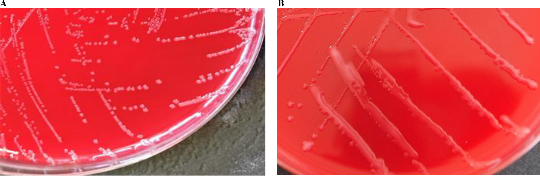
Fig. 1A and B
Growth of Pasteurella multocida colonies on 5% horse blood agar
Table 1.
Biochemical profile assessment of Pasteurella multocida strains using the API 20E
| Reaction/enzyme | Number of strains showing positive reactions (%) | ||
|---|---|---|---|
| P. multocida subsp. multocida | P. multocida subsp. multocida ornithine-negative | P. multocida subsp. septica | |
| β-galactosidase | 0 | 0 | 0 |
| Arginine dihydrolase | 0 | 0 | 0 |
| Lysine decarboxylase | 0 | 0 | 0 |
| Ornithine decarboxylase | 20(100) | 0 | 3 (100) |
| Citrate utilisation | 0 | 0 | 0 |
| Hydrogen sulphide production | 0 | 0 | 0 |
| Urease | 0 | 0 | 0 |
| Tryptophan deaminase | 0 | 0 | 0 |
| Indole production | 17(85) | 92 (100) | 1 (33) |
| Acetoin production | 0 | 0 | 0 |
| Gelatinase | 0 | 0 | 0 |
| D-glucose | 20 (100) | 92 (100) | 3 (100) |
| D-mannitol | 20 (100) | 92 (100) | 3 (100) |
| Inositol | 0 | 0 | 0 |
| D-sorbitol | 20 (100) | 92 (100) | 0 |
| L-rhamnose | 0 | 0 | 0 |
| D-saccharose | 20 (100) | 92 (100) | 3 (100) |
| D-melibiose | 0 | 0 | 0 |
| Amygdalin | 0 | 0 | 0 |
| L-arabinose | 0 | 0 | 0 |
| Cytochrome oxidase | 20 (100) | 92 (100) | 3 (100) |
| Nitrate reduction | 20 (100) | 92 (100) | 3 (100) |
| Total | 20 | 92 | 3 |
Table 2.
Biochemical profile assessment of Pasteurella multocida strains using the ID 32E
| Reaction/enzyme | Number of strains showing positive reactions (%) | ||
|---|---|---|---|
| P. multocida subsp. multocida | P. multocida subsp. multocida ornithine-negative | P. multocida subsp. septica | |
| Ornithine decarboxylase | 20(100) | 0 | 3 (100) |
| Arginine dihydrolase | 0 | 0 | 0 |
| Lysine decarboxylase | 0 | 0 | 0 |
| Urease | 0 | 0 | 0 |
| L-arabitol | 0 | 0 | 0 |
| Galacturonate | 0 | 0 | 0 |
| 5-ketogluconate | 0 | 0 | 0 |
| Lipase | 0 | 0 | 0 |
| Phenol red | 0 | 0 | 0 |
| β-glucosidase | 0 | 0 | 0 |
| Mannitol | 20 (100) | 92(100) | 3 (100) |
| Maltose | 0 | 0 | 0 |
| Adonitol | 0 | 0 | 0 |
| Palatinose | 0 | 0 | 0 |
| β-glucuronidase | 0 | 0 | 0 |
| Malonate | 0 | 0 | 0 |
| Indole production | 17 (85) | 92 (100) | 1 (33) |
| N-acetyl-β-glucosaminidase | 0 | 0 | 0 |
| β-galactosidase | 0 | 0 | 0 |
| Glucose | 20 (100) | 92 (100) | 3 (100) |
| Sucrose | 20 (100) | 92 (100) | 3 (100) |
| L-arabinose | 0 | 0 | 0 |
| D-arabitol | 0 | 0 | 0 |
| α-glucosidase | 0 | 0 | 0 |
| α-galactosidase | 0 | 0 | 0 |
| Trehalose | 7 (35) | 0 | 0 |
| Rhamnose | 0 | 0 | 0 |
| Inositol | 0 | 0 | 0 |
| Cellobiose | 0 | 0 | 0 |
| Sorbitol | 20 (100) | 92 (100) | 0 |
| α-maltosidase | 0 | 0 | 0 |
| L-aspartic acid arylamidase | 0 | 0 | 0 |
| Total | 20 | 92 | 3 |
The API 20E test showed that all P. multocida strains produced cytochrome oxidase; fermented glucose, mannitol and sucrose; and reduced nitrates to nitrites. Ornithine decarboxylase was produced by 20% of the strains, while 95.6% produced indole and 97.4% fermented sorbitol. None of the strains utilised citrate; produced galactosidase, arginine dihydrolase, lysine decarboxylase, hydrogen sulphide, urease, tryptophan deaminase, acetoin or gelatinase; nor fermented inositol, rhamnose, melibiose, amygdalin or arabinose (Table 1).
The ID 32E test results confirmed the production of ornithine decarboxylase and indole, as well as sorbitol fermentation, in the same proportion of strains. All P. multocida strains fermented mannitol, glucose and sucrose. Trehalose was fermented by 6.1% of the tested strains. It was found that the strains did not produce arginine dihydrolase, lysine decarboxylase, urease, lipase, β-glucosidase, β-glucuronidase, malonate, N-acetyl-β-glucosaminidase, β-galactosidase, α-glucosidase, α-maltosidase or L-aspartic acid arylamidase. All strains also failed to ferment D-arabitol, galacturonate, 5-ketogluconate, phenol red, adonitol, palatinose, L-arabinose, rhamnose and inositol (Table 2).
The ID 32E test results confirmed the production of ornithine decarboxylase and indole, as well as sorbitol fermentation, in the same proportion of strains. All P. multocida strains fermented mannitol, glucose and sucrose. Trehalose was fermented by 6.1% of the tested strains. It was found that the strains did not produce arginine dihydrolase, lysine decarboxylase, urease, lipase, β-glucosidase, β-glucuronidase, malonate, N-acetyl-β-glucosaminidase, β-galactosidase, α-glucosidase, α-maltosidase or L-aspartic acid arylamidase. All strains also failed to ferment D-arabitol, galacturonate, 5-ketogluconate, phenol red, adonitol, palatinose, L-arabinose, rhamnose and inositol (Table 2).
The results of sugar fermentation tests showed that none of the strains fermented dulcitol or maltose. Trehalose was fermented by 6.1% of all strains tested, and xylose by 97.4%. The results of the sugar fermentation tests are presented in Table 3.
Table 3.
Sugar fermentation of Pasteurella multocida strains isolated from rabbits
| Sugar | Number of strains showing positive reactions (%) | ||
|---|---|---|---|
| P. multocida subsp. multocida | P. multocida subsp. multocida ornithinenegative | P. multocida subsp. septica | |
| Dulcitol | 0 | 0 | 0 |
| Trehalose | 7 (35) | 0 | 0 |
| Xylose | 17(85) | 92 (100) | 3 (100) |
| Maltose | 0 | 0 | 0 |
| Total | 20 | 92 | 3 |
All strains isolated from rabbits were classified according to the criteria of Mutters et al. (24, 25) and Bisgaard et al. (1). A proportion equal to 17.4% of the isolates was identified as P. multocida subsp. multocida, and 80% as ornithine decarboxylase negative P. multocida subsp. multocida. The remaining 2.6% of the strains were classified as P. multocida subsp. septica.
The study showed that 20% of all strains produced ornithine decarboxylase. These included all strains which were P. multocida subsp. multocida and P. multocida subsp. septica. Indole production was observed in 100% of ornithine decarboxylase negative P. multocida subsp. multocida strains, 85% of P. multocida subsp. multocida strains and 33% of P. multocida subsp. septica strains. All strains of P. multocida subsp. multocida and ornithine decarboxylase negative P. multocida subsp. multocida fermented sorbitol, whereas none of the P. multocida subsp. septica strains did. Among the 20 P. multocida subsp. multocida strains, 35% fermented trehalose and 85% fermented xylose. In contrast, all ornithine decarboxylase negative P. multocida subsp. multocida and P. multocida subsp. septica strains failed to ferment trehalose but did ferment xylose.
Among the strains obtained from clinically healthy rabbits, 7.1% were P. multocida subsp. multocida, while 92.9% were ornithine decarboxylase negative P. multocida subsp. multocida. The strains from rabbits showing symptoms of rhinitis were P. multocida subsp. multocida in a 16.4% proportion, and 83.6% were ornithine decarboxylase negative P. multocida subsp. multocida. Among the strains obtained from dead animals, 25% were identified as P. multocida subsp. multocida, 64.3% as ornithine decarboxylase negative P. multocida subsp. multocida and 10.7% as P. multocida subsp. septica.
In the next stage, the capsular antigens were determined using IHA (4, 5). It was found that the capsular antigen of type A was present in 87.8% of rabbits, type D in 8.7% and type F in 3.5% of animals (Table 4). Strains possessing capsule of type A resolved all biochemical groups. Of them, 14.8% were P. multocida subsp. multocida, 84.2% ornithine decarboxylase negative P. multocida subsp. multocida and the remaining 1% were P. multocida subsp. septica. In the group of strains with capsular type D, 30% were classified as P. multocida subsp. multocida and 70% as ornithine decarboxylase negative P. multocida subsp. multocida. Among the strains with capsular type F, 50% were identified as P. multocida subsp. multocida, and 50% as P. multocida subsp. septica.
Table 4.
Distribution of Pasteurella multocida capsular antigens
| Somatic antigen number of strains (%) | Number of strains (%) | |||
|---|---|---|---|---|
| 3 | 12 | |||
| Capsular antigen | A | 38 (37.6) | 63 (62.4) | 101 (87.8) |
| D | 2 (20.0) | 8 (80.0) | 10 (8.7) | |
| F | 1 (25.0) | 3 (75.0) | 4 (3.5) | |
| Total | 41 | 74 | 115 | |
In the next stage, the capsular antigens were determined using IHA (4, 5). It was found that the capsular antigen of type A was present in 87.8% of rabbits, type D in 8.7% and type F in 3.5% of animals (Table 4). Strains possessing capsule of type A resolved all biochemical groups. Of them, 14.8% were P. multocida subsp. multocida, 84.2% ornithine decarboxylase negative P. multocida subsp. multocida and the remaining 1% were P. multocida subsp. septica. In the group of strains with capsular type D, 30% were classified as P. multocida subsp. multocida and 70% as ornithine decarboxylase negative P. multocida subsp. multocida. Among the strains with capsular type F, 50% were identified as P. multocida subsp. multocida, and 50% as P. multocida subsp. septica.
Strains isolated from both clinically healthy rabbits and those with symptoms of rhinitis belonged mainly to capsular type A. In the group of animals with symptoms of rhinitis, only 4.1% of strains had capsular antigen D. Capsule of type A also dominated among strains isolated from dead rabbits (60.7%), while type D comprised 25.0% and type F 14.3%.
Next, somatic antigens were detected using AGID (16). It was found that 35.7% of the tested strains were serotype 3, while 64.3% were classified as serotype 12 (Table 5). In addition to the clearly marked main band resulting for every one of the 41 serotype 3 strains, 28 strains showed weaker precipitation lines with sera specific for serotypes 1, 2, 5, 10 and 12. In single strains, a weak band with sera of serotypes 11 and 16 was also present. In turn, 23 serotype 12 strains showed only a clearly marked main precipitation band, while the remaining 51 strains of this serotype also showed faint precipitation lines with sera specific for one or more of serotypes 1, 2, 3, 4, 5, 6, 8, 10, 11, 14 and 16.
It emerged that 37.6% of strains in capsular antigen type A were Heddleston somatic antigen serotype 3, and 62.4% were antigen serotype 12. Among the strains with capsular antigen type D, 20% belonged to serotype 3 and 80% to serotype 12. In capsular type F, 25% of the strains had somatic antigen 3 and 75% had antigen 12 (Table 4).
Table 5.
Distribution of Pasteurella multocida somatic antigens
| Capsular antigen numer of strains (%) | Number of strains (%) | ||||
|---|---|---|---|---|---|
| A | D | F | |||
| Somatic antigen | 3 | 38 (92.7) | 2 (4.9) | 1 (2.4) | 41 (35.7) |
| 12 | 63 (85.1) | 8 (10.8) | 3 (4.1) | 74 (64.3) | |
| Total | 101 | 10 | 4 | 115 | |
The vast majority of P. multocida strains (92.7%) of serotype 3 possessed capsular antigen type A, while 4.9% had antigen type D and 2.4% F. A large majority (85.1%) of serotype 12 strains belonged to capsular type A, while 10.8% type D and 4.1% had type F (Table 5).
All strains isolated from clinically healthy animals belonged to serotype 12. Among strains from rabbits with symptoms of rhinitis, 63% belonged to serotype 12 and 37% to serotype 3. In turn, strains obtained from dead animals resolved to each serotype in 50% proportions.
Discussion
Pasteurella multocida is a Gram-negative bacterium found in many different species of domestic and wild animals, including primates, and humans. These microorganisms can be present as a commensal on mucous membranes, mainly in the respiratory system of vertebrates, or occur as primary or opportunistic pathogens (34).
The first stage of laboratory diagnosis of P. multocida involves classic bacteriological examinations. Preliminary isolation of bacterial cultures for further identification is made possible by the characteristic appearance of colonies on solid media. In our study, the characteristic colony morphology of P. multocida was noted. Large, mucoid colonies or small, round, shiny colonies with a greyish tone and regular edges were found on blood agar. Classical diagnostics relying on phenotypic characteristics, such as colony morphology, the ability to fermentation of glucose, mannitol, sucrose, trehalose, dulcitol, and sorbitol, as well as the production of indole and ornithine decarboxylase, are usually sufficient for the identification of P. multocida (13, 14). Determining biochemical properties makes it possible to establish certain biochemical profiles of bacterial strains within a given species. This method is used as a preliminary step for further classification of strains based on other techniques (33). However, as reported by Bisgaard et al. (1) and Christensen et al. (8), P. multocida may exhibit atypical biochemical features. Strains negative for mannitol or indole, or those lacking ornithine decarboxylase activity, may occasionally occur.
Many researchers have attempted to classify P. multocida biochemically. In studies conducted by Brogden et al. (2), all 48 strains isolated from rabbits fermented glucose, sucrose, mannose, mannitol and galactosidase. Most strains also fermented xylose and sorbitol, and 41 strains produced indole. Only two strains fermented lactose and trehalose, and one strain each fermented maltose and dulcitol. Lu et al. (20) isolated 42 strains from 135 nasal swabs collected from healthy rabbits, and subjected them to biochemical testing, revealing that 55% of the strains produced indole, while 24% produced ornithine decarboxylase. In our study, almost all strains classified as P. multocida fermented xylose, while none fermented arabinose. All strains produced acid from glucose, mannose, sucrose, mannitol and sorbitol. Among the 115 tested strains, 5 were indole-negative, and 92 did not produce ornithine decarboxylase. These results largely match those of Lugo-Marante et al. (21), who examined 11 strains isolated from rabbits between 1995 and 2005 and classified them as P. multocida subsp. multocida. All strains fermented sorbitol, 91% mannitol and all but one xylose. None of the strains fermented maltose, arabinose, dulcitol or trehalose. However, all the strains tested by those authors produced ornithine decarboxylase.
According to Król et al. (17), biochemical properties such as the production of acid from sorbitol, trehalose and dulcitol, as well as the production of α-glucosidase, are important for distinguishing between P. multocida subsp. multocida and P. multocida subsp. septica strains. Those authors observed that P. multocida subsp. multocida in most cases produced acid from sorbitol (94.9%) and very rarely produced acid from trehalose (2.6%) or the enzyme α-glucosidase (2.6%). In the present study, all P. multocida subsp. multocida strains utilised sorbitol, while only 35% produced acid from trehalose, and none produced α-glucosidase. Unlike the findings reported by Król et al. (17), for whom P. multocida subsp. septica fermented sorbitol in 72.4% of strains and trehalose in 65.5%, and produced α-glucosidase in 58.6% of strains, no sorbitol fermentation was observed in the present study among P. multocida subsp. septica strains.
While biochemical characteristics provide a basis for differentiation of P. multocida strains, the analysis of capsular and somatic antigens offers additional insight into their epidemiological distribution and potential association with disease. As early in 1929, Cornelius performed the first serological grouping of P. multocida based on the agglutinin absorbance test, resulting in the creation of four groups (10). In 1955, the typing system created by Carter, based on the indirect haemagglutination test, laid the foundations for the current classification of capsular antigens A, B, D and E, and in later years also capsular antigen F (4, 30). In 1972, Heddleston et al. (16) developed somatic antigen typing systems using the agar gel immunodiffusion test, which differentiates 16 somatic antigens of P. multocida. Currently, the Carter and Heddleston systems for capsular and somatic serotyping are used simultaneously. A complete serotype designation of a P. multocida strain consists of the capsular type (according to Carter) followed by the somatic type (according to Heddleston).
Accordingly, capsular and somatic antigen profiles were determined for the 115 strains of this bacterium isolated from rabbits. The capsular antigen frequencies determined were consistent with those reported by other authors from the USA, such as Lu et al. (19) and Chengappa et al. (7), who found that capsular antigens of types A and D were the most frequently isolated from rabbits. Lu et al. (20) reported that 67% of the strains possessed a capsular type A antigen and 5% a capsular type D. According to Chengappa et al. (7), among 79 tested P. multocida strains from rabbits, 74 had capsular type A, which accounted for as much as 93%, and only 5 strains, that is approximately 6%, had capsular type D. In Italy, capsular type A also predominated among examined strains, occurring in 20 strains out of 39, while types D and F were detected in 9 and 10 strains, respectively (22). Also, Mushin and Schoenbaum (23) classified 94% of the strains which they isolated from rabbits to type A and 6% to type D. Virág et al. (38) examined 32 strains obtained from both healthy rabbits and those exhibiting clinical symptoms. They detected the presence of capsular type A in 53% of the strains, type F in 28% and type D in only 9%. The authors concluded that colony morphology, biovar and capsular type showed no correlation with the health status of the rabbits.
In turn, studies on the identification of somatic antigens conducted by Brogden (2), Chengappa et al. (7), Lu et al. (19) and Percy et al. (27) showed that somatic serotype 12 was the dominant one in strains isolated from rabbits. The frequency of its detection ranged from 27% to 93%, while somatic serotype 3 was 5% to 57%. Percy et al. (27) found the serotype 12 somatic antigen in 53% of 59 P. multocida strains and the serotype 3 in 46%. Brogden (2) showed that over 66% of strains from rabbits were of somatic serotype 12 and 25% were of serotype 3, and observed serotypes 3 and 12, regardless of the place and time of a strain’s isolation. Individual strains were found to express somatic serotypes 1, 4 and 15. Also, as reported by Chengappa et al. (7), serotype 12 was the dominant serotype, while others, i.e. 1, 3, 4 and 11, were less common. Mushin and Schoenbaum (23) observed that serotype 12 was the main somatic antigen, occurring in 84% of all strains, while antigens 3 and 1 were less common. In the present study, none of the tested strains exhibited somatic serotypes other than 3 and 12. Mushin and Schoenbaum (23) also reported strains that possessed additional faint bands with one or more somatic antigens, such as 5, 7, and 12. In their studies, Chengappa et al. (7) and Lu et al. (19) found additional faint precipitation lines with sera of different types. Comparably, in addition to a clearly marked main band, we also found weak positive reactions by most strains with sera of other somatic serotypes.
Lu et al. (19) noted some interdependence between somatic serotype and strain origin. While they found serotype 12 to be dominant in both healthy and sick rabbits, they isolated serotype 3 from sick rabbits only. We also observed that somatic serotype 12 occurred in both healthy and sick animals, while serotype 3 was isolated much more frequently from sick rabbits. In turn, capsular type A was dominant among asymptomatic carriers, while types D and F were mainly found in sick animals.
Many researchers have attempted to identify relationships between P. multocida strains and their virulence. In 1924, Webster first observed that P. multocida strains were capable of persisting in the nasal cavity of rabbits. These observations suggested that P. multocida strains could differ in their behaviour and in their association with the severity of pasteurellosis (37). This concept was also supported by Okerman et al. (26), who concluded from their studies that less virulent strains had capsular type A and caused rhinitis, whereas more virulent strains had capsular type D and were associated with septicaemia.
Studies by other authors have also demonstrated similar relationships between serotypes of strains and clinical forms of the disease. Lu et al. (19) attempted to determine the connections between somatic and capsular antigens and various clinical forms of pasteurellosis. They found that both healthy and diseased rabbits were sources of serotypes A:12 and A:3. Similarly, somatic serotype 12 predominated in rabbits with rhinitis as well as in clinically healthy rabbits (12). In turn, the studies conducted by other researchers indicated that somatic serotype 3 was particularly virulent in rabbits, and that serotype A:3 was more virulent than A:12 in both naturally occurring and experimentally induced infections (19, 28). As shown by Percy et al. (27), serotypes A:3 and D:3 were associated with acute or purulent pneumonia.
Studies by Carter and Chengappa (6) indicated that serotype A:12 was the most common among P. multocida strains obtained from rabbits in the USA. A similar pattern was observed in Poland, the majority of domestic P. multocida strains isolated from rabbits were identified as serotype A:12, with A:3 occurring less frequently. The capsular antigen of type D was less prevalent, while the presence of type F was detected in only four strains. Somatic serotype 12 was also predominant within these capsular types.
In this study, phenotypic characteristics were determined, providing new and important information on the population structure of P. multocida strains isolated from rabbits in Poland. The conducted research also contributed to improving species identification and serotyping methods for P. multocida strains.
Conclusion
In summary, the analysed strains isolated from rabbits were predominantly classified as ornithine decarboxylase negative P. multocida subsp. multocida and P. multocida subsp. multocida, whereas P. multocida subsp. septica was detected only sporadically. Most strains belonged to capsular type A, while capsular types D and F were observed less frequently. With regard to somatic antigens, serotype 12 was the most commonly identified, whereas serotype 3 occurred more often among strains isolated from sick rabbits. From a practical point of view, these findings constitute baseline data that may be useful for rabbit health protection programmes in Poland and for considerations related to the selection of strains for vaccine preparation. Further research will include the genotypic characterisation of the examined strains.
Notes
[1] Conflicts of interest Conflict of Interest Statement: The authors declare that there is no conflict of interests regarding the publication of this article.
[2] Financial disclosure Financial Disclosure Statement: The study was funded as a project integral to the statutory research activity of the National Veterinary Research Institute
[3] Animal Rights Statement: None required.
[4] CRediT Authorship Contribution Statement: Sylwia Budniak: research concept and design, collection and assembly of data, data analysis and interpretation, writing the article, critical revision of the article, final approval of the article. Agnieszka Kędrak-Jabłońska: research concept and design, collection and assembly of data, data analysis and interpretation, writing the article, critical revision of the article, final approval of the article. Krzysztof Szulowski: critical revision of the article, final approval of the article.