
Fig. 1.
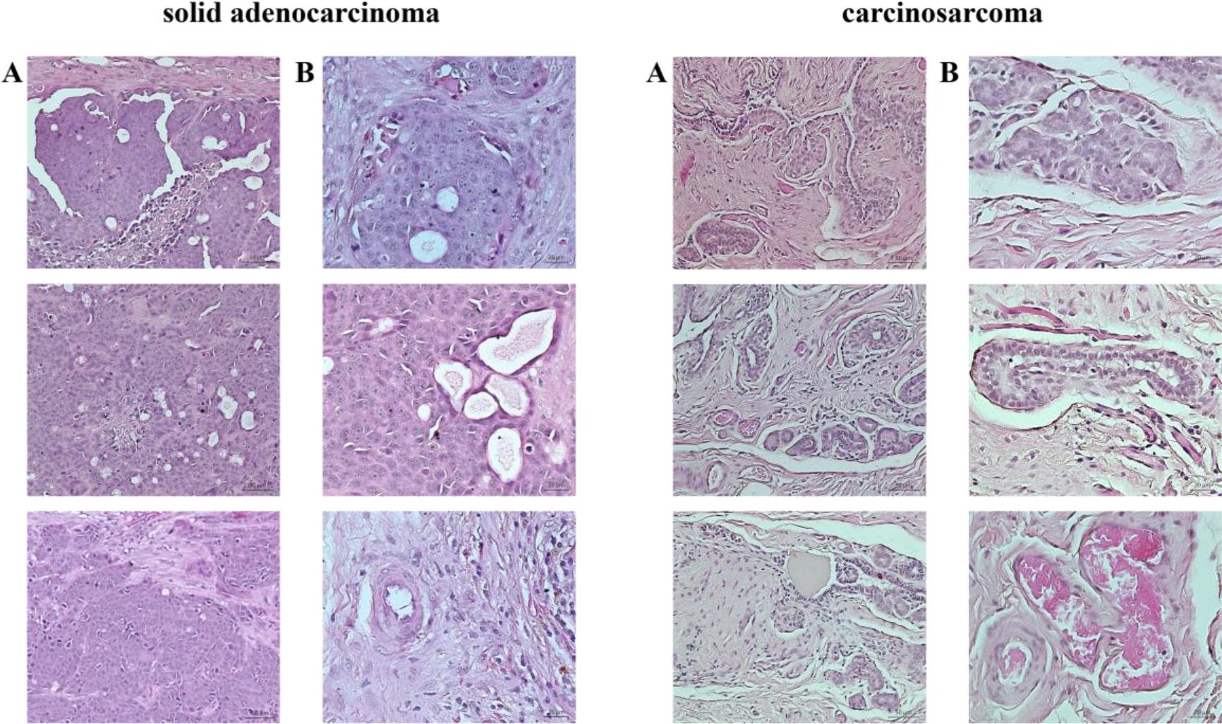
Fig. 2.
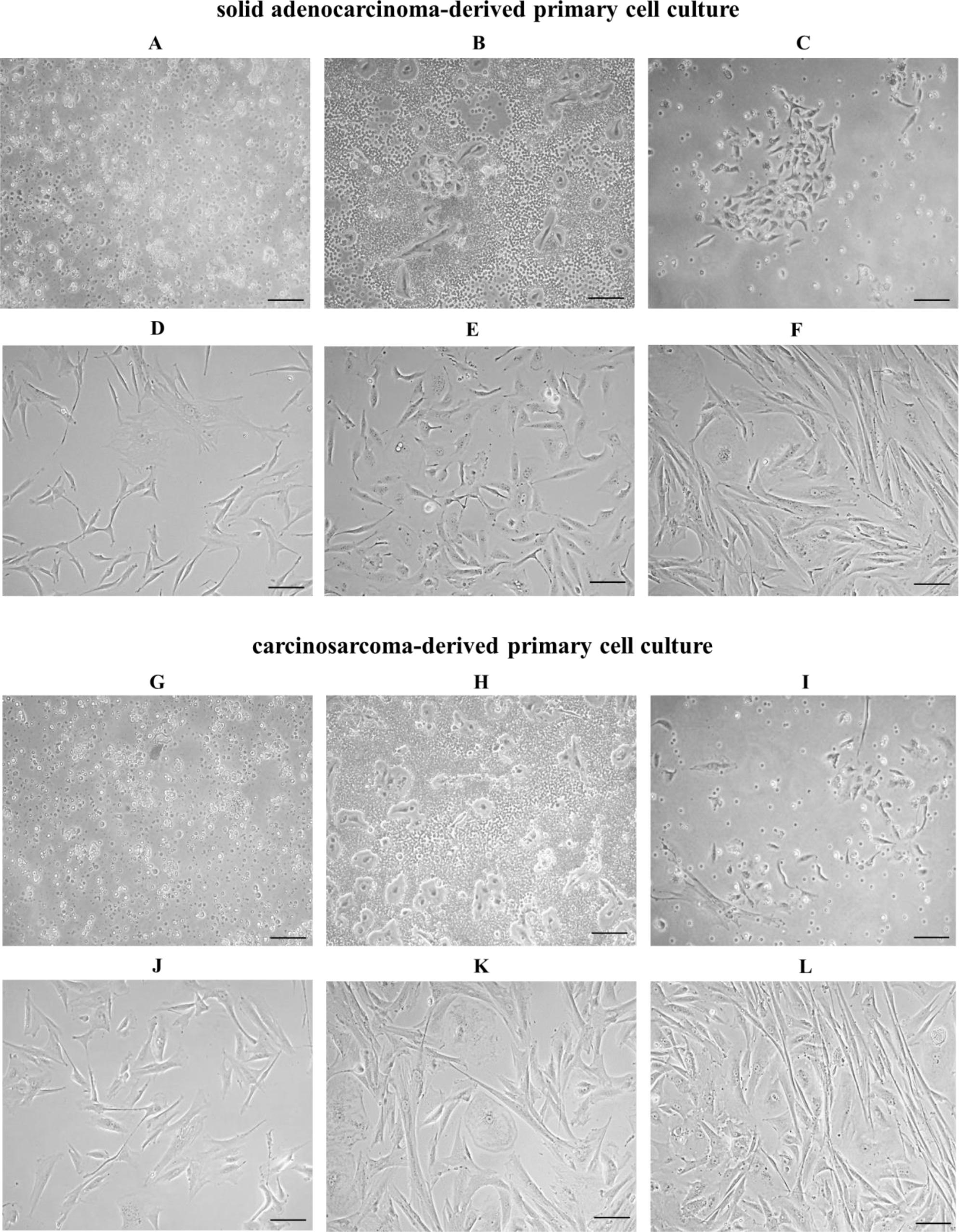
Fig. 3.
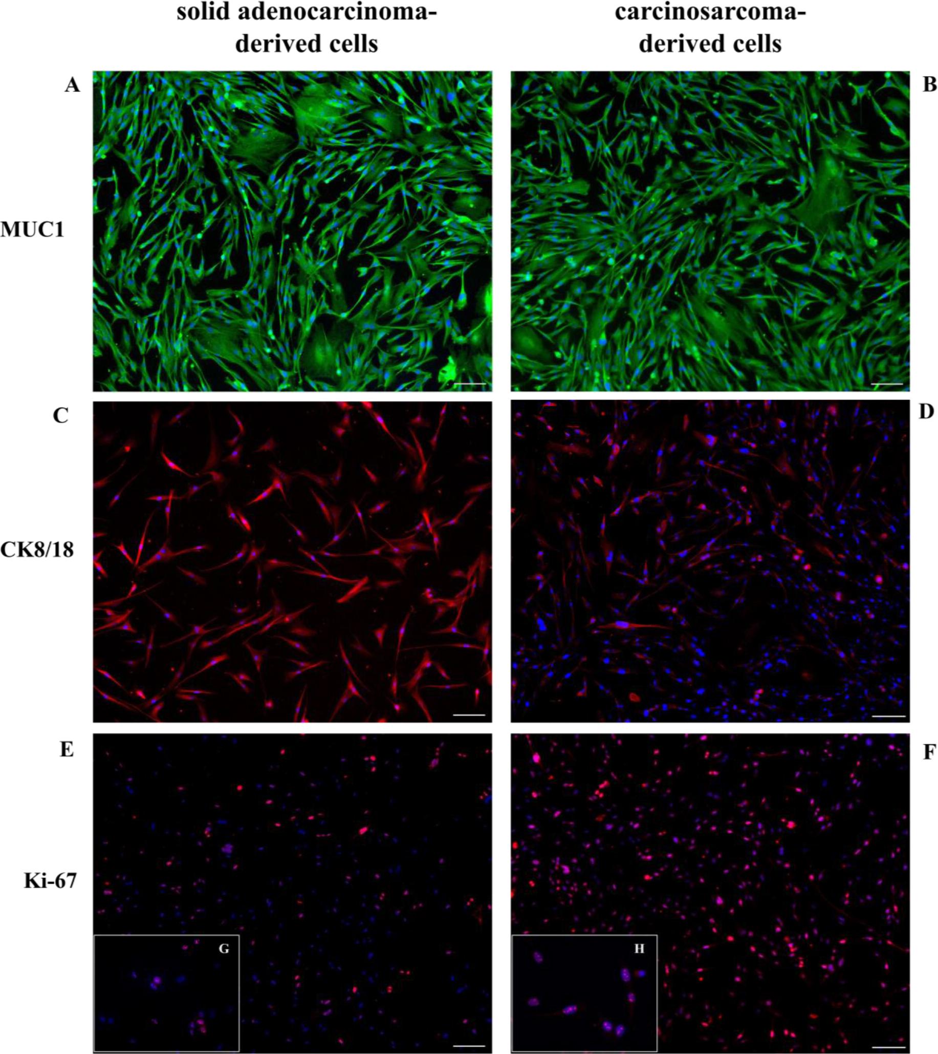
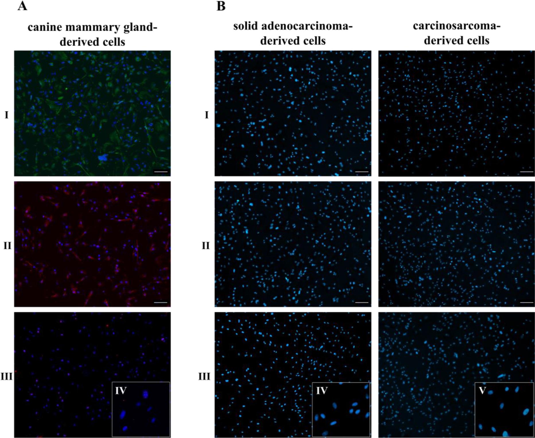
Fig. 4.
Specific primary and secondary antibodies used for immunocytochemistry
| Primary antibodies | |||||||
|---|---|---|---|---|---|---|---|
| Marker | Primary antibody | Type, clone | Host, isotype | Reactivity | Dilution | Catalogue No. | Supplier |
| mucin 1 | MUC1 | mAb, MUC1/955 | mouse, IgG | human, mouse | 1 : 150 | NBP2-44658 | Novus Biologicals, Centennial, CO, USA |
| cytokeratin 8 cytokeratin 18 | Cytokeratins 8/18 | mAb, 5D3 | mouse, IgG | human, mouse | 1 : 100 | NB120-17139 | Novus Biologicals |
| Ki-67 | Anti-Ki-67 | pAb, N/A | rabbit, IgG | human, mouse | 1 : 300 | ab15580 | Abcam, Cambridge, UK |