Fig. 1.
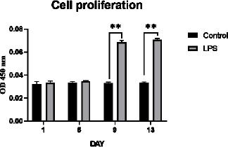
Fig. 2.
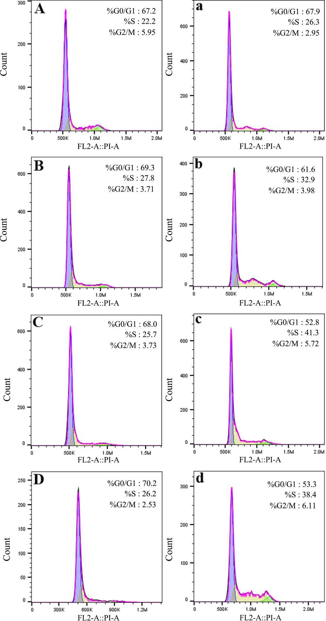
Fig. 3.
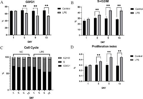
Fig. 4.
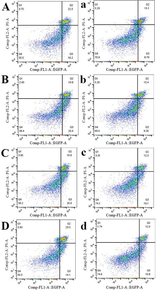
Fig. 5.
Fig. 6.

© 2024 Tingyu Yang, Guotong Zhao, Wenlu Zhu, Wanting Yu, Yijie Jiang, Yunxiao Zhou, Yong Li, published by National Veterinary Research Institute in Pulawy
This work is licensed under the Creative Commons Attribution-NonCommercial-NoDerivatives 3.0 License.