
Feline panleukopenia (FPL) is an infectious viral disease caused by the feline panleukopenia virus (FPV). This pathogen is a small, non-enveloped single-stranded DNA virus belonging, together with closely related canine parvovirus type 2 (CPV-2), to the Protoparvovirus genus and Parvoviridae family (3, 4, 20). The pair of viruses are responsible for serious infections of cats and dogs, mainly kittens and puppies, with high morbidity and mortality rates. The most common form of FPL in cats older than six weeks is the acute form accompanied by fever, depression, vomiting and anorexia (1, 13). Some cats show extreme dehydration leading to progressive weakness and depression along with the previously stated symptoms.
Feline panleukopenia virus and CPV-2 are defined as one single taxonomic entity (37). The feline virus was discovered at the beginning of the 20th century and recognised as one of the main pathogens responsible for feline viral diarrhoea (14). The canine virus emerged as a dog pathogen in the late 1970s and rapidly spread worldwide (36). It is believed that CPV-2 evolved as a host-range variant of FPV that adapted to certain other hosts among the Carnivora (minks and foxes) through changes in five or six amino acid positions in the capsid protein (34). Unlike FPV, which has exhibited a certain degree of genetic stability (11), CPV-2 has shown a high rate of genomic substitution comparable to that of RNA viruses (40). The original CPV-2 has been completely replaced by three antigenic variants (CPV-2a, 2b and 2c) with changes within the VP2 gene. The original CPV is no longer present in the Carnivora population and is only known to exist in vaccine formulations.
An estimated 90% of a protoparvovirus virion is made up of viral protein 2 (VP2), which is an integral component of the capsid protein (35). This protein is a critical component of the virion that determines the antigenic properties, host range and receptor binding of FPV and CPV-2. Sequence analysis of the VP2 gene showed the close relationship of FPV and CPV-2 strains but the differences in key amino acid residues (the original CPV-2 contains 87M, 101I, 300A, 305D, 375N, 426N and 555V; CPV-2a contains 87L, 101T, 297S, 300G, 305Y, 555I, 426N and 297A; CPV-2b contains 297S and 426D; a new CPV-2a strain contains 297A and 426N; a new CPV-2b strain contains 297A and 426D; CPV-2c contains 297A and 426E; and FPV contains 80K, 93K, 103 V, 323D and 568A) (22, 30, 31, 39).
The original CPV-2 isolates were not able to replicate in cats. However, changes in amino acids not only enhanced the binding of CPV-2-derived variants to canine cellular receptors but also affected the replication ability of the virus in cats (16, 23, 24). Moreover, CPV-2-derived strains can cause disease in cats with clinical signs similar to FPL but a generally milder course than that seen in cats infected with FPV (27, 38, 39).
Carnivore parvoviruses are likely to spread freely and rapidly in environments where only a low number of cats and dogs have been vaccinated against FPV or CPV-2. Initially, the only prophylactic intervention available against FPV or CPV-2 comprised inactivated or live attenuated virus vaccines, which proved to be ineffective long-term (33). Since CPV-2a and -2b strains seem to have advantages over conventional FPV in cats, it is possible that CPV-2a and -2b will replace FPV as the dominant parvoviruses of domestic cats even in developed countries where FPV vaccines are commonly used (18). Monitoring of parvoviruses is important because the continuous prevalence of viral infection might be associated with the emergence of new virulent strains, and the distribution of new variants poses a threat to domestic animals (dogs and cats) (2, 29). Knowledge of the current situation of pathogen occurrence is important particularly for epidemic control and preventive measures (33). This study aimed to determine the presence of CPV-2 in cats with signs of FPL in Slovakia and to investigate the suitability of a CPV antigen test for detection of FPV.
The study was conducted on samples from central Slovakia collected from October 2020 to December 2022. Cats were either from animal shelters or admitted to veterinary clinics for treatment because they presented FPL symptoms.
A total of 59 cats, 17 European shorthairs and 42 crossbreds, of different ages and both sexes were included in this study. Cats were divided into three groups, of which the first group (n = 21) consisted of cats showing symptoms of FPL (such as fever, lethargy, vomiting and diarrhoea), the second group (n = 29) comprised cats in close contact with parvovirus-infected animals but without clinical signs and the third group (n = 9) was a group of clinically healthy cats enrolled as the control group. Detailed clinical information of the animals involved in this study is presented in Table 1.
Detailed clinical information on cats from central Slovakian animal shelters or veterinary clinics with signs of feline panleukopenia
| Group | Sample ID | Breed | Sex | Age | FPV vaccination | Anamnesis |
|---|---|---|---|---|---|---|
| I. clinical signs (n = 21) | 1 | crossbreed | female | 3 months | no | vomiting, diarrhoea |
| 2 | crossbreed | male | 3 months | no | vomiting, diarrhoea, lethargy | |
| 3 | crossbreed | female | 2 months | no | vomiting, diarrhoea, fever | |
| 4 | ESH | female | 6 months | no | lethargy, anorexia, fever | |
| 5 | ESH | female | 5 months | no | lethargy, anorexia | |
| 6 | crossbreed | female | 2 months | no | vomiting, diarrhoea | |
| 7 | crossbreed | male | 3 months | no | vomiting, lethargy | |
| 8 | crossbreed | female | 3 months | no | lethargy, fever | |
| 9 | crossbreed | female | 3 months | no | vomiting, diarrhoea, fever | |
| 10 | ESH | female | 3 years | no | lethargy, fever | |
| 11 | crossbreed | female | 4 months | yes | vomiting, lethargy, fever | |
| 12 | ESH | female | 3 years | no | vomiting, lethargy, anorexia | |
| 13 | crossbreed | male | 7 months | no | vomiting, lethargy, fever | |
| 14 | crossbreed | male | 6 months | no | diarrhoea, fever, vomiting | |
| 15 | crossbreed | male | 5 years | yes | diarrhoea | |
| 16 | ESH | male | 3 months | no | vomiting, lethargy, fever | |
| 17 | ESH | male | 5 months | no | vomiting, lethargy, fever, diarrhoea | |
| 18 | crossbreed | male | 5 months | no | vomiting, lethargy, fever | |
| 19 | ESH | male | 6 months | no | vomiting, lethargy, fever | |
| 20 | crossbreed | male | 1 year | no | vomiting, diarrhoea | |
| 21 | crossbreed | male | 3 months | no | diarrhoea, vomiting | |
| II. close contact with parvovirus-infected animal (n = 29) | 1 | crossbreed | female | 4 years | yes | healthy |
| 2 | crossbreed | female | 6 months | no | healthy | |
| 3 | crossbreed | male | 4 months | no | healthy | |
| 4 | crossbreed | female | 4 years | yes | healthy | |
| 5 | crossbreed | female | 5 years | yes | healthy | |
| 6 | ESH | female | 2 years | no | healthy | |
| 7 | ESH | female | 1 year | no | healthy | |
| 8 | ESH | female | 4 years | yes | healthy | |
| 9 | crossbreed | male | 5 years | yes | healthy | |
| 10 | ESH | female | 4 months | no | diarrhoea | |
| 11 | crossbreed | female | 3 months | no | diarrhoea | |
| 12 | crossbreed | male | 2 months | no | diarrhoea | |
| 13 | crossbreed | female | 3 months | no | diarrhoea, vomiting | |
| 14 | crossbreed | male | 3 months | no | healthy | |
| 15 | ESH | female | 1 year | no | healthy | |
| 16 | crossbreed | male | 5 months | no | healthy | |
| 17 | crossbreed | male | 2 years | yes | healthy | |
| 18 | crossbreed | male | 2 years | yes | intermittent diarrhoea | |
| 19 | crossbreed | female | 5 months | no | diarrhoea, overcame FPL | |
| 20 | ESH | female | 5 years | no | healthy | |
| 21 | crossbreed | male | 2 years | yes | intermittent diarrhoea | |
| 22 | crossbreed | female | 4 months | no | healthy | |
| 23 | ESH | female | 2 years | no | healthy | |
| 24 | crossbreed | female | 1 year | no | healthy, overcame FPL | |
| 25 | crossbreed | male | 1 year | yes | healthy | |
| 26 | crossbreed | female | 5 months | no | diarrhoea, overcame FPL | |
| 27 | crossbreed | male | 4 months | no | vomiting | |
| 28 | crossbreed | female | 3 years | yes | healthy | |
| 29 | crossbreed | female | 6 months | no | healthy, overcame FPL | |
| III. clinically healthy (n = 9) | 1 | ESH | male | 1 year | yes | healthy |
| 2 | ESH | female | 2 years | yes | healthy | |
| 3 | crossbreed | male | 7 months | yes | healthy | |
| 4 | crossbreed | female | 3 years | yes | healthy | |
| 5 | crossbreed | male | 3 months | yes | healthy | |
| 6 | crossbreed | male | 5 months | yes | healthy | |
| 7 | crossbreed | female | 1 year | yes | healthy | |
| 8 | crossbreed | female | 2 years | yes | healthy | |
| 9 | ESH | male | 1 year | yes | healthy |
ESH – European shorthair; FPL – feline panleukopenia
Swabs were collected in duplicate directly from the rectum using cotton swabs. One swab was used immediately for rapid antigen testing and the other was stored at room temperature for further processing.
Cats were initially screened for the presence of parvovirus antigens using a chromatographic immunological antigen (Ag) test for CPV and canine coronavirus (CCV) (Rapid CPV/CCV Ag test; Bionote, Gyeonggi-do, South Korea) according to the manufacturer’s instructions.
All samples were investigated for parvovirus infection by PCR targeting the VP2 gene (1,755 base pairs). Rectal swabs were suspended in 400 μL of phosphate-buffered saline and viral DNA was extracted using a DNeasy Blood and Tissue Kit (Qiagen, Hilden, Germany). The PCR assay was performed using primers designed for full-length CPV-2 as described by Hu et al. (15) (CPV-F: 5′-AGA GACAATCTTGCACCAAT-3′ and CPV-R: 5′-ATG TTAATATAATTTTCTAGGGTG CT-3′) and PPP Master Mix (Top-Bio, Vestec, Czech Republic) according to the manufacturer’s instructions, in a final volume of 25 μL comprising 12.5 μL of PPP Master Mix (2×), 1 μL of each primer (10 μM), 1 μL of template DNA and 9.5 μL of PCR water. The PCR conditions were set as an initial denaturation step at 98°C for 3 min; 34 cycles of denaturation at 98°C for 30 s, annealing at 55°C for 30 s and extension at 72°C for 1 min and 48 s; and a final extension at 72°C for 10 min (Biometra Tone 96G thermocycler; Analytik Jena, Jena, Germany). As a positive control, laboratory-confirmed CPV-2 was used. The PCR products were separated in 1% agarose gel with a GelRed Nucleic Acid Stain (Biotinum, Fremont, CA, USA) and visualised using a UV transilluminator (MiniBIS, DNR Bio-Imaging Systems, Jerusalem, Israel). The positive samples were then subjected to Sanger sequencing for the partial VP2 gene. The sequences of all positive samples being identical, a representative nucleotide sequence was submitted to the GenBank database under accession number PP209372. This sequence was used for National Center for Biotechnology Information BLASTn analysis.
A total of 23 rectal samples (38.9%, 23/59) were confirmed to be antigen positive in the antigen test (Table 2). The tests indicated 100% positivity in all cats with clinical signs of parvovirus infection (21/21). Among the group of cats in close contact with parvovirus-infected animals, parvovirus infection was confirmed in two individuals (6.89%, 2/29). No cat from the clinically healthy group was detected to be antigen positive (0/9). A summary is presented in Table 3.
Antigen-based detection of parvovirus infection in rectal swab samples of European shorthair and crossbred cats in central Slovakia grouped by feline panleukopenia status
| Group | Sample ID | CPV antigen test |
|---|---|---|
| I. clinical signs (n = 21) | 1–21 | + |
| II. close contact with parvovirus-infected animal (n = 29) | 1–9; 11–26; 28, 29 | - |
| 10, 27 | + | |
| III. clinically healthy (n = 9) | 1–9 | - |
CPV – canine parvovirus; + – positive result; - – negative result
Summary of parvovirus infection findings in rectal swab samples of European shorthair and crossbred cats in central Slovakia grouped by feline panleukopenia status
| Groups | CPV antigen test | PCR test | Total positive | ||
|---|---|---|---|---|---|
| Ag+ | Ag- | PCR+ | PCR- | ||
| I. clinical signs (n = 21) | 21 | 0 | 21 | 0 | 21 (100%) |
| II. close contact with parvovirus-infected animals (n = 29) | 2 | 27 | 2 | 27 | 2 (6.89%) |
| III. clinically healthy (n = 9) | 0 | 9 | 0 | 9 | 0 |
Ag+ – parvovirus infection clinically confirmed by commercial antigen test for canine parvovirus (CPV) and canine coronavirus Ag- – parvovirus infection clinically excluded by commercial antigen test; PCR+ – parvovirus infection confirmed by PCR PCR- – parvovirus infection excluded by PCR
Samples were also examined for parvovirus infection by PCR based on the presence of the VP2 gene. All antigen-positive samples (n = 23) were confirmed for the presence of the VP2 gene (Fig. 1 and Table 3). None of the antigen-negative samples tested positive.
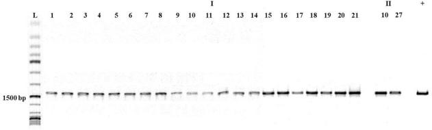
Amplicons of the full-length VP2 gene of canine parvovirus 2 (CPV-2) (1,755 bp) resolved on agarose gel as confirmation of parvovirus infection in European shorthair and crossbred cats in central Slovakia based on the presence of this gene in rectal swab samples. L – GeneRuler 1 kb DNA Ladder; I – group with clinical signs of parvovirus infection; II – group with close contact with parvovirus-infected animals; + – positive control (CPV-2-positive sample)
Analysis of the representative FPV sequence (PP209372) in BLASTn revealed 99.78–100% pairwise identity with FPV and excluded CPV-2 infection (Table 4).
Sequences producing significant alignments with the VP2 gene amplicons of investigated rectal swab samples from European shorthair and crossbred cats in central Slovakia. Sequence analysis used the National Center for Biotechnology Information BLASTn algorithm
| Scientific name | Pairwise identity (%) | Query coverage (%) | GenBank accession number |
|---|---|---|---|
| Feline panleukopenia virus | 100.00 | 100 | MK671185.1, MK671183.1, MK671171.1, MK671170.1, MK671156.1, MK425504.1, MK425500.1, MK425499.1, MK425498.1, MK425497.1, KP019621.2, MG764511.1, MG764510.1, MF541133.1, MF541132.1, MF541119.1, KX685354.1, KX900570.1, KT240130.1, KT357491.1, OP796716.1, OP796714.1, KP682520.1, KP019617.1, OM638042.1, MW091486.1, MW091487.1, MZ712026.1, MZ322607.1, MZ357122.1, MZ357120.1, MZ357119.1, MW017627.1, MT270583.1, MW331496.1, MN419003.1, MT274378.1, HQ184201.1, FJ936171.1, DQ474238.1, DQ474237.1, DQ474236.1, EU221281.1, AY606131.1, DQ099430.1, AY955826.1, DQ003301.1, AB054227.1, AB054226.1, AB054225.1 |
| Feline parvovirus | 100.00 | 100 | MK266790.1, MK266788.1, MK295775.1, OM885375.1, ON646210.1, MZ442303.1, OR211675.1 |
| Feline panleukopenia virus | 99.78 | 100 | OQ863619.1, OQ863618.1, OQ863617.1, OQ863615.1, MZ508524.1 |
| Feline parvovirus | 99.78 | 100 | OR198066.1, OQ869254.1, OQ868569.1, OQ868568.1, OQ868566.1, OQ868565.1, OQ868564.1, OQ868562.1, OQ868554.1, OQ868553.1, OQ868552.1, OQ868550.1, OQ868548.1, OQ868547.1, OQ868542.1, OQ868536.1, OQ868535.1, OQ868534.1, OR194134.1, OR194132.1, OR194130.1, OR194129.1, OR194128.1, OR194125.1, OR194122.1, OQ535514.1, OQ535513.1, OQ535511.1, OQ535510.1, OQ535509.1 |
Disease killed 2 of the 23 animals which were antigen and PCR positive for parvovirus infection, which represents an 8.69% mortality rate.
Although cats were not the host for the original CPV-2, the newer CPV-2 antigenic types have acquired the ability to replicate in cats (18). Feline disease caused by CPV-2-derived variants manifests clinical signs similar to those of disease caused by FPV. This implies that more studies are needed to know the true prevalence and significance of CPV-2 in cats worldwide. Many studies demonstrated the prevalence of CPV-2 infection in cat populations over a wide geographical range. In Vietnam and Taiwan, the virus was found in more than 80% of cats presumed to be infected with FPV (17), whereas in Germany, it was only detected in 10% of parvovirus-carrying cats (38). In contrast, other authors have detected CPV-2 strains in the faeces of clinically healthy animals (9, 26, 28). A study by Balboni et al. (5) found equal prevalence of FPV and CPV-2 in examined samples from asymptomatic cats. In general, CPV-2 is not a common causative agent of feline panleukopenia, and only a small number of cats that showed signs of this disease tested positive for the presence of CPV-2. This study aimed to gather much-needed data on CPV-2 feline infection and focused on its detection in central Slovakia. Our survey showed that 23 out of 59 cats were positive for parvovirus infection by both the rapid antigen test and conventional PCR test, which meant a prevalence of 38.9%. As expected, all cats with clinical signs of feline panleukopenia were positive for parvovirus infection. On the other hand, only two cats in close contact with parvovirus-infected animal tested antigen and PCR positive. The mortality rate in our study was 8.69% (2/23), which is a significantly lower rate than that in other studies, where it ranged from 50% to 90% (6, 8, 19). Sequencing of positive samples and their subsequent BLASTn analysis excluded CPV-2 infection and confirmed the presence of FPV. Our results are consistent with those of several studies. In a study by Byrne et al. (7) performed in Australia in 2018, CPV-2 was not detected in any of 218 faecal samples collected from cats and kittens living in shelters. Infection with CPV-2 was also not confirmed in a Portuguese population of cats by Miranda et al. (25). In Italy but with a small part of the samples from the UK, Decaro et al. (11) characterised 39 out of 39 parvovirus strains as FPV at molecular level in an investigation of pathogens isolated from cats with feline panleukopenia. These results demonstrate the dominance of FPV over CPV-2 as the causative agent of feline panleukopenia and the results of the present study indicate the predominant status of FPV in feline panleukopenia infections in Slovakia.
The relatively high observed parvovirus prevalence rate (38.9%, 23/59) in a relatively small group of animals could be connected to the lack of vaccination against FPV infection, since 91.3% of the positive-testing cats were unvaccinated (21/23). Neither of two cats in contact with a parvovirus-infected animal was vaccinated. However, two cats with confirmed feline panleukopenia were vaccinated. This can be explained by reports showing that protective immunity is not achieved in a significant proportion of cats (12, 32). A further example of immunisation failing in a notable proportion of kittens is the research by Dawson et al. (10), in which 25% and 39% of kittens were observed to be without antibody response after two or three vaccinations, respectively.
According to Jacobson et al. (21), the IDEXX SNAP Parvo enzyme immunoassay, designed to detect CPV-2, is also suitable for the detection of FPV in faeces and achieves a specificity of 96%–100%. In our study, all 23 samples positive in antigen testing were positive in subsequent testing by conventional PCR. Since none of the antigen-negative samples were confirmed as PCR positive, our results indicate that the commercial antigen test designed for the diagnosis of CPV-2 infection is sensitive enough and suitable for the clinical diagnosis of FPL.
This study provides baseline epidemiological data for future prevention and control measures against parvovirus infection and highlights the need for cat vaccination programmes against feline panleukopenia. However, further studies performed on a larger number of animals are necessary to confirm the data’s implications.