
Table 1.
Primers used in this experiment
| Primer name | Primer type | Primer sequence (5′ to 3′) | Product size (base pairs) |
|---|---|---|---|
| Pig β-actin | Forward | CGGGACATCAAGGAGAAGC | 132 |
| Reverse | CTCGTTGCCGATGGTGATG | ||
| Pig TRPML1 | Forward | CAGTTACAAGAACCTCAC | 111 |
| Reverse | CAGTCAGGGATTTCATTG | ||
| Pig TRPML2 | Forward | AATCAGTATCATCGTCTA | 140 |
| Reverse | ATATTCAGTGTCTCATTAG | ||
| Pig TRPML3 | Forward | TTACTTGGTCTGGCTGTT | 129 |
| Reverse | TCCTCTTGGTAATGCTTAATTG | ||
| Monkey GAPDH | Forward | TCAACGACCACTTTGTCAAGCTCA | 97 |
| Reverse | GCTGGTGGTCCAGGGGTCTTACT | ||
| Monkey TRPML1 | Forward | CGGATGACACCTTCGCAGCCTAC | 108 |
| Reverse | ACGCATACCGGCCCAGTGACAC | ||
| Monkey TRPML2 | Forward | CTGTGGCTGGATTGTCTTAGG | 121 |
| Reverse | CTGGATTTGGGCAAAGGTTG | ||
| Monkey TRPML3 | Forward | CCAGAAATTGAAACTGAGTGTT | 205 |
| Reverse | ATGTTATAGTCAGAGTAAAGTC | ||
| PRRSV ORF5 | Forward | GAAATGCTTGACCGCGGGCT | 261 (16) |
| Reverse | GTGTCAAGGAAATGGCTTGT |
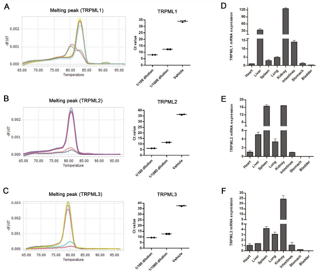
Fig. 1.
Identification of primer specificity for transient receptor potential mucolipin (TRPML) genes in quantitative reverse-transcription PCR (qRT-PCR) and their distribution in peripheral porcine tissues. A – Melting curves of qRT-PCR for TRPML1 using the designed primers, and relative threshold cycle (Ct) values of TRPML1 obtained with different templates; 1/100(0) dilution – dilution of the constructed TRPML1 plasmid; vehicle – negative control containing no template; B – Melting curves of qRT-PCR for TRPML2 using the designed primers, and relative Ct values of TRPML2 obtained with different templates; C – Melting curves of qRT-PCR for TRPML3 using the designed primers, and relative Ct values of TRPML3 obtained with different templates; D–F – levels of porcine TRPML1, TRPML2 and TRPML3 messenger RNA (mRNA) in different porcine tissues determined by qRT-PCR. Data are presented as the means ± standard deviation
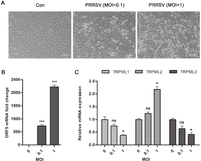
Fig. 2.
Effects of porcine reproductive and respiratory syndrome virus (PRRSV) infection of Meat Animal Research Center 145 (MARC-145) monkey kidney cells on the expression of three TRPML proteins. A – Cell morphology with increased multiplicity of infection (MOI) of PRRSV infection. Morphology of MARC-145 cells at a magnification of 100× (scale bars 100 μm); B – Messenger RNA level of PRRSV open reading frame 5 assessed by quantitative reverse-transcription PCR analysis; C – Differential intracellular expression of three TRPML proteins regulated by PRRSV infection. For quantitative analyses, all data were obtained from at least three independent experiments and data are presented as the means ± standard deviation; Con – MARC-145 cells without virus infection; * – P-value < 0.05; ** – P-value < 0.01; *** – P-value < 0.001; ns – no significance
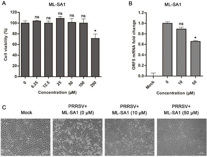
Fig. 3.
Inhibitory activity of mucolipin synthetic agonist 1 (ML-SA1) against porcine reproductive and respiratory syndrome virus (PRRSV) in Meat Animal Research Center 145 (MARC-145) monkey kidney cells. A – Cytotoxicity of ML-SA1 in MARC-145 cells. Viability of cells treated with concentrations of ML-SA1 from 0 μM to 200 μM measured by a cell-counting kit 8 assay; B – Messenger RNA level of PRRSV open reading frame 5 with different concentrations of ML-SA1 treatment in MARC-145 cells incubated with 10 μM or 50 μM of ML-SA1; C – MARC-145 cell morphology in PRRSV infection after addition of different concentrations of ML-SA1 and incubation with 10 μM or 50 μM ML-SA1. For quantitative analyses, all data were obtained from at least three independent experiments and data are presented as the means ± standard deviation; Mock –MARC-145 cells without virus or ML-SA1 treatment; * – P-value < 0.05; ** – P-value < 0.01; *** – P-value < 0.001; ns – no significance