
Fig. 1.
Fig. 2.
Fig. 3.
Fig. 4.
Fig. 5.
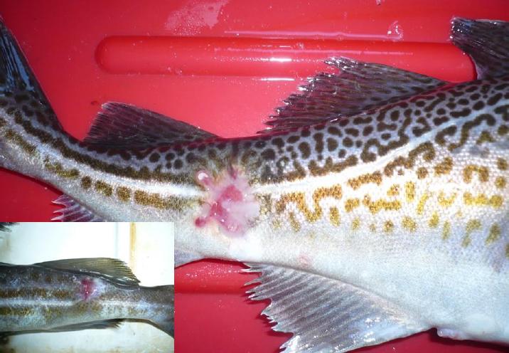
Fig. 6.
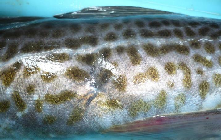
Fig. 7.
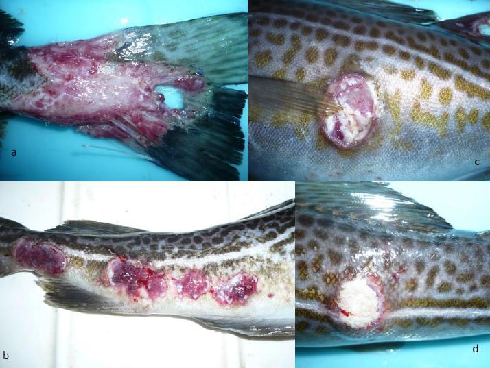
Fig. 8.
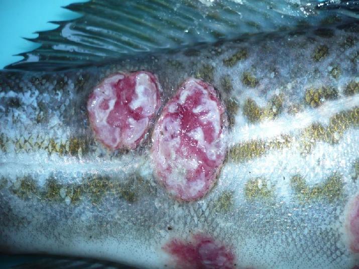
Fig. 9.
Fig. 10.
Fig. 11.
Fig. 12.
Groups of bacteria isolated from skin ulcers of Baltic cod in correlation with the fishing area
| Fishing area | Bacteria |
|---|---|
| Gdańsk Bay | Acinetobacter spp., Aeromonas spp., Alcaligenes faecalis, Chryseobacterium indologenes, Enterococcus faecalis, Microbacterium sp., Pseudomonas spp., |
| Słupsk Furrow | Acinetobacter spp., Aeromonas spp., Chryseobacterium indologenes, Delftia spp., Micrococcus sp., |
| Kołobrzeg-Darłowo | Acinetobacter spp., Aeromonas spp., Chryseobacterium indologenes, Pseudomonas spp., Serratia spp., |
| Bornholm North | Acinetobacter spp., Aeromonas sp., Pseudomonas spp., Shewanella putrefaciens group, Stenotrophomonas maltophilia |
| Bornholm South | Acinetobacter spp., Citrobacter freundii, Pseudomonas spp., |