
Table 1
Primers used in the study
| Primer name | Primer sequences (5′→3′) | Product size (bp) |
|---|---|---|
| rli106F | GCCTCTTTGCAATCGAAA | |
| rli106R | ACTACTATTACTTTAGACTAGTTTTCAATT | 225 |
| P1 | GGTACC AAATAGCCGACTGCTCCAGC | |
| P2 | AAATTCGTGTTATAATAAAATTGGTGAAATTAGATAAGTG | 351 |
| P3 | CACTTATCTAATTTCACCAATTTTATTATAACACGAATTT | |
| P4 | CTGCAG GATCTCTAGCATTGGAAAGCCA | 383 |
| P5 | AAATAGCCGACTGCTCCAGC | |
| P6 | GATCTCTAGCATTGGAAAGCCA | 722 |
| P7 | CCGTGCTTGATTGCCGTTAC | |
| P8 | AACTAGGCCGCCTGCACC | 1391/1166 |
| CF | AAGCTTGCCTCTTTGCAATCGAAA | |
| CR | CGGGATCCACTACTATTACTTTAGACTAG | 239 |
| rli106F1 | GGATCCCAGTTGGCATATTAATATC | |
| rli106R1 | CGGGGTACCGGTTAAGCGGTTTTGC | 271 |
| DegUF | CAAGCTTAAAAGAGAAATGTTTAATT | |
| DegUR | GGTACCACTGTTTTTTCACTAATGA | 721 |
| 16S rRNAF | GATGCATAGCCGACCTGAGA | |
| 16S rRNAR | TGCTCCGTCAGACTTTCGTC | 116 |
| hemCF | CGGATCCATGACGTTATCTAAAGGA | |
| hemCR | CCAAGCTTTTAAATTCGTTCTTCTACA | 1122 |
| DegUF1 | GGATCCATGGCACTCAAAATCATGATTG | |
| DegUF2 | CAAGCTTTTAGCGAATGTATACCCAGCC | 687 |
[i] F – forward; R – reverse
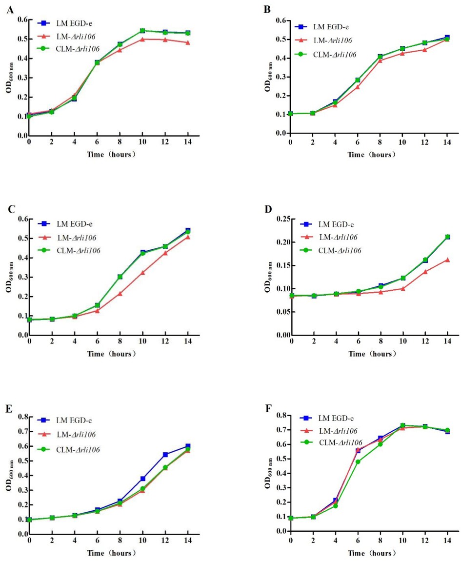
Fig. 1
Growth curves of LM strains under different environmental stresses. LM EGD-e – parental strain of Listeria monocytogenes; LM-△rli106 – rli106 deletion strain of Listeria monocytogenes; CLM-△rli106 – rli106 complementation strain of Listeria monocytogenes
A–F – Growth curves of LM EGD-e, LM-△rli106 and CLM-△rli106 under the conditions of 42°C, pH 9, 5% NaCl and 8% NaCl, 3.8% ethanol and 5 mM H2O2. Bars indicate the standard error of the mean
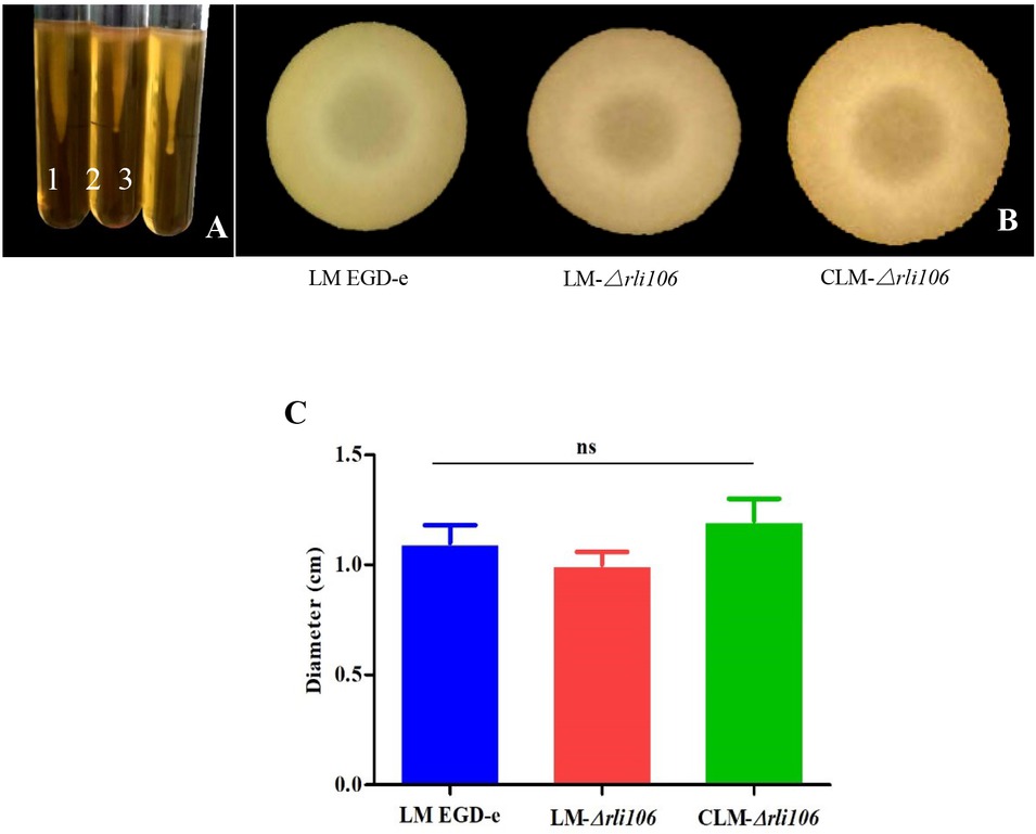
Fig. 2
Determination and comparisons of the motility of different strains of LM. LM EGD-e – parental strain of Listeria monocytogenes; LM-△rli106 – rli106 deletion strain of Listeria monocytogenes; CLM-△rli106 – rli106 complementation strain of Listeria monocytogenes
A – Kinetics of LM EGD-e (1), LM-△rli106 (2) and CLM-△rli106 (3) in semi-solid medium; B – Kinetics in tablets;
C – Diameter size of the bacterial colony on the plate; ns – not significant, P > 0.05. Bars indicate the standard error of the mean
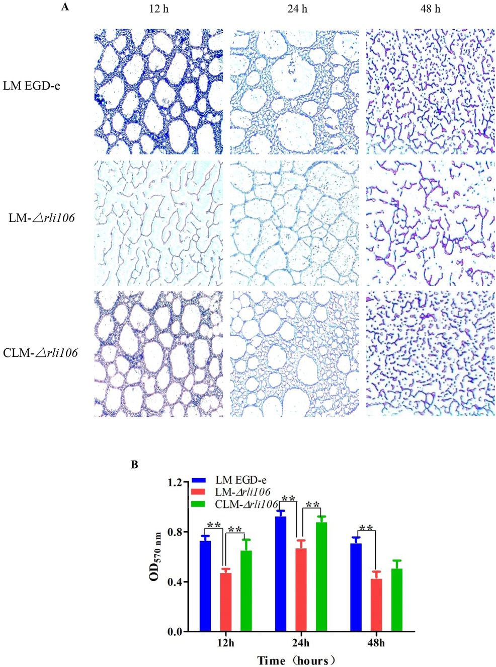
Fig. 3
Effects of rli106 gene deletion on biofilm formation of LM
A – Formation of biofilm by the parental strain of Listeria monocytogenes (LM EGD-e), rli106 deletion strain of Listeria monocytogenes (LM-△rli106) and rli106 complementation strain of Listeria monocytogenes (CLM-△rli106) at 12 h, 24 h and 48 h; B – Comparison of biofilm formation at 12 h, 24 h and 48 h. Bars indicate the standard error of the mean (SE); ** – P < 0.01
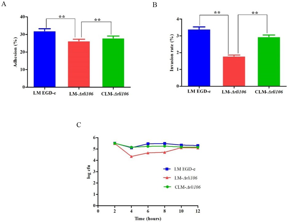
Fig. 4
Determination of adhesion, invasion and intracellular viability in RAW264.7 macrophages for the parental strain of Listeria monocytogenes (LM EGD-e), rli106 deletion strain of Listeria monocytogenes (LM-△rli106) and rli106 complementation strain of Listeria monocytogenes (CLM-△rli106). A – Adhesion rate of LM EGD-e, LM-△rli106 and CLM-△rli106 in RAW264.7 cells; B – Invasion rate of LM EGD-e, LM-△rli106 and CLM-△rli106 in RAW264.7 cells; C – Determination of intracellular viability in macrophages. Bars indicate the standard error of the mean (SE); ** – P < 0.01
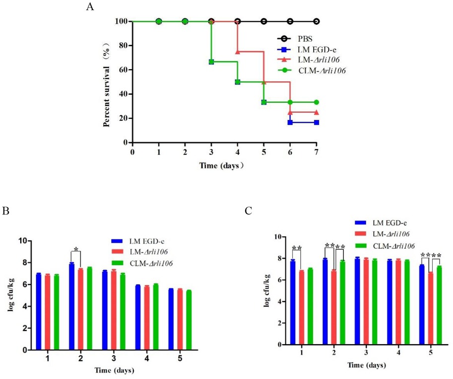
Fig. 5
Effects of rli106 gene deletion on virulence of LM. A – Survival curves of mice infected with the parental strain of Listeria monocytogenes (LM EGD-e), rli106 deletion strain of Listeria monocytogenes (LM-△rli106) and rli106 complementation strain of Listeria monocytogenes (CLM-△rli106); B and C – Bacterial loads in the livers and spleens of mice infected by LM EGD-e, LM-△rli106 and CLM-△rli106; bars indicate the standard error of the mean (SE); * – P < 0.05; ** – P < 0.01 (LM EGD-e)
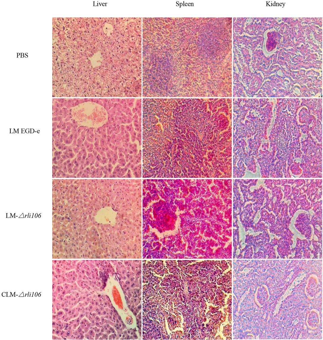
Fig. 6
Histopathological examination of liver, spleen and kidney of mice infected by LM strains (haematoxylin and eosin staining, ×400). LM EGD-e – parental strain of Listeria monocytogenes rli106; LM-△rli106 – rli106 deletion strain of Listeria monocytogenes; CLM-△rli106 – rli106 complementation strain of Listeria monocytogenes
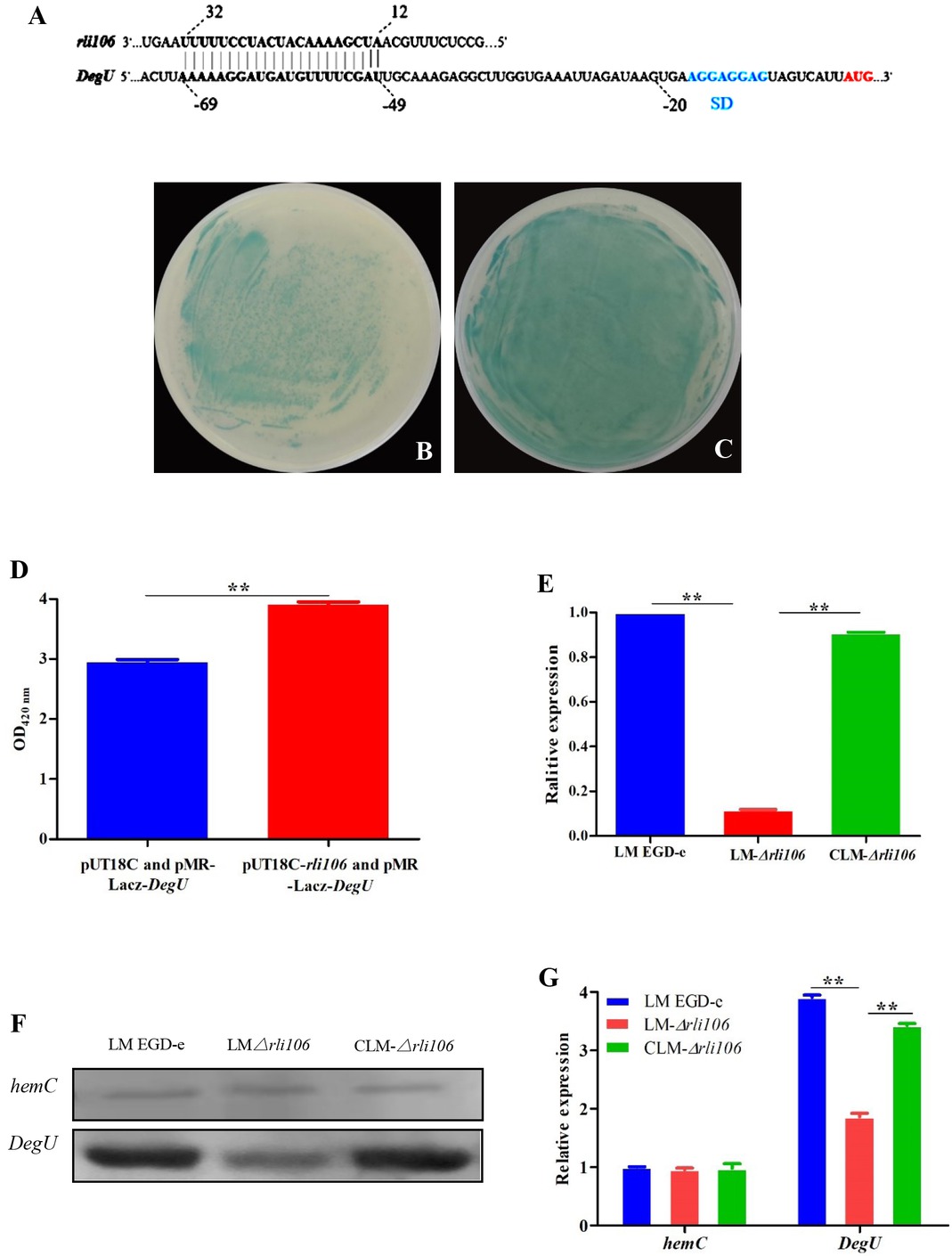
Fig. 7
Verification of interaction between rli106 sRNA and mRNA of target gene DegU. A – Complementary pairing position of DegU target gene and rli106; B and C – Bacterial lawns of BTH101 co-transformed by pUT18C and pMR-LacZ-DegU, and BTH101 co-transformed by pUT18C-rli106 and pMR-LacZ-DegU; D – OD420 nm of bacterial lawns; E – mRNA level of DegU target gene in the parental strain of Listeria monocytogenes (LM EGD-e), rli106 deletion strain of Listeria monocytogenes (LM-△rli106) and rli106 complementation strain of Listeria monocytogenes (CLM-△rli106); F and G – protein level of DegU in LM EGD-e, LM-△rli106 and CLM-△rli106