
Fig. 1
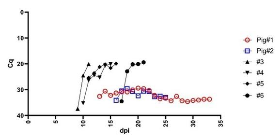
Fig. 2
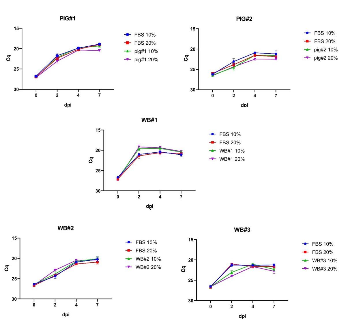
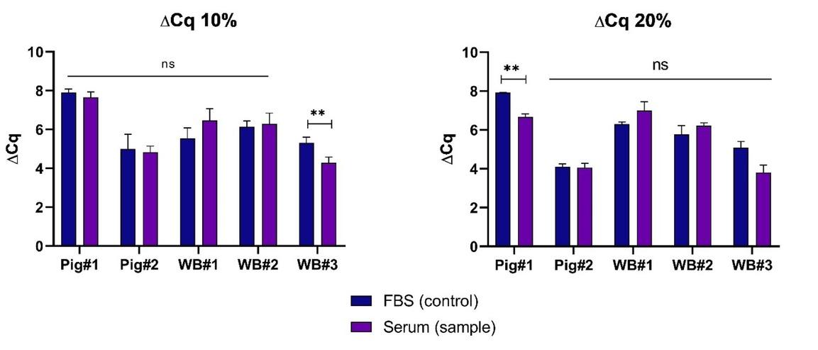
Fig. 3
ASFV DNA detection and anti-ASFV antibody detection and titre in selected samples
| Serum/Sample | Blood qPCR Cq (±SD) | Serum qPCR Cq | Antibodies (ELISA) | Antibody titre (log10/mL) |
|---|---|---|---|---|
| Pig#1 | 32.3 (±1.7) | NEG | POS | 5.01 |
| Pig#2 | 31.7 (±1.4) | 34.7* | POS | 5.31 |
| WB#1 | NEG | NEG | POS | 5.51 |
| WB#2 | NEG | NEG | POS | 5.51 |
| WB#3 | NEG | NEG | POS | 5.21 |
Characteristics of the animals from which the sera used in the study were obtained
| Serum/Sample | Species | Sex | Age | Clinical symptoms | Gross lesions |
|---|---|---|---|---|---|
| Pig#1 | Domestic pig | Male | 9 weeks | Fever, dyspnoea joint swelling, | n/d |
| Pig#2 | Domestic pig | Male | 9 weeks | Moderate fever | Enlargement/hyperaemia of submandibular lymph nodes |
| WB#1 | Wild boar | Male | 18 months | n/a | n/d |
| WB#2 | Wild boar | Female | 18 months | n/a | n/d |
| WB#3 | Wild boar | Male | 24 months | n/a | n/d |
Haemadsorption assay results in the presence of selected sera at 20% concentration
| dpi | |||||||
|---|---|---|---|---|---|---|---|
| Serum/sample | 1 | 2 | 3 | 4 | 5 | 6 | 7 |
| FBS | + | + | ++ | ++ | ++ | ++ | ++ |
| Pig#1 | − | − | − | − | − | − | − |
| Pig#2 * | − | + | + | ++ | ++ | ++ | ++ |
| WB#1 | − | − | − | − | − | − | − |
| WB#2 | − | − | − | − | − | − | − |
| WB#3 | − | − | − | − | − | − | − |