
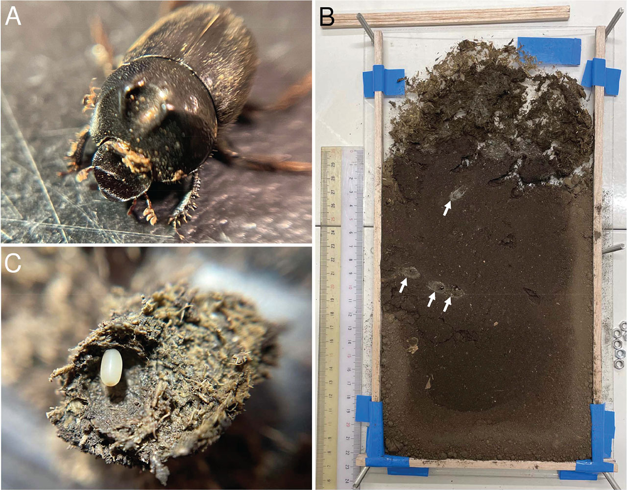
Figure 1:
Onthophagus atripennis and their brood balls. (A) An adult male of Onthophagus atripennis. (B) A rearing case of Onthophagus atripennis and their brood balls (white arrow). (C) A brood ball and egg of Onthophagus atripennis.
Table 1:
Onthopahgus atripennis collected by pit-hole trap in this study and the associated nematodes. All beetles were collected in Kawasaki, Kanagawa.
| Date | Site | Associated nematode | Strain |
|---|---|---|---|
| 2021.5.14 | Meiji University | - | - |
| 2021.5.14 | Meiji University | Tokorhabditis atripennis | SHR9 |
| 2021.5.14 | Meiji University | - | - |
| 2021.5.14 | Meiji University | - | - |
| 2021.5.14 | Meiji University | - | - |
| 2021.5.15 | Meiji University | - | - |
| 2021.5.15 | Meiji University | - | - |
| 2021.8.2 | Meiji University | - | - |
| 2021.8.2 | Meiji University | - | - |
| 2021.8.2 | Meiji University | - | - |
| 2021.8.2 | Meiji University | Onthodiplogaster japonica | SHR18 |
| 2021.8.4 | Meiji University | Tokorhabditis atripennis | SHR21 |
| 2021.8.4 | Meiji University | - | - |
| 2021.9.30 | Kurokawa Field Science Center | Onthodiplogaster japonica | SHR154 |
| 2021.9.30 | Kurokawa Field Science Center | Tokorhabditis atripennis | SHR16 |
| 2021.9.30 | Kurokawa Field Science Center | - | - |
| 2021.9.30 | Kurokawa Field Science Center | - | - |
| 2021.9.30 | Kurokawa Field Science Center | - | - |
| 2021.9.30 | Kurokawa Field Science Center | - | - |
| 2021.9.30 | Kurokawa Field Science Center | - | - |
| 2021.9.30 | Kurokawa Field Science Center | Onthodiplogaster japonica | SHR22 |
Table 2:
The details of beetles’ rearing cases and samples isolated from the cases.
| ID | Date of setting | Date of dissection | Number of beetles | Number of Brood balls | |
|---|---|---|---|---|---|
| Male | Female | ||||
| 1 | 2023.7.14 | 2023.7.21 | 2 | 2 | 2 |
| 2 | 2023.7.14 | 2023.7.21 | 1 | 2 | 4 |
| 3 | 2023.7.16 | 2023.7.24 | 1 | 2 | 0 |
| 4 | 2023.7.18 | 2023.7.25 | 2 | 2 | 0 |
Table 3:
The nematode strains isolated from samples of rearing cases. The ID number described in “Isolated from” is the “ID” used in Table 2.
| Strain | Isolated from | Estimated taxon | Sequence region (Primer) | Accession No. |
|---|---|---|---|---|
| SHR88 | Feces from ID 1 | T. atripennis | 28S rDNA (D1F/D4R), 18S | PV022059 |
| rDNA (18S-CL-F3/18S-CL-R1) | PV022047 | |||
| SHR142 | Tunnel from ID 1 | T. atripennis | 28S rDNA (D1F/D4R), 18S | PV022060 |
| rDNA (18S-CL-F3/18S-CL-R1) | PV022048 | |||
| SHR133 | Feces from ID 2 | Pelodera sp. | 28S rDNA (D2a/D4R), 18S | PV022061 |
| rDNA (988F/F22/R13/2646R) | PV022049 | |||
| SHR132 | Feces from ID2 | T. atripennis | 28S rDNA (D1F/D4R), 18S | PV022062 |
| rDNA (18S-CL-F3/18S-CL-R1) | PV022050 | |||
| SHR93 | Tunnel from ID2 | T. atripennis | 28S rDNA (D1F/D4R) | PV022051 |
| SHR120 | Tunnel from ID 2 | Oscheius sp. | 18S rDNA (18S-CL-F3/18S-CL-R1) | PV022063 |
| SHR108 | Brood ball from ID 2 | T. atripennis | 28S rDNA (D1F/D4R), 18S | PV022064 |
| rDNA (18S-CL-F3/18S-CL-R1) | PV022052 | |||
| SHR107 | Tunnel from ID 3 | T. tauri | 28S rDNA (D1F/D4R), 18S | PV022065 |
| rDNA (18S-CL-F3/18S-CL-R1) | PV022053 | |||
| SHR143 | Feces from ID 3 | Mononchoides sp. | 28S rDNA (D1F/D2a/D3b/D4R), 18S | PV022066 |
| rDNA (18S-CL-F3/18S-CL-R1) | PV022054 | |||
| SHR106 | Feces from ID 4 | T. atripennis | 28S rDNA (D1F/D4R), 18S | PV022067 |
| rDNA (18S-CL-F3/18S-CL-R1) | PV022055 | |||
| SHR150 | Feces from ID 4 | Pelodera sp. | 28S rDNA (D1F/D4R), 18S | PV022068 |
| rDNA (18S-CL-F3/18S-CL-R1) | PV022056 | |||
| SHR114 | Tunnel from ID 4 | T. atripennis | 28S rDNA (D1F/D4R), 18S | PV022069 |
| rDNA (18S-CL-F3/18S-CL-R1) | PV022057 |
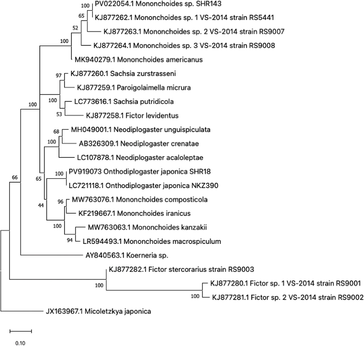
Figure 2:
Phylogenetic tree of Mononchoides sp. (SHR143) and Onthodiplogaster japonica (SHR18) with 20 Diplogastrid taxa inferred from the 28S rDNA gene by using the Maximum Likelihood method under Tamura-Nei model. Numbers at the nodes represent support values in percent for 1000 bootstrap replicates.
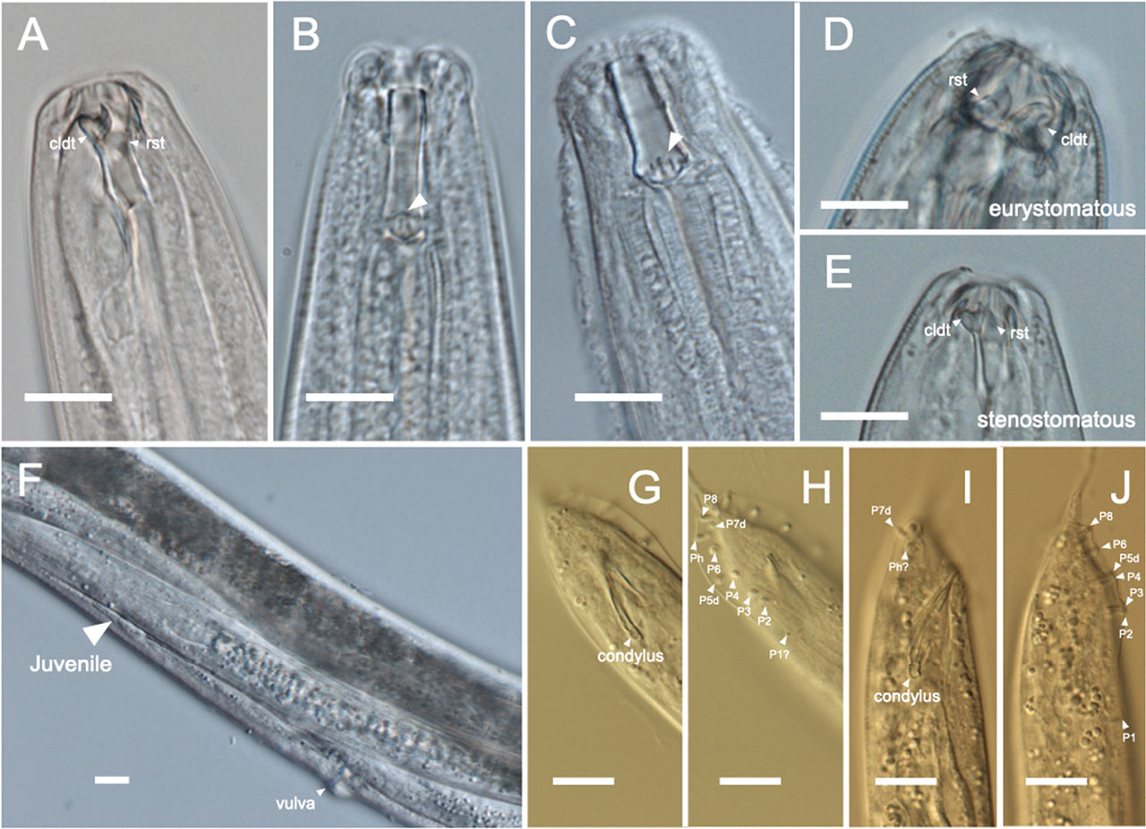
Figure 3:
Characteristic parts of nematode isolated in this study. Head regions of (A) Onthodiplogaster japonica (SHR18), (B) Pelodera sp. (SHR133), (C) Pelodera sp. (SHR150), and (D) eurystomatous form and (E) stenostomatous form of Mononchoides sp. (SHR143). (F) Ventral part and a juvenal of Tokorhabditis atripennis (SHR108). Mail tail regions of (G, H) Tokorhabditis atripennis (SHR108), and (I, J) Tokorhabditis tauri (SHR107). The white bars in these images are scale of 10 μm. cldt: claw-like dorsal tooth. rst: right subventral tooth.
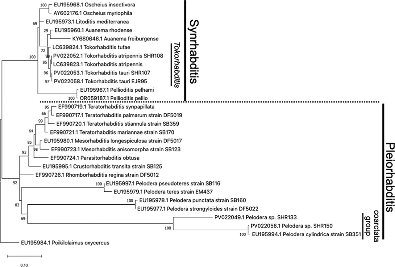
Figure 4:
Phylogenetic tree of Tokorhabditis spp. with 7 syn. Rhabditis taxa and Pelodera spp. with 9 Pleiorhabditis taxa inferred from the 28S rDNA gene by using the maximum likelihood method under GTR + G + I model. Numbers at the nodes represent support values in percent for 1,000 bootstrap replicates.
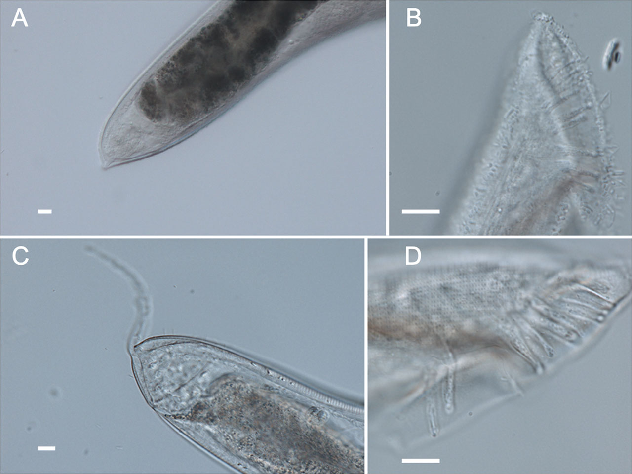
Figure 5:
Tail regions of Pelodera (coarctata group) isolated in this study. (A) The female tail of SHR133. (B) The male tail of SHR133. (C) The female tail of SHR150. (D) The male tail of SHR150. The white bars in these images are scale of 10 μm.
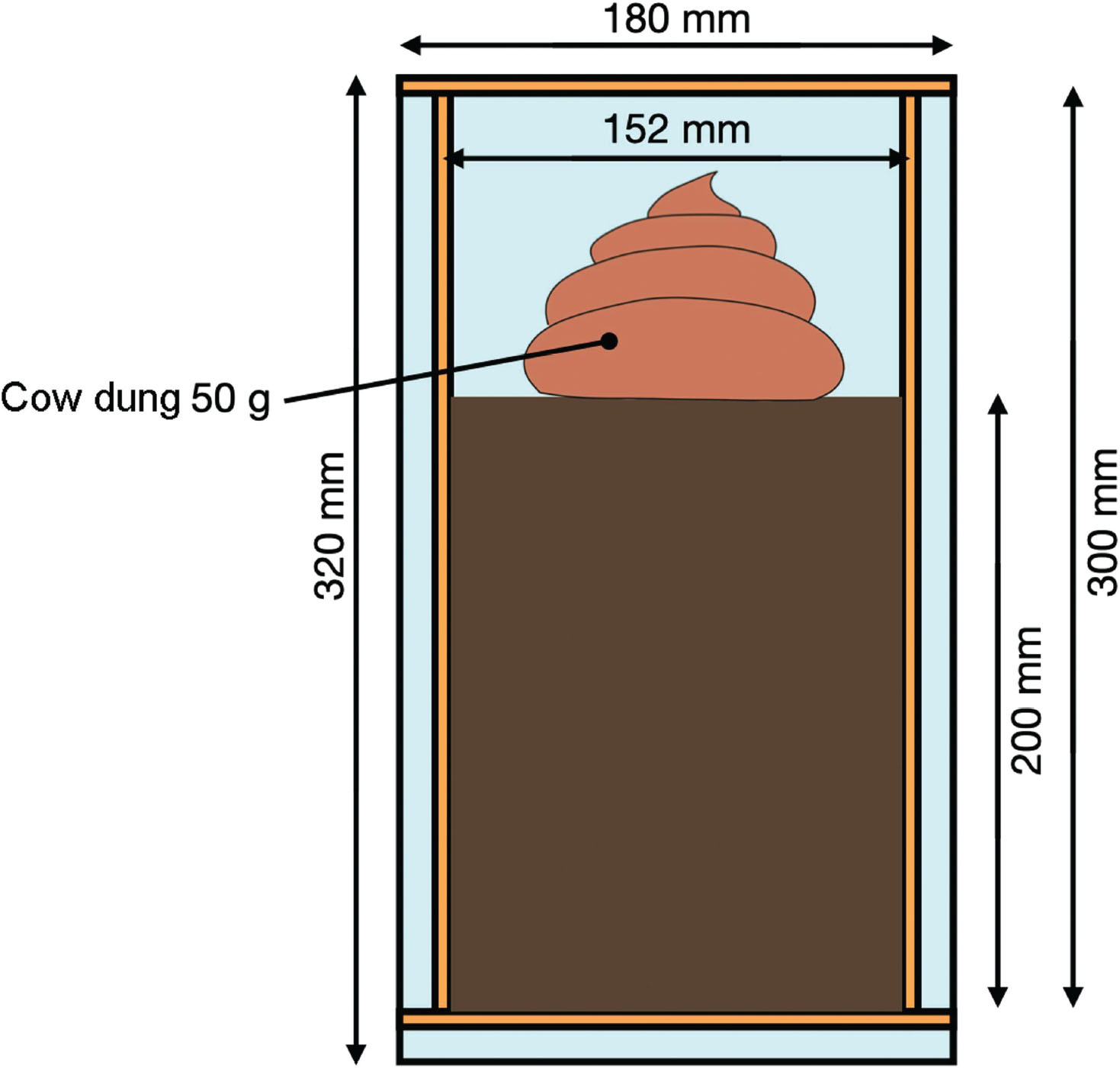
Figure S1:
The dimension of dung beetles’ rearing case.
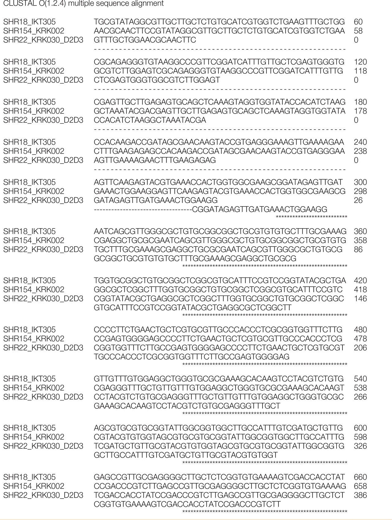
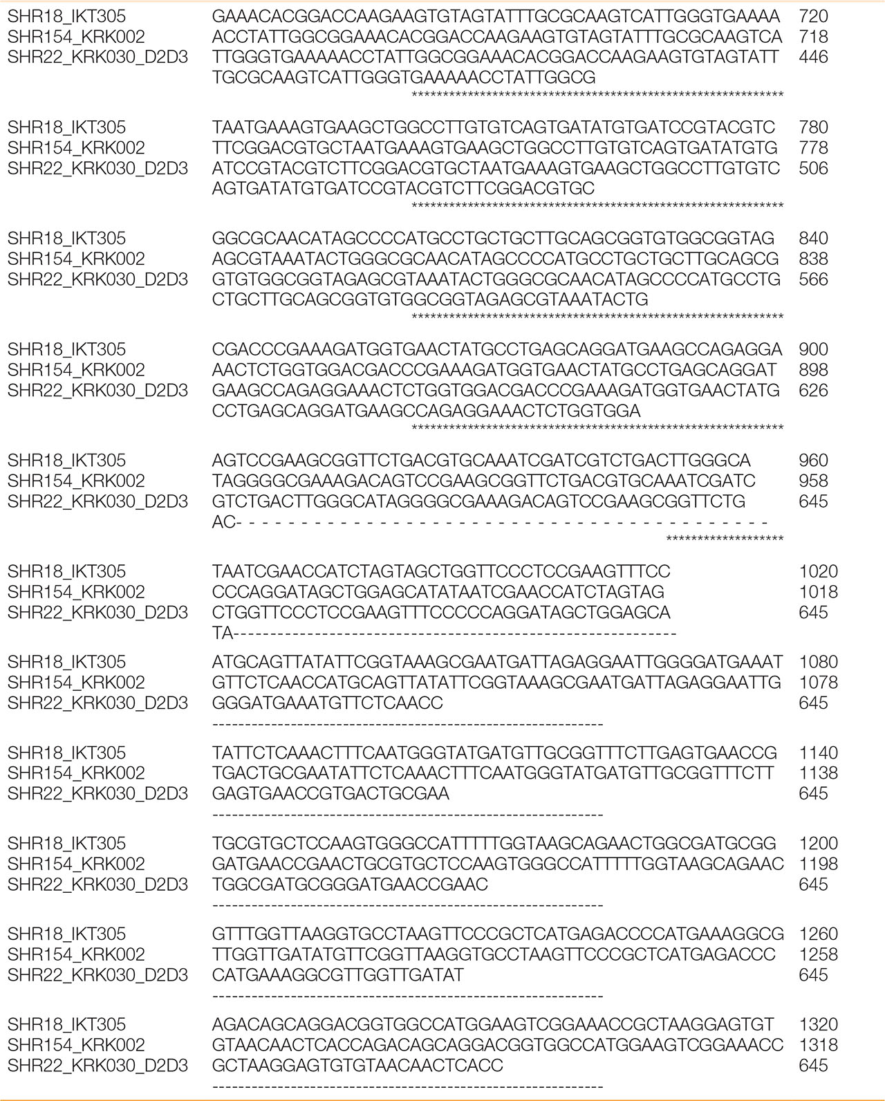

Figure S2:
The alignment result of partial 28S rDNA region of the three strains, SHR18, 27, and 154.
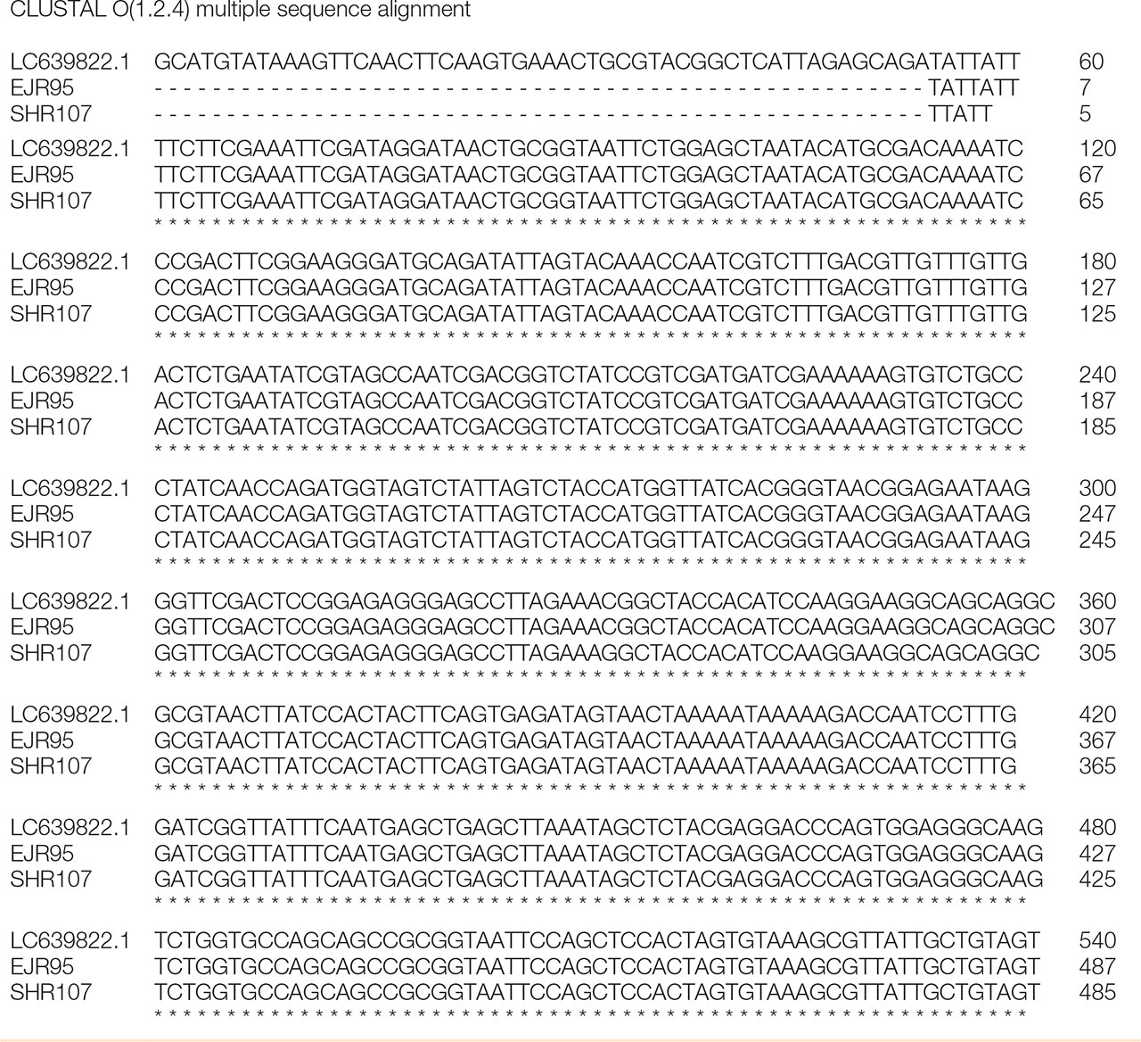
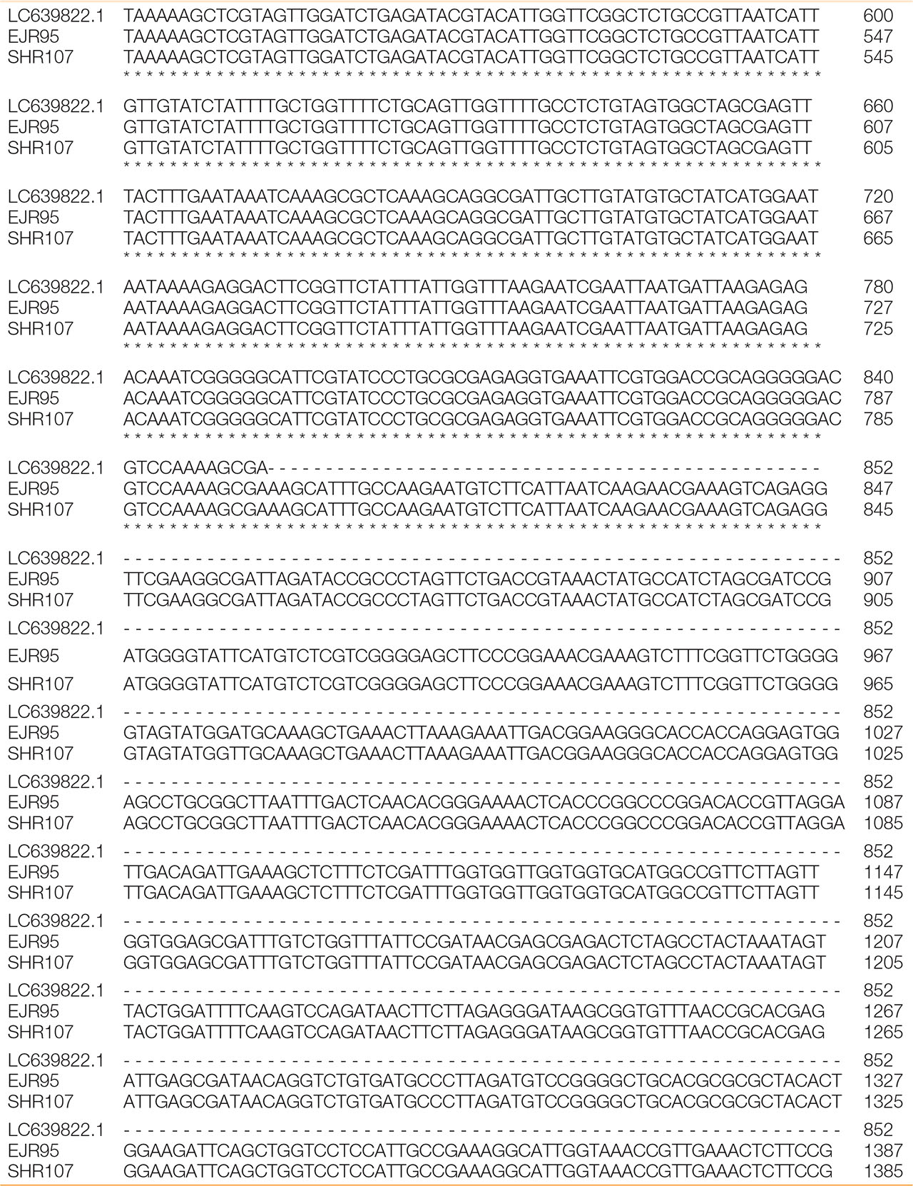
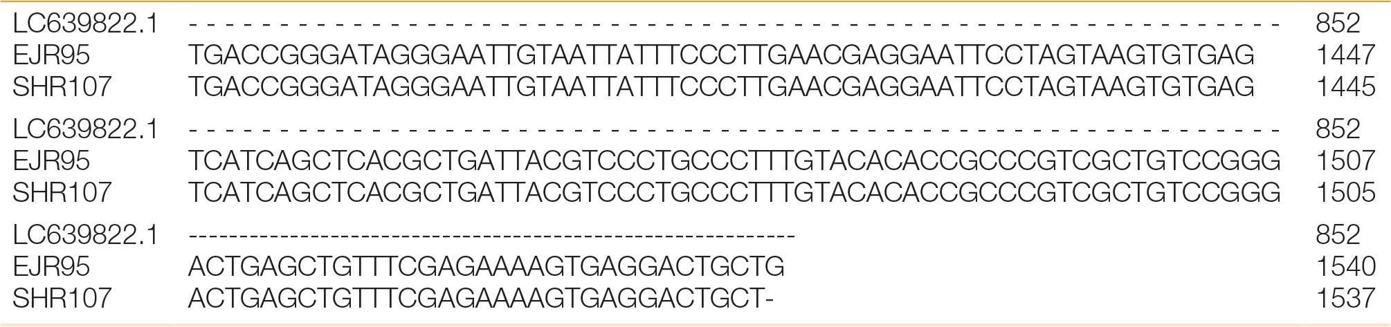
Figure S3:
The alignment result of partial 18S rDNA region of the three strains, EJR13, 95, and SHR107.
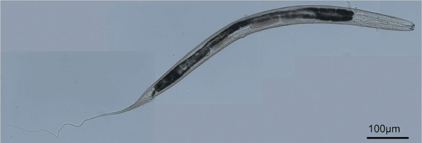
Figure S4:
The overall view of Mononchoides sp., strain SHR143.