
Figure 1:
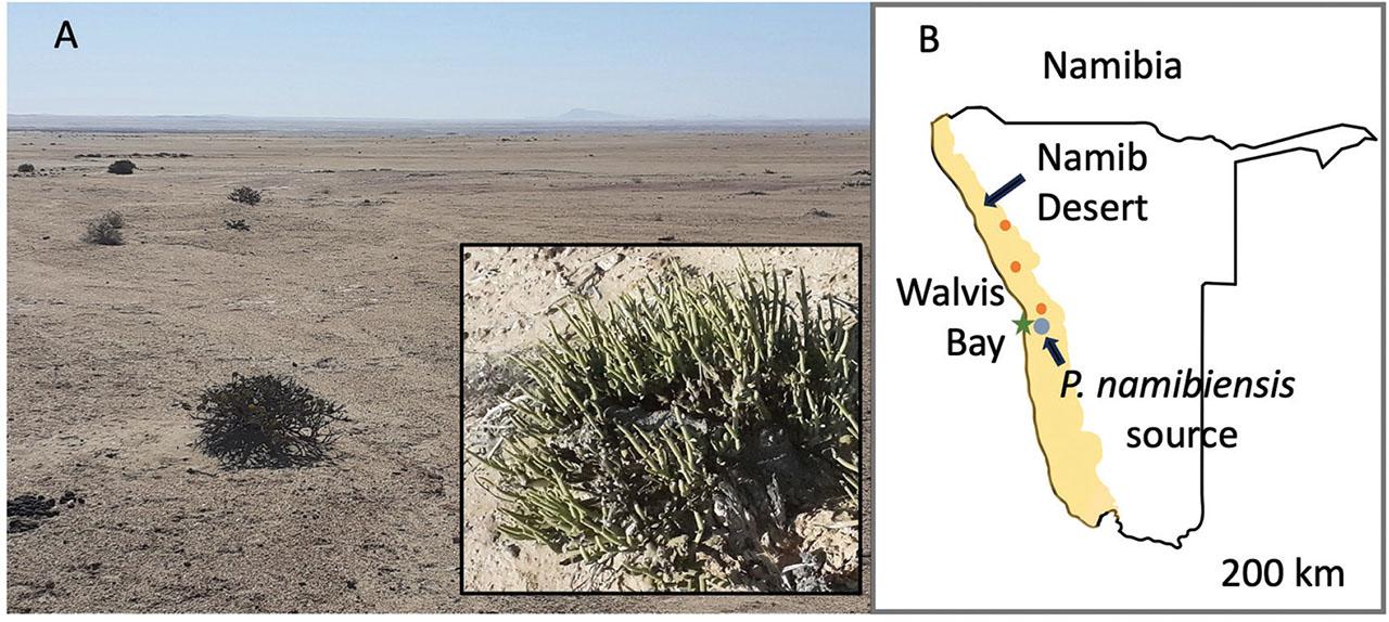
Figure 2:
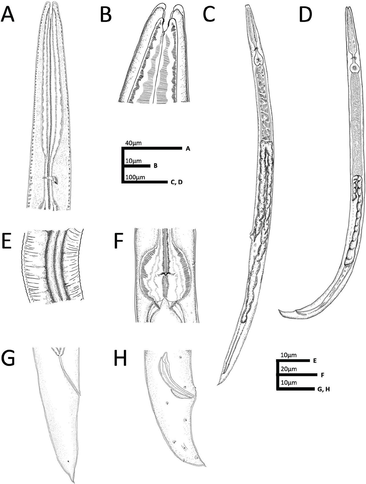
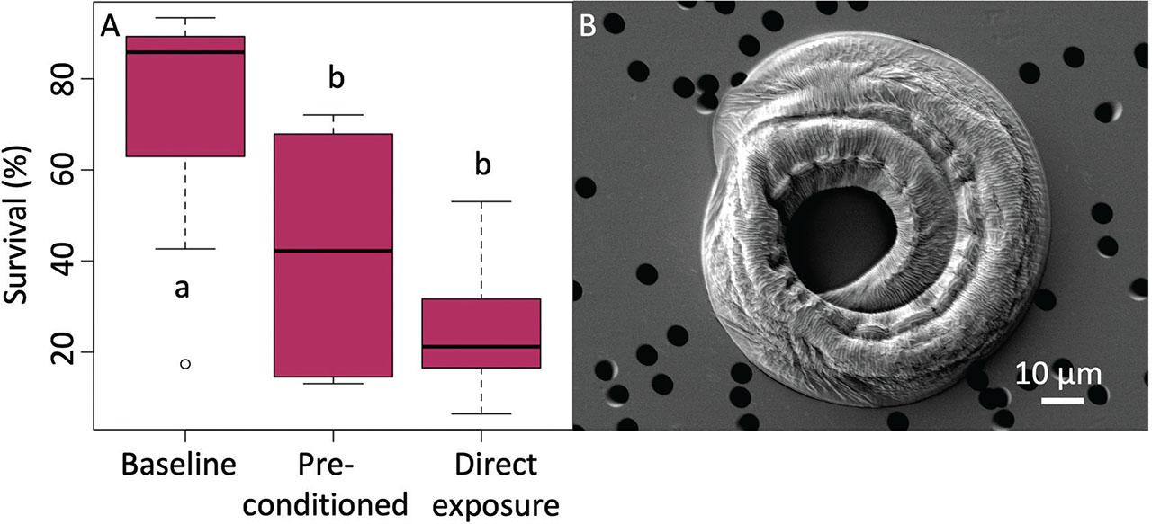
Figure 3:
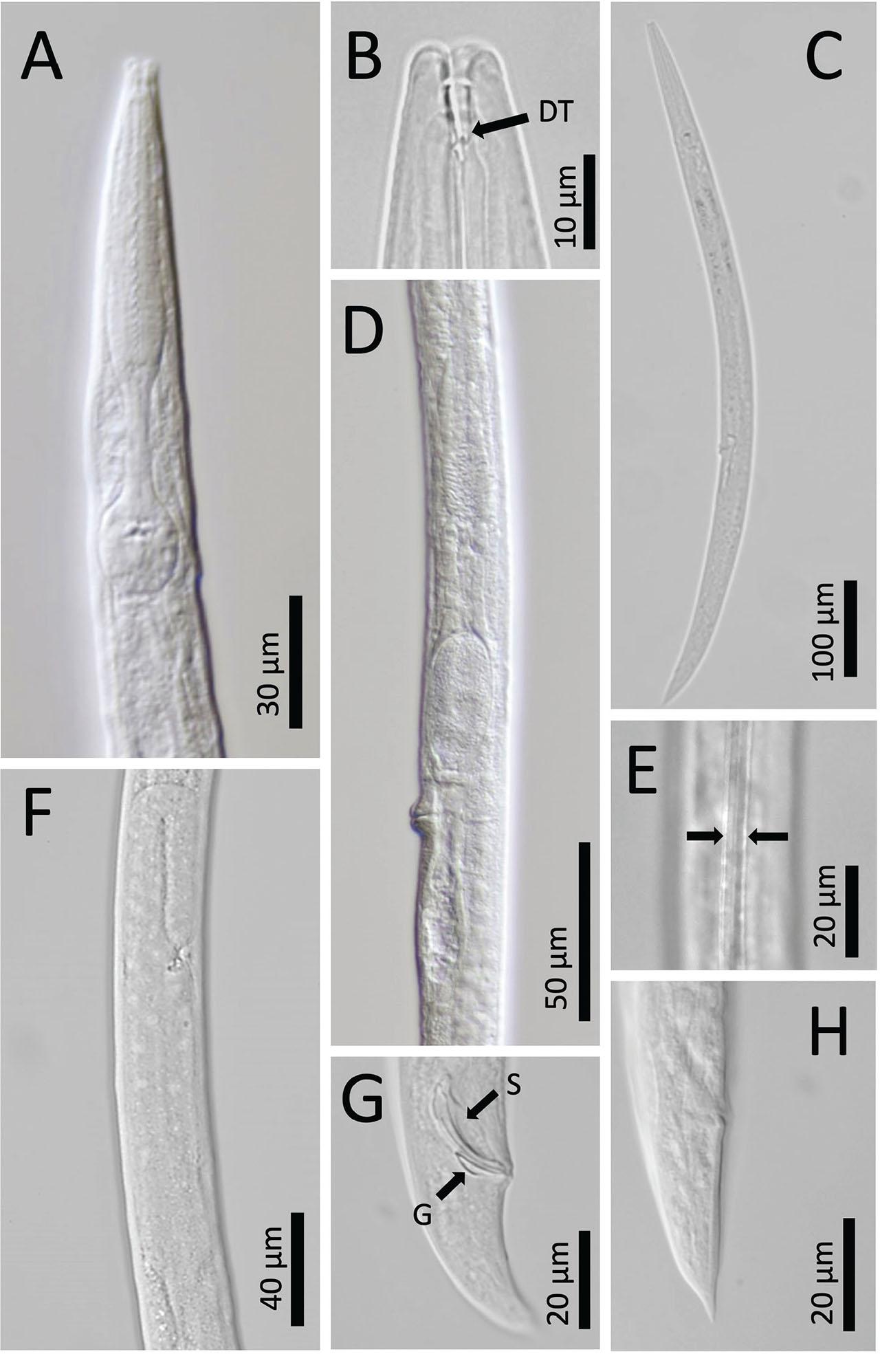
Figure 4:
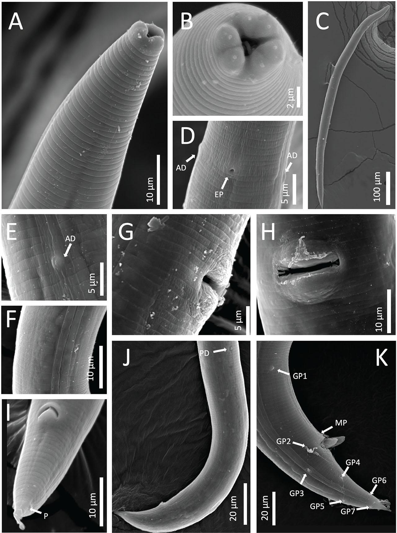
Figure 5:
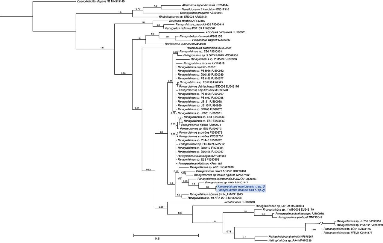
Figure 6:
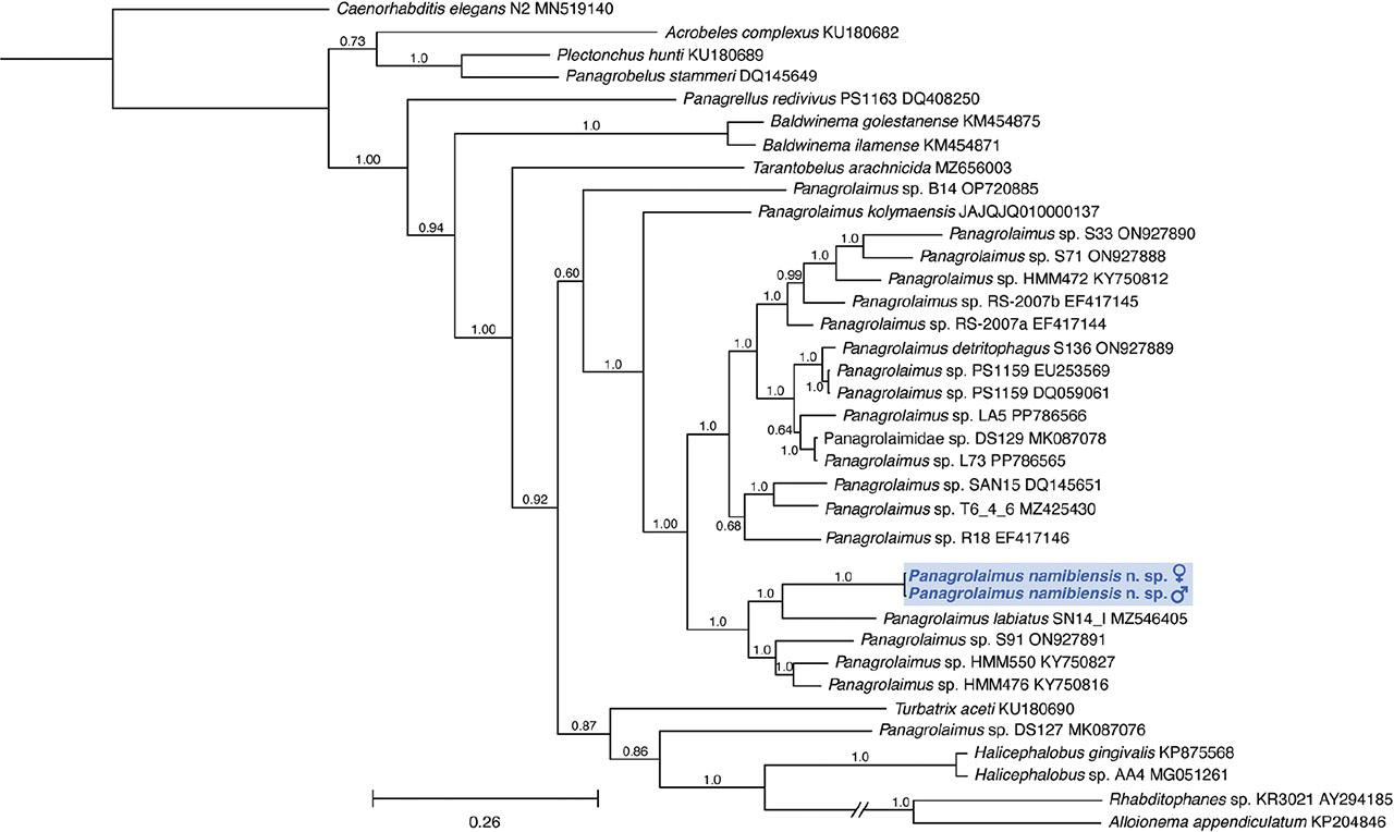
Figure 7:
j_jofnem-2024-0039_tab_002
| SSU rDNA | LSU rDNA D2-D3 | ||
|---|---|---|---|
| Taxon Name | Accession Number | Taxon Name | Accession Number |
| Acrobeles complexus | KU180671 | Acrobeles complexus | KU180682 |
| Alloionema appendiculatus | KP204844 | Alloionema appendiculatum | KP204846 |
| Baldwinema ilamense | KM454870 | Baldwinema golestanense | KM454875 |
| Baujardia mirabilis | AF547385 | Baldwinema ilamense | KM454871 |
| Caenorhabditis elegans N2 | MN519140 | Caenorhabditis elegans N2 | MN519140 |
| Halicephalobus gingivalis | KP875567 | Halicephalobus gingivalis | KP875568 |
| Halicephalobus sp. AA4 | MF470238 | Halicephalobus sp. AA4 | MG051261 |
| Neoalloionema tricaudatum | KR817916 | Panagrellus redivivus PS1163 | DQ408250 |
| Panagrellus redivivus PS1163 | AF083007 | Panagrobelus stammeri | DQ145649 |
| Panagrobelus stammeri | AF202153 | Panagrolaimidae sp. DS129 | MK087078 |
| Panagrolaimidae sp. DS129 | MK087064 | Panagrolaimus detritophagus S136 | ON927889 |
| Panagrolaimus sp. ES6 | FJ590961 | Panagrolaimus kolymaensis | JAJQJQ010000137 |
| Panagrolaimus artyukhovskii | MK636578 | Panagrolaimus labiatus SN14 | MZ546405 |
| Panagrolaimus davidi AC_Pd2 | HQ270131 | Panagrolaimus sp. B14 | OP720885 |
| Panagrolaimus davidi | FJ590981 | Panagrolaimus sp. DS127 | MK087076 |
| Panagrolaimus detritophagus | FJ590980 | Panagrolaimus sp. HMM472 | KY750812 |
| Panagrolaimus detritophagus BS0008 | EU543176 | Panagrolaimus sp. HMM476 | KY750816 |
| Panagrolaimus facetus | KY119819 | Panagrolaimus sp. HMM550 | KY750827 |
| Panagrolaimus kolymaensis | JAJQJQ010000793 | Panagrolaimus sp. L73 | PP786565 |
| Panagrolaimus labiatus SN14 | MW412913 | Panagrolaimus sp. LA5 | PP786566 |
| Panagrolaimus paetzoldi 452 | FJ040414 | Panagrolaimus sp. PS1159 | EU253569 |
| Panagrolaimus paetzoldi | ON713642 | Panagrolaimus sp. PS1159 | DQ059061 |
| Panagrolaimus sp. isolate rigidus1 | MK547102 | Panagrolaimus sp. R18 | EF417146 |
| Panagrolaimus rigidus | FJ590974 | Panagrolaimus sp. RS-2007a | EF417144 |
| Panagrolaimus sp. 10 ARA-2018 | MH399798 | Panagrolaimus sp. RS-2007b | EF417145 |
| Panagrolaimus sp. 4164 | MK301117 | Panagrolaimus sp. S33 | ON927890 |
| Panagrolaimus sp. AS01 | KC522708 | Panagrolaimus sp. S71 | ON927888 |
| Panagrolaimus sp. DL0117 | FJ590986 | Panagrolaimus sp. S91 | ON927891 |
| Panagrolaimus sp. DL0128 | FJ590987 | Panagrolaimus sp. SAN-15 | DQ145651 |
| Panagrolaimus sp. DL0139 | FJ590989 | Panagrolaimus sp. T6_4_6 | MZ425430 |
| Panagrolaimus sp. ES1 | FJ590960 | Plectonchus hunti | KU180689 |
| Panagrolaimus sp. ES2 | FJ590962 | Rhabditophanes sp. KR3021 | AY294185 |
| Panagrolaimus sp. ES3 | FJ590963 | Tarantobelus arachnicida | MZ656003 |
| Panagrolaimus sp. ES5 | FJ590972 | Turbatrix aceti | KU180690 |
| Panagrolaimus sp. 3 GVDU-2019 | MN082330 | ||
| Panagrolaimus sp. JB051 | FJ590971 | ||
| Panagrolaimus sp. JB115 | FJ590969 | ||
| Panagrolaimus sp. JB131 | FJ590968 | ||
| Panagrolaimus sp. JU765 | FJ590956 | ||
| Panagrolaimus sp. PS1159 | U81579 | ||
| Panagrolaimus sp. PS1159 | FJ590977 | ||
| Panagrolaimus sp. PS1162 | FJ590958 | ||
| Panagrolaimus sp. PS1579 | FJ590976 | ||
| Panagrolaimus sp. PS1732 | FJ590959 | ||
| Panagrolaimus sp. PS1806 | FJ590957 | ||
| Panagrolaimus sp. PS3966 | FJ590983 | ||
| Panagrolaimus sp. PS443 | KC522712 | ||
| Panagrolaimus sp. PS443 | FJ590978 | ||
| Panagrolaimus sp. SN103 | FJ590970 | ||
| Panagrolaimus subelongatus | AY284681 | ||
| Panagrolaimus superbus | KC522707 | ||
| Panagrolaimus superbus | FJ590973 | ||
| Panagrolaimus trilabiatus | KF011487 | ||
| Plectonchus wyganti | KJ636307 | ||
| Procephalobus sp. 1 WB-2008 | EU543179 | ||
| Propanagrolaimus sp. LC91 | KJ434175 | ||
| Propanagrolaimus sp. WTM1 | KJ434176 | ||
| Rhabditophanes sp. KR3021 | AF202151 | ||
| Strongyloides procyonis | AB205054 | ||
| Tarantobelus arachnicida | MZ655999 | ||
| Turbatrix aceti | KU180673 | ||
Measurements of Panagrolaimus namibiensis n_ sp_ from the gravel plains of the Namib Desert of Namibia_ Measurement units are μm ± the standard deviation, with the range in parentheses_
| Character | Females | Males | |
|---|---|---|---|
| Holotype | Paratypes | Paratypes | |
| n | - | 12 | 13 |
| L | 900.8 | 897.8 ± 44.9 (831.5–976.7) | 764.6 ± 49.3 (717.7–853.0) |
| a | 22.0 | 21.1 ± 3.0 (16.5–25.2) | 22.0 ± 1.9 (18.0–25.3) |
| b | 5.9 | 6.2 ± 0.4 (5.7–7.0) | 5.7 ± 0.3 (5.2–6.3) |
| c | 17.5 | 21.5 ± 1.3 (19.3–23.6) | 19.5 ± 1.6 (17.5–22.4) |
| c’ | 1.9 | 1.8 ± 0.2 (1.4–2.1) | 1.6 ± 0.2 (1.3–1.9) |
| V (%) | 60.9 | 59.3 ± 1.5 (57.2 – 61.6) | - |
| Lip region width | 8.9 | 8.5 ± 0.5 (7.7–9.3) | 7.5 ± 0.5 (7.0–8.6) |
| Stoma length | 11.4 | 12.1 ± 0.8 (10.9–13.3) | 11.5 ± 0.9 (10.4–12.8) |
| Stoma width | 2.2 | 2.4 ± 0.2 (2.1–2.8) | 2.2 ± 0.2 (1.9–2.7) |
| Corpus length | 89.7 | 88.3 ± 3.4 (81.7–92.6) | 79.9 ± 5.6 (69.8–90.9) |
| Corpus width | 19.7 | 19.7 ± 1.8 (17.5–23.6) | 15.8 ± 2.6 (12.4–21.1) |
| Isthmus | 27.7 | 28.9 ± 2.0 (25.3–32.6) | 29.0 ± 1.9 (25.5–32.9) |
| Basal bulb | 27.2 | 27.1 ± 2.1 (24.8–31.3) | 24.7 ± 2.3 (22.5–29.4) |
| Pharynx length | 144.6 | 144.9 ± 4.9 (135.2–152.2) | 133.6 ± 6.8 (123.9–149.0) |
| Neck | 152.8 | 154.8 ± 6.1 (145.9–166.1) | 140.6 ± 7.2 (131.0–155.9) |
| Neck-base body diameter | 36.4 | 38.7 ± 4.6 (32.9–47.8) | 33.2 ± 4.5 (25.0–40.4) |
| Nerve ring - ant. end | 115.0 | 115.3 ± 7.1 (102.2–127.4) | 105.7 ± 9.1 (91.7–123.2) |
| Excretory pore - ant. end | 141.0 | 141.1 ± 11.3 (115.1–157.6) | 136.0 ± 12.2 (115.4–157.0) |
| Deirid - ant. end | 141.0 | 141.7 ± 11.3 (115.4–157.1) | 136.5 ± 12.6 (115.4–157.6) |
| Mid-body diameter | 40.9 | 43.4 ± 5.8 (34.6–55.4) | 35.1 ± 4.2 (29.5–41.9) |
| Cuticle annuli thickness | 1.2 | 1.2 ± 0.1 (1.1–1.4) | 1.2 ± 0.1 (1.0–1.4) |
| Lateral field | 3.7 | 3.7 ± 0.4 (3.2–4.3) | 3.5 ± 0.4 (3.1–4.2) |
| Vulva - ant. end | 548.5 | 532.3 ± 26.5 (499.0–572.8) | - |
| Vulval body diameter | 41.6 | 44.1 ± 5.1 (35.9–50.5) | - |
| Vulva-anus distance | 298.2 | 302.6 ± 14.8 (275.2–321.1) | - |
| Vagina length | 13.7 | 14.2 ± 1.5 (11.3–16.5) | - |
| Ovary length | 342.3 | 389.7 ± 30.0 (326.8–430.3) | - |
| Post-vulval sac | 25.5 | 21.9 ± 3.4 (16.5–27.2) | - |
| Rectum length | 27.9 | 25.8 ± 2.8 (20.1–30.3) | - |
| Testes length | - | - | 207.8 ± 22.3 (184.1–254.9) |
| Spicule length | - | - | 29.3 ± 2.0 (24.5–31.6) |
| Gubernaculum length | - | - | 12.6 ± 1.5 (10.9–15.4) |
| Tail | 51.4 | 42.3 ± 2.3 (39.2–45.9) | 39.4 ± 3.6 (32.5–45.9) |
| Phasmid-anus distance | 23.1 | 18.5 ± 3.5 (13.3–24.2) | 18.9 ± 3.7 (14.3–28.7) |
| Anal body diameter | 26.8 | 23.4 ± 2.9 (20.4–31.0) | 24.7 ± 2.9 (20.8–29.3) |