
Figure 1:
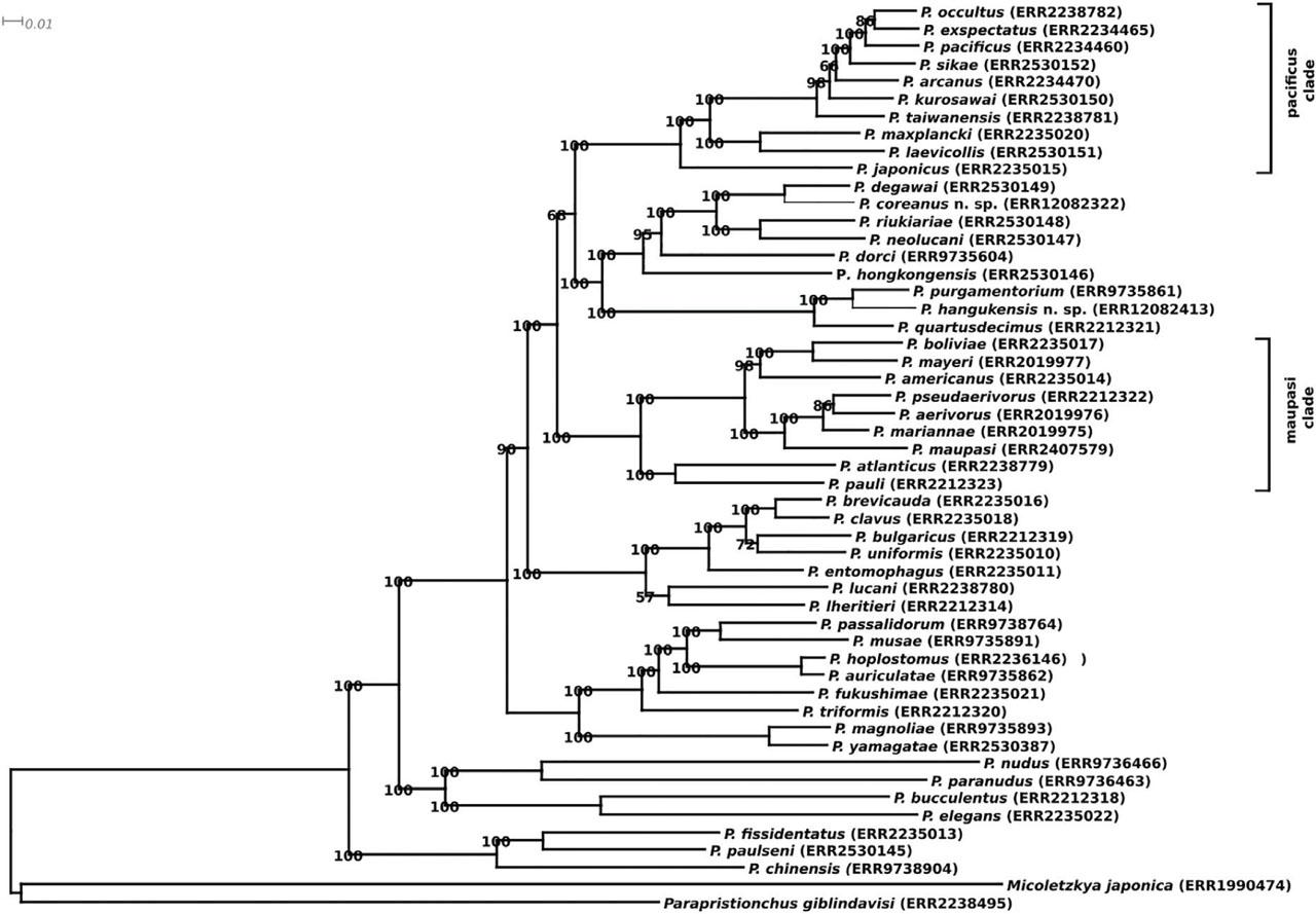
Figure 2:
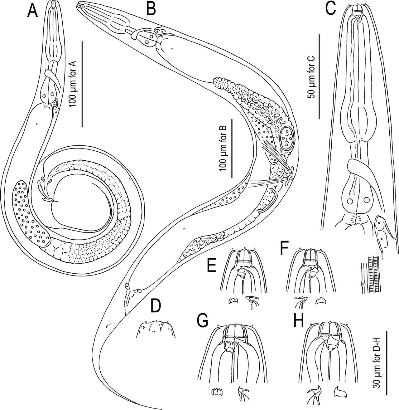
Figure 3:
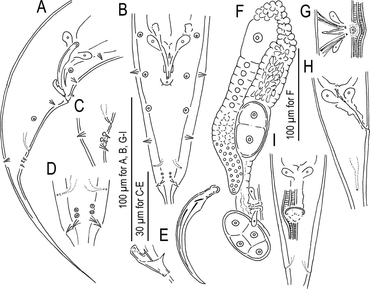
Figure 4:
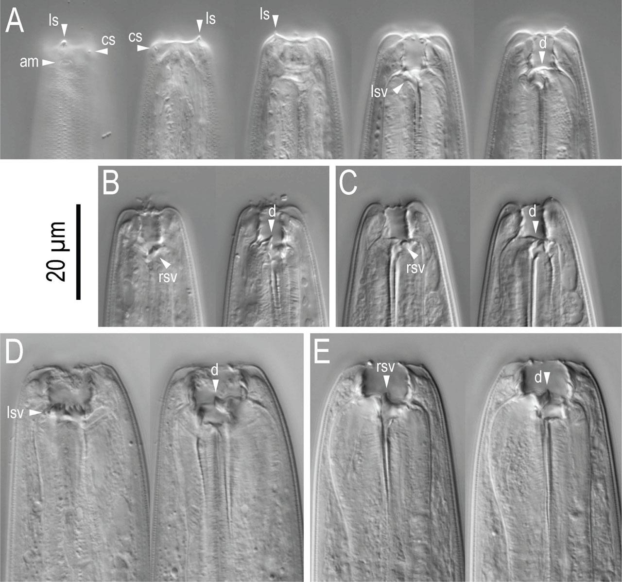
Figure 5:
Figure 6:
Figure 7:
Figure 8:
Figure 9:
Figure 10:
Supplemental Figure 1:
Morphological measurements of the two new species_
| Character | Pristionchus coreanus n.sp. RS6268 | Pristionchus hangukensis n.sp. RS6291 | ||
|---|---|---|---|---|
| stenostomatous male | stenostomatous female | stenostomatous male | stenostomatous female | |
| n | 10 | 10 | 10 | 10 |
| L | 1002 ± 94.3 (872 – 1158) | 1448 ± 126.0 (1312 – 1661) | 928 ± 54.8 (858 – 1001) | 1336 ± 90.2 (1248 – 1542) |
| L’ | 851 ± 93.3 (721 – 1025) | 1211 ± 120.4 (1067 – 1442) | 764 ± 41.0 (685 – 820) | 1078 ± 76.5 (993 – 1249) |
| a | 13 ± 2.2 (9.1 – 16) | 12 ± 1.5 (11 – 16) | 13 ± 1.3 (11 – 16) | 13 ± 0.7 (11 – 14) |
| b | 6.0 ± 0.4 (5.5 – 6.7) | 7.6 ± 0.5 (6.8 – 8.4) | 7.0 ± 0.4 (6.5 – 7.6) | 9.0 ± 0.7 (8.0 – 10.0) |
| c | 6.7 ± 0.9 (5.7 – 8.5) | 6.1 ± 0.5 (5.4 – 7.1) | 5.8 ± 0.4 (5.3 – 6.6) | 5.3 ± 0.5 (4.9 – 6.3) |
| c’ | 3.4 ± 0.5 (2.5 – 4.0) | 4.4 ± 0.6 (3.8 – 5.5) | 4.4 ± 0.5 (3.9 – 5.3) | 6.4 ± 0.6 (5.5 – 7.7) |
| Ant. stoma length (cheilo- + gymnostom) | 5.1 ± 0.8 (4.0 – 6.7) | 5.7 ± 1.4 (4.0 – 7.8) | 6.9 ± 1.1 (5.0 – 9.3) | 7.7 ± 0.7 (6.7 – 9.1) |
| Total stoma length | 11 ± 1.0 (9.3 – 12) | 12 ± 1.1 (11 – 14) | 10 ± 1.1 (8.3 – 11.6) | 13 ± 1.0 (11 – 14) |
| Stoma width | 5.8 ± 0.4 (5.3 – 6.5) | 7.5 ± 0.9 (6.0 – 8.7) | 4.9 ± 0.9 (3.6 – 6.5) | 5.4 ± 0.9 (4.3 – 7.5) |
| Ant. pharynx (pro + metacorpus) | 95 ± 7.6 (83 – 111) | 109 ± 8.2 (99 – 130) | 69 ± 5.6 (63 – 82) | 79 ± 6.0 (72 – 89) |
| Post. pharynx (isthmus + basal bulb) | 67 ± 7.0 (56 – 78) | 77 ± 5.1 (69 – 87) | 57 ± 2.9 (54 – 61) | 61 ± 3.6 (55 – 66) |
| Total pharynx length | 162 ± 13.6 (142 – 183) | 186 ± 12.1 (176 – 217) | 126 ± 7.6 (120 – 143) | 140 ± 7.4 (131 – 152) |
| Ant./total pharynx % | 59 ± 1.7 (57 – 62) | 58 ± 1.5 (56 – 62) | 55 ± 1.6 (52 – 57) | 57 ± 2.2 (54 – 60) |
| Median bulb diameter | 27 ± 2.5 (24 – 32) | 35 ± 2.9 (31 – 40) | 23 ± 1.2 (21 – 24) | 27 ± 2.0 (25 – 31) |
| Terminal bulb diameter | 28 ± 3.9 (22 – 36) | 36 ± 4.9 (31 – 46) | 22 ± 1.1 (20 – 23) | 27 ± 2.3 (24 – 33) |
| Ant. end to cardia | 167 ± 13.2 (151 – 191) | 192 ± 8.8 (185 – 212) | 132 ± 8.4 (123 – 148) | 148 ± 7.5 (139 – 160) |
| Ant. end to S-E pore | 149 ± 15.5 (125 – 170) | 195 ± 15.5 (171 – 214) | 114 ± 11.3 (99 – 139) | 153 ± 12.0 (137 – 172) |
| Ant. end to nerve ring | 118 ± 9.2 (109 – 133) | 136 ± 8.9 (124 – 155) | 97 ± 6.2 (90 – 108) | 111 ± 6.5 (104 – 124) |
| Testis length | 703 ± 83.8 (571 – 827) | - | 630 ± 59.4 (529 – 718) | - |
| Ant. end to vulva distance | - | 691 ± 66.1 (600 – 821) | - | 608 ± 40.2 (563 – 703) |
| Vulva to anus distance | - | 525 ± 60.0 (454 – 628) | - | 473 ± 37.3 (420 – 543) |
| T or V | 70 ± 4.3 (66 – 80) | 48 ± 1.5 (46 – 50) | 68 ± 6.7 (53 – 77) | 46 ± 1.1 (43 – 47) |
| Max. body diameter | 78 ± 19.1 (63 – 127) | 121 ± 16.4 (91 – 141) | 71 ± 5.1 (64 – 81) | 105 ± 8.1 (93 – 115) |
| Cloacal or anal body diameter | 45 ± 5.3 (40 – 54) | 54 ± 5.3 (43 – 63) | 36 ± 2.2 (33 – 39) | 39 ± 3.5 (33 – 45) |
| Tail length | 149 ± 10.8 (129 – 161) | 237 ± 23.7 (208 – 296) | 159 ± 13.9 (132 – 178) | 252 ± 27.0 (204 – 297) |
| Spicule curve | 50 ± 4.3 (40 – 54) | - | 46 ± 2.3 (43 – 50) | - |
| Spicule chord | 42 ± 2.9 (36 – 47) | - | 38 ± 2.6 (35 – 43) | - |
| Gubernaculum length | 18 ± 2.4 (13 – 21) | - | 18 ± 1.6 (16–22) | - |